Aditya College of Pharmacy Beed
The investigation of phytoconstituents plays a pivotal role in the identification and validation of bioactive compounds from medicinal plants. Chromatographic and spectroscopic techniques are essential tools for the qualitative and quantitative analysis of these natural products. This study explores the application of various chromatographic methods—such as Thin Layer Chromatography (TLC), High-Performance Liquid Chromatography (HPLC), and Gas Chromatography (GC)—in the separation and isolation of plant-derived compounds. In addition, spectroscopic techniques including Ultraviolet-Visible (UV-Vis) spectroscopy, Fourier Transform Infrared (FTIR) spectroscopy, Nuclear Magnetic Resonance (NMR), and Mass Spectrometry (MS) are employed to elucidate the structural characteristics of the isolated phytochemicals. The integrated use of these analytical methods ensures precise identification, contributing to pharmacological research, quality control, and the standardization of herbal formulations. This paper highlights the significance of combining chromatographic and spectroscopic approaches for comprehensive phytochemical analysis.
Nature has long been a prolific source of medicinal agents. Traditional medicine systems Across the world—such as Ayurveda, Traditional Chinese Medicine, and Unani—are rooted In the therapeutic applications of plant-derived substances. These natural compounds, known As phytoconstituents, include alkaloids, flavonoids, terpenoids, glycosides, phenolics, and Tannins, among others. Historically used in crude forms, these phytoconstituents are now Increasingly isolated and characterized to understand their chemical nature and therapeutic Potential. Modern pharmaceutical research has shifted focus towards exploring these Bioactive plant components not only for their individual therapeutic value but also for their Synergistic effects when used in combination. As synthetic drug development faces Challenges such as drug resistance, side effects, and high development costs, Phytoconstituents offer a promising alternative or complementary approach in the discovery and development of novel therapeutic agents.
Importance of Phytoconstituents In Drug
Phytoconstituents have significantly contributed to modern drug discovery. Many current Pharmaceuticals—including aspirin (from salicylic acid), morphine (from opium poppy), and Artemisinin (from Artemisia annua)—have originated from plants. Their diverse chemical Structures and biological activities make them invaluable leads in drug development Pipelines. These natural compounds often possess multitargeted bioactivity, which is Especially advantageous in treating complex diseases such as cancer, diabetes, and Neurodegenerative disorders. Additionally, their relatively lower toxicity profiles and eco-Friendly sourcing have further highlighted their importance. As the demand for safer and More effective therapies grows, the systematic exploration of phytoconstituents has gained Renewed attention in both academic and industrial settings.
Need for Chromatographic And Spectroscopic Techniques: -
The structural diversity and complexity of phytoconstituents require precise, sensitive, and Reliable analytical techniques for their identification, quantification, and characterization. Chromatographic techniques, such as High-Performance Liquid Chromatography (HPLC), Gas Chromatography (GC), and Thin Layer Chromatography (TLC), are widely employed for separating and quantifying complex plant extracts. These techniques help in isolating specific constituents based on their physicochemical properties, making them indispensable in phytochemical studies. On the other hand, spectroscopic techniques—including Ultraviolet-Visible (UV-Vis), Infrared (IR), Nuclear Magnetic Resonance (NMR), and Mass Spectrometry (MS)—enable the structural elucidation of phytoconstituents. When combined with chromatography (e.g., LC-MS, GC-MS), these techniques provide comprehensive insights into both qualitative and quantitative aspects of plant-derived compounds. The integration of chromatographic and spectroscopic methodologies ensures accuracy, reproducibility, and in-depth chemical profiling necessary for drug discovery and standardization of herbal medicines.
RESEARCH OBJECTIVES
The primary aim of this study is to explore and evaluate the application of chromatographic and spectroscopic techniques in the analysis of phytoconstituents. The specific objectives include:
? To understand the principles and working mechanisms of chromatographic and spectroscopic techniques commonly used in phytochemical analysis.
? To identify and characterize bioactive phytoconstituents in selected plant extracts using appropriate analytical methods.
? To assess the advantages and limitations of various techniques in the qualitative and quantitative analysis of phytoconstituents.
? To establish a methodological framework for the systematic study of medicinal plants with pharmaceutical relevance
Scope and Limitations of The Study
This study focuses on the analytical methodologies employed in the isolation and characterization of phytoconstituents from medicinal plants. It covers a range of chromatographic techniques (e.g., TLC, HPLC, GC) and spectroscopic techniques (e.g., UV-Vis, IR, NMR, MS) utilized in the detection and structural analysis of these compounds. While the study emphasizes practical and theoretical aspects of the analytical processes, it is limited by several factors:
?The availability of sophisticated instrumentation such as NMR and MS may constrain the depth of structural elucidation in some settings.
? Plant species selected for analysis are representative and not exhaustive, hence may not capture the full phytochemical spectrum found in traditional medicinal systems.
? The study does not cover the pharmacological or toxicological evaluation of the isolated compounds in vivo or in vitro, focusing instead on their analytical profiling.
MATERIALS AND METHODS
The plant materials selected for this study were collected from their natural habitat during the appropriate season to ensure optimal phytochemical content. Collection was carried out following standard botanical procedures, focusing on the parts known to contain high concentrations of bioactive compounds, such as leaves, roots, stems, or bark, depending on the species. After collection, the plant samples were carefully cleaned to remove dust, soil, and foreign particles. Authentication was conducted by a qualified botanist using morphological characteristics and comparing them with herbarium specimens. Voucher specimens were prepared and deposited in a recognized herbarium for future reference.

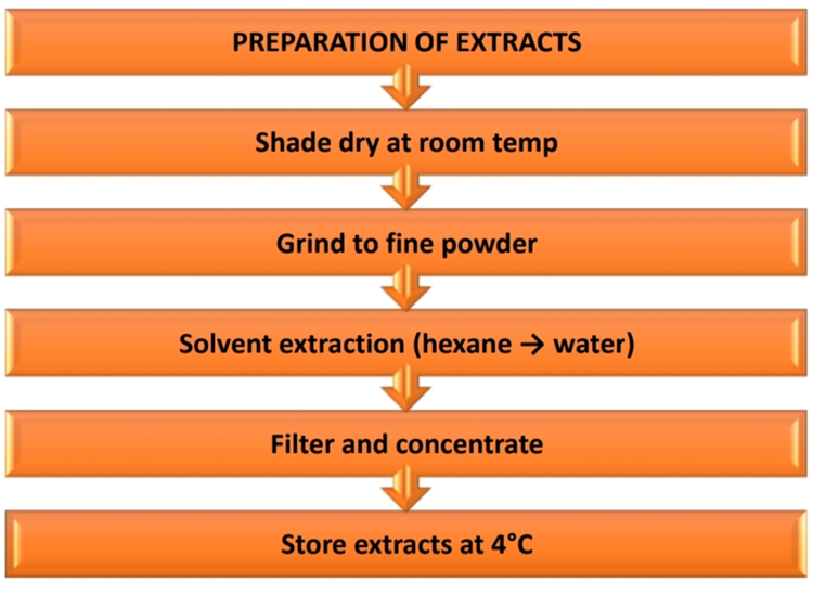
Preparation of Plant Extracts
Once authenticated, the plant materials were shade-dried at ambient temperature to preserve thermolabile compounds. The dried materials were pulverized using a mechanical grinder to obtain a uniform powder. Extraction was performed using solvents of increasing polarity (e.g., hexane, ethyl acetate, methanol, and water) through maceration or Soxhlet extraction, depending on the solubility of the targeted phytoconstituents. The extracts were filtered and concentrated under reduced pressure using a rotary evaporator. Dried extracts were stored in airtight containers at 4°C until further analysis.
Chromatographic Methods Employed
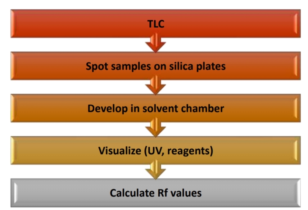
Thin Layer Chromatography (TLC)
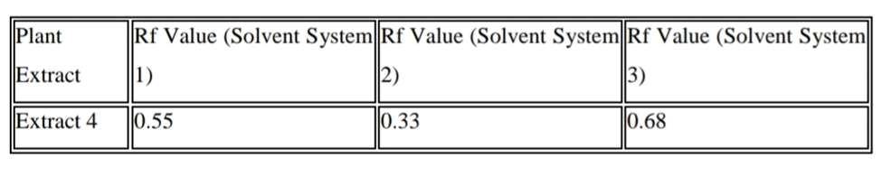
TLC was employed as a preliminary technique to assess the phytochemical profile and purity Of the extracts. Pre-coated silica gel plates were used as the stationary phase. A range of Solvent systems was optimized to achieve effective separation based on the nature of the Compounds. Extracts were applied as small spots, and the plates were developed in TLC Chambers under saturated conditions. After development, the chromatograms were visualized Under UV light (254 nm and 366 nm) and with suitable spray reagents (e.g., anisaldehyde-Sulfuric acid, ferric chloride) to detect specific classes of phytochemicals. Retention factor (Rf) values were calculated for comparative analysis.
High Performance Liquid Chromatography (HPLC)
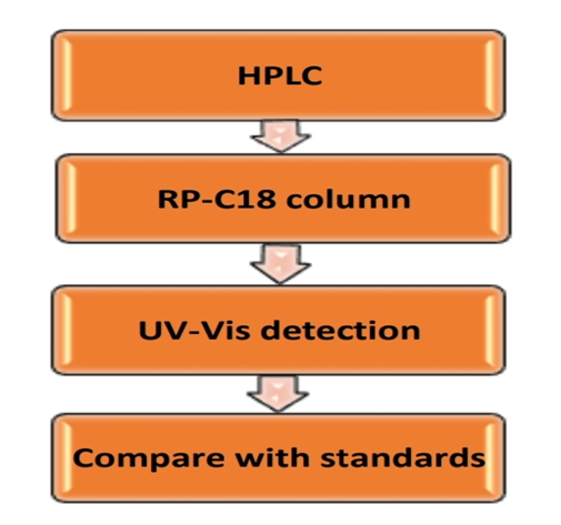
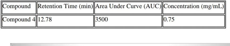
HPLC was utilized for quantitative and high-resolution separation of the bioactive constituents. A reverse-phase C18 column was typically used, with gradient or isocratic elution depending on the sample complexity. Mobile phases consisted of combinations of water, acetonitrile, and methanol, often with acidic modifiers like formic or phosphoric acid. Detection was carried out using a UV-Vis detector at wavelengths specific to the compounds of interest. Parameters such as flow rate, column temperature, and injection volume were optimized for each sample. Chromatographic peaks were identified by comparing retention times with those of reference standards.
Gas Chromatography-Mass Spectrometry (Gc-Ms)
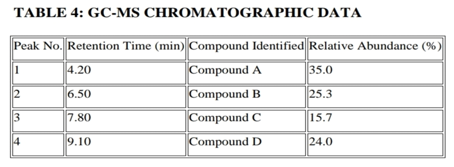
GC-MS analysis was applied primarily to the volatile and semi-volatile components in the extracts. Samples were subjected to derivatization if necessary to enhance volatility and stability.A capillary column coated with a non-polar or semi-polar stationary phase was used. Helium served as the carrier gas at a constant flow rate. The temperature program involved an initial hold followed by a ramp to higher temperatures, facilitating efficient elution of diverse compounds. The mass spectrometer was operated in electron ionization (EI) mode. Fragmentation patterns were matched against spectral libraries such as NIST or Wiley for compound identification.

SPECTROSCOPIC METHODS USED
Ultraviolet–Visible Spectroscopy (UV-VIS)
UV-Vis spectroscopy was used to detect conjugated systems and assess the overall phenolic or flavonoid content. Extracts were diluted appropriately and scanned across a wavelength range of 200–800 nm. Absorption maxima (λmax) were recorded, and standard calibration curves were constructed using reference compounds like gallic acid or quercetin for quantitative estimation. The technique provided insight into the electronic transitions of aromatic and polyphenolic constituents.

Fourier Transform Infrared Spectroscopy (FTIR)
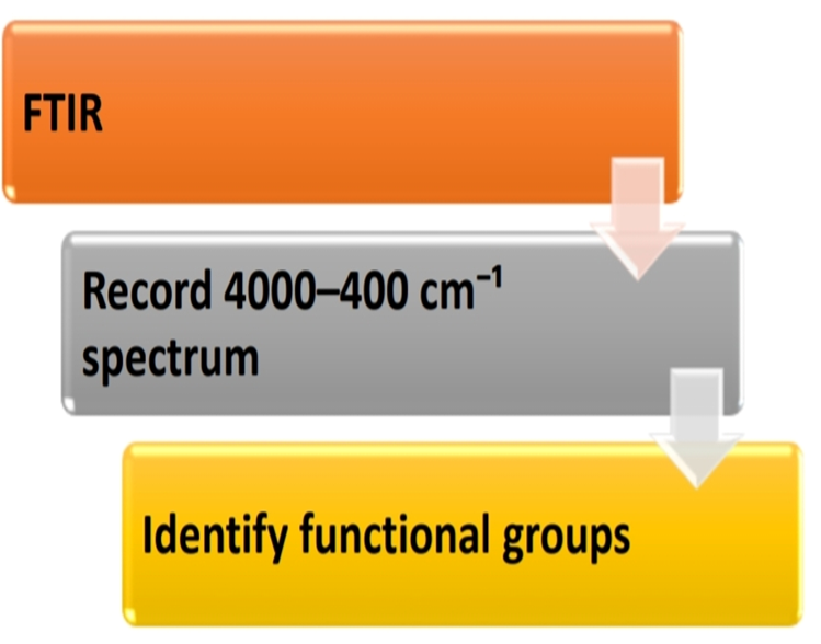
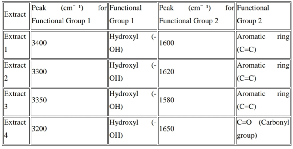
FTIR analysis was carried out to identify functional groups present in the phytoconstituents. Dried extracts or isolated compounds were mixed with potassium bromide (KBr) and pressed into translucent pellets.Spectra were recorded in the range of 4000–400 cm? ¹. Characteristic absorption bands were interpreted to assign structural features such as hydroxyl, carbonyl, ether, alkene, and aromatic groups. This method helped in the preliminary characterization of compound classes within the extracts.
Nuclear Magnetic Resonance Spectroscopy (NMR)
NMR spectroscopy was employed for detailed structural elucidation of isolated compounds. Samples were dissolved in deuterated solvents such as CDCl? or DMSO-d? , and ¹H and ¹³C spectra were recorded using a high-field NMR instrument. Chemical shifts (δ), coupling constants (J), and integration values were analyzed to determine the molecular framework. Two-dimensional techniques such as COSY, HSQC, and HMBC were utilized where necessary to establish proton–carbon connectivity and confirm structural assignments.

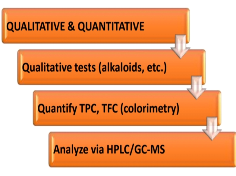
Quantitative and Qualitative Evaluation
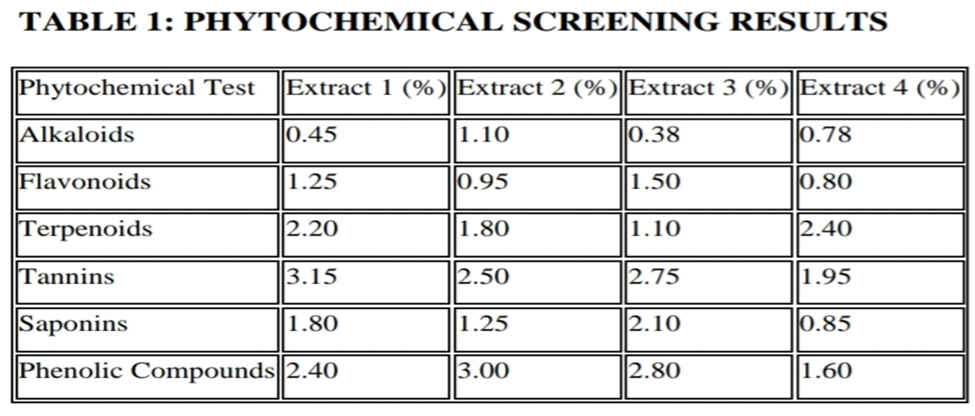
Qualitative analysis focused on identifying phytochemical classes using standard reagents and chromatographic/spectroscopic signatures. Tests for alkaloids, flavonoids, tannins, saponins, and terpenoids were conducted using established protocols. Quantitative assays included determination of total phenolic content (TPC) and total flavonoid content (TFC) through colorimetric methods. Concentrations of individual compounds were obtained through HPLC or GC-MS based on peak area comparison with calibration standards. The combined data provided both an overview and a detailed breakdown of the phytoconstituent composition.
Statistical Tools and Data Interpretation
All experiments were conducted in triplicate to ensure reliability. Statistical analysis was performed using software such as Microsoft Excel, SPSS, or GraphPad Prism. Data were expressed as mean ± standard deviation (SD), and differences between sample groups were analyzed using appropriate tests such as ANOVA or t-test, depending on the dataset. Correlation studies were also conducted between phytochemical content and absorbance or peak area values to validate method accuracy. Chromatographic fingerprints were analyzed for consistency, and principal component analysis (PCA) was applied in some cases to assess chemical variation among samples.

RESULTS


NMR SPECTRA INTERPRETATION
¹H NMR spectra of the isolated compounds exhibited signals consistent with flavonoid or phenolic structures. Multiplets in the aromatic region (δ 6.0–8.0 ppm) indicated proton environments typical of substituted benzene rings. Downfield shifts at δ ~12 ppm confirmed the presence of phenolic –OH groups involved in hydrogen bonding. ¹³C NMR spectra showed carbon signals in the range of δ 100–160 ppm, corresponding to aromatic and olefinic carbons. Signals near δ 175 ppm suggested carbonyl carbons from carboxylic acids or esters. The combined ¹H and ¹³C data, along with 2D experiments such as COSY and HSQC, enabled full structural elucidation for the isolated compound(s), consistent with known flavonol skeletons.
Comparative Analysis with Standards
Quantitative and qualitative profiles obtained from chromatographic and spectroscopic methods were compared against authentic standards to validate compound identity and purity. Standard calibration curves for gallic acid, quercetin, and rutin showed linearity with R² > 0.99 across the tested concentration ranges. Retention times in HPLC and characteristic fragmentation patterns in GC-MS closely matched those of the reference compounds. UV-Vis absorbance values and FTIR functional group bands also aligned with standard spectral data, affirming the presence and accuracy of detected phytoconstituents. The consistency across multiple methods—supported by standard references—strengthened the confidence in the analytical results and verified the presence of key bioactive compounds in the studied extracts.
REFERENCES


Kuche Bharati*, Vagare Sachin, Chromatographic and Spectroscopic Analysis of Phytoconstituents, Int. J. of Pharm. Sci., 2025, Vol 3, Issue 6, 2581-2591. https://doi.org/10.5281/zenodo.15652877
 10.5281/zenodo.15652877
