1,2,35 Research scholar, Department of Pharmaceutics, Dr. Shivajirao Kadam College of Pharmacy, Kasabe Digraj, Maharashtra, India
4 Research scholar, Department of Pharmaceutics, Maratha Vidya Prasarak Samaj College of Pharmacy, Nashik, Mahashtra, India
Regenerative medicine is being revolutionized by the combination of cellular therapies and 3D bioprinting, which permits the precise creation of intricate, particular tissue for each patient constructs for tissue repair and organ replacement. Cell-laden bioinks can be precisely deposited using methods like inkjet and extrusion-based bioprinting (EBB), which promotes functional tissue-like constructs with excellent cell viability and spatial fidelity. Because of their biocompatibility and gelation ability, hydrogel-based bioinks—especially those made from sodium alginate (SA)—are frequently utilized. However, physical and chemical modifications are required due to SA's intrinsic mechanical instability, rapid degradation, and lack of bioactivity. Chemical techniques that improve mechanical strength, biodegradability, and biological interactions include oxidation, esterification, sulfation, amidation, methacrylation, and polymer grafting. Physical techniques that can enhance rheology, printability, structural integrity, and cell adhesion include blending with natural or synthetic polymers such as PEGDA, chitosan, pectin, CMC, & carbopol introducing nanomaterials. In multi-head bioprinting, polycaprolactone works as a structurally stable synthetic framework; hydrophobicity and bioinertness can be controlled by surface treatments and polymer blending. Ionic crosslinking using the "egg-box" paradigm stabilizes the structure of hydrogels made of alginate. The suitability of bioink for extrusion printing is confirmed by characterization methods such as FTIR, XRD, DSC, SEM, and thorough rheological evaluations. MTT experiments for in vitro cytocompatibility show >70?ll viability in all formulations. The development of stable, biologically active & adjustable tissue constructions is made possible by advancements in chemical and physical modification techniques, enhanced bioink formulations, and bioprinting methods. These improvements have the potential to significantly accelerate the clinical application of biofabrication for tissue restoration, facilitating medication delivery and effective tissue regeneration
In regenerative medicine, the use of combining bioprinting technology & 3D printing in conjunction using cell transplantation draws considerable interest. The integration of cellular therapies with 3D bioprinting emerges as a promising strategy that plays a crucial role in current and future medical interventions. With improved methods for organ replacement and tissue repair, these advancements are transforming the field of regenerative medicine.[1]Tissue engineering was recently transformed by 3D printing by the fabrication of intricate materials tailored to patients materials & providing precise control over the spatial arrangement of cells & biomaterials. Conversely, cellular treatments include the use of living cells for tissue replacement, repair, regeneration. Combining cell therapy & 3D printing enables the creation of customized, cell-filled constructions that replicate the intricate structure and functionality of natural tissues. This method promotes the formation of functional tissue-like structures as well as interactions between cells and matrices.[2] Inkjet bioprinting is a revolutionary technology that employs printing principles in tissue engineering and biomedical domains. By carefully applying cells and biomaterials (bioinks) onto culture substrates, it makes it possible to develop intricate three-dimensional (3D) structures, such as living tissues & organs. As the development of appropriate bioinks & biopapers, this technique has great promise to advance tissue biofabrication and medical applications. Continuous and on-demand inkjet printing are the two primary types of inkjet printing systems. Inkjet technology is an effective tool for developing specialized drug delivery systems, such as microparticles, nanoparticles, microneedles, with customized release profiles & targeted therapeutic effects because it allows specific control over particle geometry and drug loading.[3]The first commercially available 3D printing method was stereolithography, a subtype of additive manufacturing. Several industries widely employ stereolithography due to its effectiveness, adaptability, and superior quality of designed parts. Stereolithography apparatus (SLA) technologies have become widely adapted for therapeutic engineering applications because of their widespread commercialization & the growing availability of biocompatible, bioactive, and biodegradable photosensitive polymers. In order to selectively polymerize photosensitive materials into complicated three-dimensional structures, conventional stereolithography employs ultraviolet light. Most commercially available SLA-fabricated materials are made by constantly tracing 2D cross-sections of a 3D model using a UV laser. As a result, parts are constructed sequentially, layer by layer, starting at the bottom. An alternative to this conventional technique is projection stereolithography.[4]Extrusion-based bioprinting is a rapidly evolving technology that has improved remarkably over the past ten years. Several biologics, including cells, tissues, tissue constructs, organ modules, and microfluidic devices, may be printed by it for applications in various fields from basic research & pharmaceutics to clinical settings. Although printing a variety of bioinks, such as tissue spheroids, tissue strands, cell pellets, decellularized matrix components, microcarriers & cell laden hydrogels, offers many advantages and flexibilities, the technique currently confronts a number of restrictions and difficulties. Extrusion-based bioprinting (EBB) is the most popular bioprinting technique due to its simplicity of use, affordability, the ability to dispense extremely viscous hydrogels, and compatibility with constructs containing cells.[5]
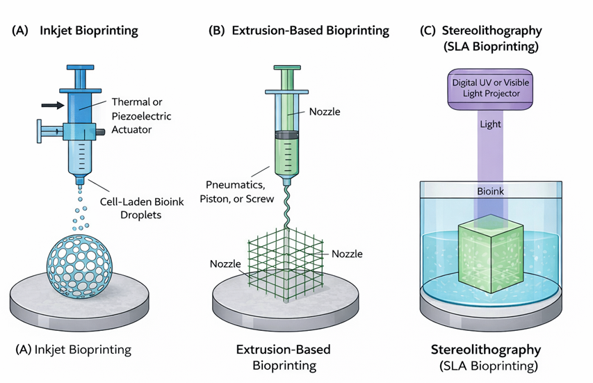
Figure no.1: (A)Inkjet bioprinting (B)Extrusion based bioprinting (C) Stereolithography
Bioinks are biomaterials that can be used to integrate biomolecules and enclose cells. Because hydrogels contain a large amount of water, which it is favorable for the cell survival & protects the fabrication cells caused by manufacture, cell laden bioinks are hydrogel based. The viscosity, gelation, and crosslinking characteristics of bioinks are the primary factors that must be taken into consideration prior to printing. These characteristics can have a significant effect on cell viability, proliferation & morphology following using print, as well as print fidelity construct stability & print deviation from computer aided designs.[6]However, the bioink's mechanical and rheological characteristics are essential for successful extrusion printing. In addition to biocompatibility & post-print mechanical stability, the ideal bioink should have an appropriate balance between low viscosity under shear (to enable smooth extrusion) & high structural fidelity after deposition.[7] Brown algae yield alginate, a negatively charged natural polymer that gels mildly via divalent cations like calcium. Researchers favor it for biomedical uses due to safety, affordability & compatibility with living tissues.[8 SA forms a linear chain composed of β-D-mannuronic acid (M) blocks and α-L-guluronic acid (G) segments connected by 1-4 glycosidic linkages. It can rapidly form a three-dimensional crosslinked network by chelating with different cations like Ca²?, Ba²?, or other polyvalent ions due to chemical structure, which is abundant with -COOH & -OH functional groups. The gelation properties are significantly influenced by ratio of M to G units: SA with a high G unit concentration often forms high-strength gels, whereas larger M unit content produces better elasticity. Sodium alginate gels demonstrate superior swelling and hydration capacity capabilities due to their 3D crosslinked network. These hydrogels also demonstrate good biocompatibility. Because of these qualities, SA anideal biomedical material is used extensively for regenerative matrices & wound healing materials & controlled release platforms.[9] Due to many -COOH and -OH moieties along its polymer chain cause high water affinity alginate experiences massive swelling and uncontrollable degradation, which results poor structural integrity in physiological solutions & severel limits its usefulness in the biomedical field.[10] Alginate is frequently blended with other natural or synthetic polymers to improve its viscoelastic behavior, bioactivity, and structural stability in order to overcome these limitations.[11] Natural polymers such as gelatin/GelMA, collagen, hyaluronic acid, chitosan & nanocellulose are frequently blended with sodium alginate to improve its bioactivity, cell-adhesion properties as well as viscoelastic behavior—all of which are essential for extrusion bioprinting.[12] Similarly, alginate can be blended with synthetic polymers including PEG/PEGDA, PVA, Pluronic F127 & nanoclay (Laponite) to enhance the mechanical stability, shear-thinning behavior, shape fidelity, and long-term structural integrity of printed constructs.[13] In recent years, class of nontoxic, cell-compatible, lightly networked polyacrylic acid polymers is known by trade name "carbopol." Carbopol as rheology modifier that develops the low properties of the inks and the printability range, a cross-linking polymer that controls final printed structure's physical & (bio)chemical properties. Carbopol serves as the viscosity control agent which demonstrates required thixotropic rheological characteristics & shear thinning. The ability of CBP to maintain its favorable rheological characteristics at extremely low concentrations and when combined with other bioink constituents such as polymers, proteins, tiny molecules & cells is a significant benefit. We found that the intrinsic rheological characteristics of CBP produced good extrudability at low pressures and enabled the creation of smooth, stable filaments when we used an extrusion printing method to a mixture of water and CBP.[14]
1.0 MODIFICATION METHOD
Chemical, physical, and biological techniques are used to modify sodium alginate (SA), with chemical modification being the most widely used to modify its characteristics for several applications. In order to add new functions or enhance properties like biocompatibility, biodegradability, mechanical strength & delivery capability, chemical modification procedures for SA mostly include reactions that target its hydroxyl and carboxyl functional groups.
1.1 Chemical Modification:
Table 1:- Chemical Modification Strategies For Sodium Alginate
|
Modification Method |
Functional Group Targeted |
Key Effects |
Biomedical Applications |
|
Oxidation (OSA) |
Vicinal diols → aldehydes |
Enhanced degradability and flexibility |
Biodegradable scaffolds |
|
Esterification |
–COOH / –OH |
Increased hydrophobicity and mechanical strength |
Drug delivery, wound dressings |
|
Sulfation |
–OH → –OSO?H |
Anticoagulant and antiviral activity |
Tissue regeneration, biomaterials |
|
Amidation |
–COOH + amines |
Improved stability and self-assembly |
Carriers, hydrogels |
|
Methacrylation |
–OH → C=C groups |
Photo-crosslinking and tunable mechanics |
3D bioprinting bioinks |
|
Polymer grafting |
Backbone modification |
Dual crosslinking and improved rheology |
Load-bearing tissue scaffolds |
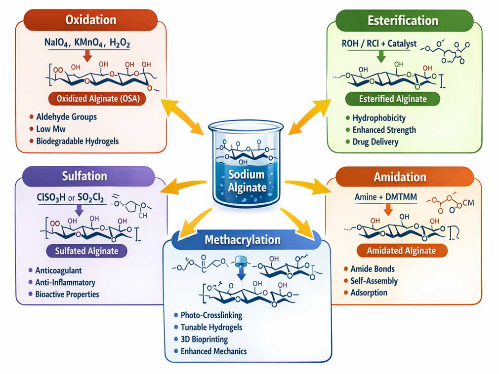
Figure no.2:- Chemical Modification Strategies For Sodium Alginate
1.1.1 Oxidation
Oxidized sodium alginate (OSA), produced by periodate cleavage of vicinal diols, ranks among the most widely adopted chemically altered alginate variants. Exposure to an oxidants enable decrease molecular weight of sodium alginate & improve its rheological characteristics. Greater oxidation extent produces enhanced dissolvability, lower chain length & accelerated breakdown for Ca2+ cross-linked SA hydrogels. Therefore, oxidized sodium alginate has several benefits while building biodegradable scaffolding. However, at low sodium alginate concentrations, this oxidative process generally works well.[15] A large number studies have revealed a significant decrease in the molecular weight of SA after being oxidized by NaIO?, resulting in the production of aldehyde groups with greater reactivity. The resulting OSA showed good biodegradability and improved molecular flexibility in addition to maintaining the low water solubility and biocompatibility of alginate.[16] Oxidizing agents such as periodate, ozone, potassium permanganate, and hydrogen peroxide can be used to oxidize SA. Under acidic conditions, potassium permanganate may oxidize SA; this degradation of SA increased as KMnO? concentration increased & the pH of the solution decreased.[17] The fundamental OSA parameter, oxidation degree (OD), has significant influence on the chemical and physical characteristics of OSA-based materials such hydrogels,[18] microspheres[19] & electrospinning.[20] Owing of these increased OD, OSA-based materials may have low network density and crosslinking degree. Additionally, the degree of OD is related to the alginate's gelling properties.
1.1.2 Esterification
Esterification proceeds via connecting hydrocarbon chains to a carboxyl function. This approach has been effectively applied to the modification of native alginate, due to which the hydrophobic character of the material increased. Alginates can be modified via direct esterification by treatment with selected alcohols in the presence of a suitable catalyst. In organic solvents such as DMSO or DMF, alkyl halides react with TBA-alginate salts frequently with tetrabutylammonium fluoride (TBAF) for chemoselectivity.[21] Sodium alginate (SA) is modified by esterification through acid catalyzed dehydration condensation between carboxyl & hydroxyl groups, which forms ester linkages & permits alkyl chain attachment to improve hydrophobicity and functionality. The methods are divided into surface modification of products (e.g., maleic anhydride on Ca²-alginate beads for improved oil adsorption) and pre-product molecular alteration, which introduces ester groups that increase mechanical rigidity, tensile strength, and bioadhesion, forming hydrogels ideal for drug delivery and wound healing.[22]
1.1.3 Sulfation
Sulfated alginate derivatives are created by chemically adding -OSO?H moieties to -OH sites on the polysaccharide chain in sodium alginate. Alginate is typically reacted with either one,three-benzene sulfonyl chloride or chlorosulfonic acid in formamide solvent. Because it is controllable and repeatable in the process, chlorosulfonic acid is frequently used as a sulfating agent. The sulfation promotes molecular rearrangements of the alginate chain's three dimensional structure, which improves or imparts biological activities such as anticoagulant, anti-inflammatory, antiviral & immunomodulatory properties. Sulfated alginates are helpful in biomedical, functional food, and biomaterial applications because of their qualities.[23]
1.1.4 Amidation
Alginate carboxyl and amine groups create amide bonds by amidation, which incorporates hydrophobic tails to allow micelles and vesicles to self-assemble. DMTMM-mediated reactions with amines like furanamine, amino acids, or peptides optimize at pH 5–6, improving enzymatic resistance (e.g., against alginate lyase) and breaking hydrogen bonding for enhanced spinnability. As an adsorbent, modified alginate showed better dye binding affinity and selectivity along with decreased surface tension and conductivity. It is suitable for functional carriers and food-compatible when using natural amines (such as amino acids); synthetic versions are intended for biomedical applications.[24]
1.1.5 Methacrylation
Methacrylation has been shown to be one of the most effective chemical modification method for improving the mechanical strength and crosslinking versatility of alginate. Methacrylation introduces reactive methacrylate (C=C) functional groups onto the alginate backbone, commonly via hydroxy esterification using methacrylic anhydride under controlled pH conditions.[25]The resulting methacrylated alginate works as a photo-crosslinkable macromer, facilitating covalent network formation upon exposure to ultraviolet or visible light in the presence of appropriate initiators. This change from basically ionic to hybrid covalent-ionic or entirely covalent crosslinking significantly enhances hydrogel stability, minimizes dissolution under physiological ionic circumstances & greatly widens tunability over stiffness, elasticity, degradation kinetics, and mesh structure.[25,26,27] Alginate also converted into a very elastic bioink precursor for 3D bioprinting by the addition of methacrylate groups. By splitting extrusion (or droplet deposition) from hydrogel solidification, photocrosslinkable Alg-MA enables printing at viscosities that promote cell viability and extrusion smoothness while achieving rapid post-printing stabilization and shape retention through regulated photopolymerization.[28]
1.1.6 Grafting
This includes covalently attaching (grafting) synthetic polymer chains (or other polymers) to the backbone of alginate, resulting in a "graft copolymer" with alginate as the backbone and the synthetic polymer as the side chain. This alters the properties of alginate (e.g., hydrophilicity, mechanical strength, responsiveness, swelling, and degradation) while maintaining some of its natural characteristics, such as ionic crosslinking capacity. Hydrogels with dual-mechanism networks (ionic + hydrophobic associations) have been created by grafting thermoresponsive poly(N-isopropylacrylamide) (PNIPAM) to alginate (Alg-g-PNIPAM), resulting in improved mechanical stiffness & 3D printability.[29,30] Natural alginate lacks cell-adhesive ligands; a common technique for increasing bioactivity is to chemically graft short peptides containing the RGD motif onto it. cell adhesion receptors on many different types of cells identify the RGD motif, which promotes cell adhesion, spreading, survival & signaling. Grafting is commonly done using carbodiimide chemistry (or oxidation + reductive amination if using oxidized alginate) to generate stable covalent connections between alginate carboxyls (or aldehydes on oxidized alginate) and the amino groups of the peptide.[31] Alginate can be grafted with synthetic polymers (like PNIPAM) to create hybrid hydrogels that differ extensively from pure alginate in terms of mechanical, thermal, or stimuli-responsive properties. For instance, Alg–g–PNIPAM hydrogels coupled with methylcellulose displayed increased mechanical strength and retained 3D-printed shape fidelity, even at physiological temperature & after crosslinking.[32]The bioinert feature of plain alginate is overcome by RGD-grafted alginate scaffolds, which greatly improve cell adhesion, spreading, viability, proliferation, and enable differentiation in a variety of cell types such as mesenchymal stem cells & endothelial cells.[33] Design composite hydrogels and bioinks optimized for printing performance and biological functionality is made possible by combining polymer grafting (for mechanical and rheological tuning) with RGD-grafting (for bioactivity). This results in constructs that are printable, stable, and supportive of cell behavior. For example, recent work developing composite hydrogels with oxidized alginate grafted with RGD (“ADA-RGD”) and reinforcing polymers/nanocells generated IPN hydrogels with good pore structure, improved dynamics & higher cell adhesion/proliferation – beneficial for bone or tissue scaffolds.[34]The effects of graft copolymerization on swelling, porosity, and network hydrophilicity/hydrophobicity can be used to regulate nutrition transport, degradation rates & the release of loaded components (drugs, growth hormones). This is useful when designing scaffolds for drug delivery or controlled tissue regeneration.[35]
1.2. Physical Modification
Table 2:- Physical Modification Approaches For Alginate-Based Bioinks
|
Strategy |
Materials Used |
Improved Properties |
Outcome |
|
Polymer blending |
Gelatin, PEGDA, chitosan, pectin, CMC |
Improved rheology and bioactivity |
Enhanced printability |
|
Nanocomposites |
Nanoclay, graphene, carbon nanotubes (CNTs) |
Increased mechanical strength and conductivity |
Smart biomaterials |
|
Rheology modifiers |
Carbopol |
Shear-thinning behavior and thixotropy |
High-fidelity extrusion |
|
Hybrid networks |
PEGDA, interpenetrating polymer networks (IPNs) |
Mechanical robustness |
Long-term structural stability |
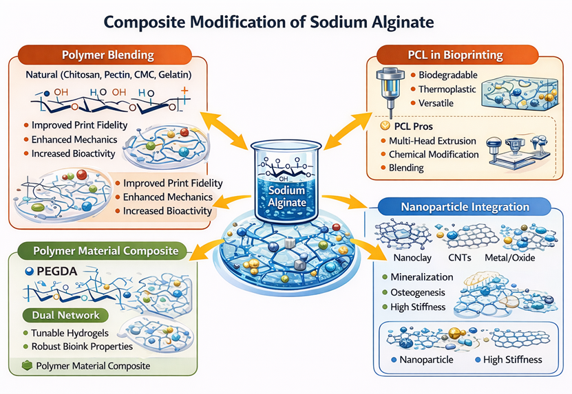
Figure no.3:- Composite modification of sodium alginate
1.2.1 Composite Modification
The wide range of performance control and excellent tunability of SA hybrid networks deliver increasing interest for smart materials development. By using hybrid material combinations leverage, diverse materials can effectively be incorporated, surpassing the limitations of pure alginate systems in terms of mechanical strength & functional responsiveness. Recent studies emphasize three main approaches as polymer mixing, inorganic reinforcement & nanoparticle integration. This involves integration of nanostructured components as graphene, nanoclay, carbon nanotubes, metal or oxide nanoparticles within alginate matrices generates combined boundary interactions like improved mechanical strength, conductivity, or barrier performance. Typical manufacturing processes, including as homogeneous blending & in situ synthesis, commonly use sol to gel phase transition of SA to immobilize functional components inside three dimensional network structures. Mechanical qualities, biocompatibility, and environmental responsiveness are all synergistically improved as a result. These multi-scale composite techniques offer a useful method for creating cutting-edge functional materials like flexible sensors and smart drug carriers.[36]
1.2.2 Polymer Material Composite
Polyethylene glycol diacrylate modification of SA offers improved material properties, shows strong potential in biomedical uses. Adding PEGDA segments efficiently reduces viscosity & increases flow characteristics of alginate mixtures, flow characteristics of alginate mixtures, enhancing spinning performance & enabling the creation of varied dual network structures, such as microspheres & thin films, via modified molding techniques.[37]The sodium alginate or polyethylene glycol diacrylate gel solution with a semi-interpenetrating network structure is prepared synthetically using the "one-pot" method. This method was used to create fibers with excellent mechanical qualities.[38]
1.2.3 Physical Blending
A frequently employed approach for overcoming the intrinsic limitations of pure alginate—such as poor print fidelity, low mechanical strength, limited bioactivity & lack of cell-adhesive motifs—is to physically blend sodium alginate (SA) with natural or synthetic polymers. Physical blending improves hydrogel functionality by generating polymer-polymer interactions such as hydrogen bonding, electrostatic interactions, entanglement, or network interpenetration, whereas chemical modification changes the backbone structure of alginate.[39,40] These changes greatly enhance alginate-based bioinks' mechanical characteristics, cell compatibility, degradation profile & rheological behavior, making them more appropriate for extrusion and freeform 3D bioprinting[41]
1.2.4 Natural Blending
Blend With Chitosan
Sodium alginate (SA) and chitosan (CH), a naturally occurring polycationic polysaccharide, is often blended to create polyelectrolyte complexes (PECs) through electrostatic interactions between the negatively charged carboxylate groups of alginate and the positively charged amino groups of chitosan.[42] When compared to individual polymers alone, this interaction produces hydrogel networks and films with significantly improved tensile strength, thermal stability & structural integrity.[43] According to studies, the presence of chitosan results in hydrogels that are smoother, more cohesive, with fewer pores, improved yield stress, and greater spreadability—all of which increase in directly proportion to the concentration of chitosan.[35,36] In alginate-chitosan bioinks, the ideal chitosan concentrations (typically ~2–3% w/v, depending on molecular weight and deacetylation) induce a uniform gel microstructure with remarkable stability.[44,45] Additionally, because chitosan delays alginate contraction and promotes prolonged matrix hydration, chitosan cross-linked or chitosan-coated alginate microparticles show improved swelling control and mucoadhesive qualities, which are advantageous for controlled and sustained drug release applications.[46,47] Chitosan-based printed platforms showed biological activity, stable biocompatibility, and biomolecule adaptation even after several kinds of post-printing alterations.[48,49]
Advantages of Chitosan
The FDA has approved few 3D-bioprintable bio-inks for use in medicine, although biomaterials based on chitosan are approved for tissue engineering and medication delivery.[50]Chitosan based bioinks can be designed to be extrudable with high fidelity due to wide viscosity window & diverse crosslinking options methods, & adjustable mechanical properties, which are accomplished by adding a variety of reinforcing chemicals.[51]Chitosan based hydrogels exhibit excellent cell viability & high bioactivity, with their versatile, adjustable parameters positioning these bioinks at the top for 3D bioprinting applications.[52]
Blend With Pectin
When combined with sodium alginate, pectin, a naturally occurring polysaccharide high in galacturonic acid units, improves the mechanical integrity and ionic conductivity of composite films and hydrogels.[53] Due to SA–pectin blends have complementary functional groups that can form hydrogen bonds and interact with ions when divalent cations like calcium are present, they create stable networks.[54] This mixture is helpful for tissue engineering scaffolds and drug delivery systems that need high water absorption and regulated release profiles because it modifies swelling behavior and breakdown rate. It is a viable option for food packaging and biomedical applications because of the synergistic action of alginate and pectin, which also supports film integrity and inhibits rapid disintegration.[55]
Blend With CMC
In the presence of calcium ions, carboxymethyl cellulose (CMC), an anionic cellulose derivative, physically blends with sodium alginate primarily through hydrogen bonding and ionic crosslinking. As a result, hydrogels with improved mechanical behavior, pH sensitivity, and nanoparticle stabilization properties are generated. Bioactive substances like turmeric extract are frequently encapsulated in polymers with controlled swelling and release properties due to the CMC-SA combination. The hydroxyl and carboxyl groups of both polymers produce intermolecular hydrogen bonds, which strengthen the gel's strength and durability. These mixtures maintain their inherent biodegradability and bioactivity, which is useful for designing smart release systems in the food and pharmaceutical industries, where the response to the pH of the surrounding environment initiates specific delivery or preservation effects.[56]
Blend With Carbopol
The restricted printability range defined by arises due to requirements for bioinks with rheological characteristics permit smooth filament deposition alongside robust cell survival in bioink formulations before & after printing is one of the main obstacles to the widespread application of this technology. In this study, they use Carbopol enables printability of low viscosity hydrogels usually not extrudable or have poor extrusion fidelity. They explained how minimal carbopol levels enable provide appropriate flow behavior characteristics for a wide range of formulations, enabling incorporation of polymers exhibiting various crosslinking methods as well as the addition of additives & cells. Across diverse hydrogel compositions, trace carbopol amounts enable provide optimal flow characteristics, allowing extrusion of polymers featuring various crosslinking methods as well as the introduction of additives & cells. Poly(ethyleneglycol) diacrylate based ink formulations with extrusion three dimensional printing create compliant yet durable gels featuring adjustable structural reinforcement. Surface seeded cells on cured gels & embedded cells in extruded bioinks, cell laden constructs produced using these bioink systems showed excellent cell survival, studies confirm material biocompatibility with structural integrity maintained through 14 days. By ensuring the necessary rheological characteristics & improving diversity of compatible components could achieve high fidelity extrusion, CBP incorporation enables high resolution deposition bioinks could facilitate rapid progress of filament deposition bioprinting. Focusing on biochemical, cellular & mechanical requirements of the intended applications rather than the rheology required to attain optimal printability, such approaches enable investigators to engineer advanced bioink formulations.[14]
1.2.5 PCL in Bioprinting
Poly-caprolactone (PCL) is a biodegradable synthetic polymer that has wide use in 3D printing, particularly in tissue engineering and regenerative medicine, because of its remarkable mechanical and biocompatible properties. PCL was initially used as a filament in conventional 3D printing processes like fused deposition melting (FDM), but its usage in bioprinting— which prints cells directly is a more recent idea.
PCL's Advantages and Characteristics as a Biomaterial:
Biomedical researchers like PCL because of a number of important features: The biodegradability and low melting point This polymer is biodegradable and has a low melting point of 55–60°C, which is consistent with the temperatures at which 3D bioprinters operate. Biocompatibility and Physicochemical Properties: PCL is very compatible with biological tissues, has a slow rate of degradation (1-4 years), and is well soluble. It has great physicochemical properties, cheap & easy to customize.
Versatility in Fabrication:
PCL is a thermoplastic polymer suitable for FDM and may be produced utilizing a variety of 3D printing methods, such as stereolithography & selective laser sintering.
PCL's Restrictions and Challenges in Bioprinting:
PCL faces many difficulties for direct application in bioprinting, Issues with Cell
Encapsulation.
PCL is not appropriate for direct cell encapsulation since its melting temperature can kill cells.
Additionally, extrusion require high pressure, which might cause cell rupture from extreme strain.
Viscosity and Hydrophobicity: PCL's high viscosity and hydrophobic properties restrict from being used directly in many bioprinting techniques.
Absence of Bioactivity and Conductivity: PCL by itself might not be able to provide a strong enough cellular reaction or have the conductivity required for some tissue engineering applications.
Techniques for Overcoming Restrictions:
Multi-Head Extrusion Bioprinting (EBP):
This technique enables the co-printing of cell-laden bioinks from different print heads with PCL as a supporting framework. This dual-layer method preserves excellent cell survival while enhancing mechanical characteristics and scaffold homogeneity.
Chemical Modification and Blending:
To improve its qualities, PCL can be altered or combined with other biomaterials.
Examples:
To increase hydrophilicity and porosity, add hydrophilic substances like alginate.
Tetrafluoromethane or oxygen plasma surface treatment to enhance cell adhesion.
Adding graphene or using NaOH to improve adhesion, hydrophilicity, and cell viability.
Using bioactive polymers (such as Halomonas levan) or calcium phosphate microparticles to promote cell proliferation and osteoinduction.
Chemical alteration of conductive polymers for nerve tissue engineering, such as PPy-b-PCL
EBP that is cryogenic PCL can now be printed in a liquid state due to recent developments in extrusion-based cryogenic printing.[57]
1.3 Ionic Crosslinking
Ionic crosslinking is one of the simplest and most effective method to crosslink alginate. Alginate films, fibers, hydrogels, nanoparticles, and microparticles have all been created using ionic crosslinking. When cations are present, the G blocks in alginate chains firmly bind together at their intersections. High G alginates produce stronger gels. Alginate's affinity, however, also has a significant impact on how well they crosslink. Along with details about mechanical stability, the affinity constant for gelling provides us with a summary of the crosslinking process's spatial uniformity. In former mechanism, alginate solution diffuses from an external reservoir; in the latter, ion release is facilitated by the solution pH or divalent cation availability. The majority of titratable sites along the alginate backbone are created by negatively charged carboxylate groups, which are essential for both crosslinking and metal binding processes. Because of their geometric order, sections of alginate chains filled with G subunits generate zigzag structures with voids of the right size that are particular to Ca2+. Numerous oxygen atoms are also involved in this arrangement (darkened circles). This configuration is referred to as a "egg-box" model, because Ca2+ aligns itself in the guluronate block structure like an egg box. High efficiency and the appropriate degree of crosslinking are provided by such a setup.[58]
2.0 Characterization of Hydrogel:
2.1 FTIR
Fourier Transform Infrared spectroscopy is renowned analytical method for characterizing unique functional groups inside a given substance, as each group has distinct absorption bands at specific wavenumbers in the infrared region. FTIR is particularly suitable for qualitative characterization of organic and polymeric samples, as the intensity and position of IR peaks correlate directly with the type of chemical bonds present.[59-60] The sample was prepared for measurement using the traditional KBr pellet procedure, which involves compressing a small amount of the material under high pressure to produce a transparent pellet after it has been finely powdered and homogenized with spectroscopic-grade potassium bromide.[61]FTIR analysis was then performed on the created pellets. For the sake of good signal-to-noise ratio and spectral clarity, 64 scans at a resolution of 4.0 cm?¹ were used to record the spectra. The measurement range, which covered the basic vibrational modes of the majority of functional groups are important for structural characterisation from 4000 to 400 cm?¹.[62]
2.2 XRD
A key method for the primary characterization of material characteristics, such as crystal structure, crystal defects, crystalline stresses, and molecular-level distortions, is X-ray diffraction (XRD). The interaction of X-rays with the periodic atomic planes of crystalline material is the foundation of XRD. The atoms in the crystal lattice scatter or diffract the incident X-rays in particular directions when a monochromatic X-ray beam is aimed at the specimen. Constructive and destructive interference, which rely on the angle of incidence and the lattice plane spacing, cause this scattering. In this study, sodium periodate was analyzed using the Bruker D8 Advance X-ray diffractometer, which allowed for accurate determination of its crystalline characteristics and confirmation of its structural integrity. A detector, appropriately placed around the specimen, records the intensity and position of the diffracted X-rays, generating a diffraction pattern that provides detailed information on the crystalline structure, phase composition, degree of crystallinity, and any structural distortions present in the material.[63]
2.3 DSC
DSC is the most often used method for figuring out the glass transition temperature (Tg) of amorphous solid dispersions. Differential scanning calorimetry (DSC) is divided into two types: heat-flux DSC and power-compensation. Heat-flux DSC uses symmetrically placed thermocouples to monitor the sample and reference pans, allowing for quantitative heat flow measurement and thermal transition determination. Although power-compensation DSC allows for significantly higher heating and cooling rates, heat-flux designs are still the most popular due to their durability and ease of operation. Thermal events such as crystallisation, curing, and degradation use Arrhenius-based kinetics, which allows the measured heat flow to be correlated to the reaction rate by incorporating proportionality constants and heat capacity contributions. Melting and glass transition are the most commonly studied thermal transitions in pharmaceutical systems. Since Tg represents molecular mobility and physical stabilityThe glass transition is a second-order phase transition characterised by a step change in heat capacity (Cp) over a restricted temperature range. Vibrational motion dominates Cp below Tg, and as the temperature rises, rotational and translational mobility gradually become more significant. Although both conventional DSC and modulated DSC (MDSC) may measure Tg, MDSC offers superior sensitivity and resolution, particularly for systems with subtle transitions.[64,65]
2.4 SEM
Scanning electron microscopy (SEM) is commonly utilised in pharmaceutical research for product development and quality control, notably for determining particle size, shape, and surface properties.[66] Unlike optical systems, SEM images are produced by complicated electron-specimen interactions that yield various signals—secondary electrons (SE), backscattered electrons (BSE), Auger electrons, and distinctive X-rays—which are detected by specifically designed sensors. These signals are the result of elastic and inelastic collisions, with BSE providing compositional contrast (greater in materials with higher atomic numbers) and SE providing high-resolution imaging. Additional signals, such as Bremsstrahlung radiation, enhance material identification. SEM is useful for evaluating processing impacts (e.g., hot melt extrusion, spray drying, electrospinning) and, when combined with energy-dispersive X-ray spectroscopy (EDS), allows for elemental mapping and better interpretation of miscibility and material properties.[67]
2.5 Rheological Study
Various rheological properties including shear thinning performance, frequency & temperature induced shifts in storage modulus (G′) & loss modulus (G″), the linear viscoelastic region , thixotropy & the long-term stability of pre-crosslinked hydrogel inks under ambient and physiological conditions were assessed to confirm the development of a 3D printable ink with optimal printability. Rheological measurements employed an Anton Paar MCR 302 rheometer equipped with a 25 mm parallel-plate geometry. Hydrogel inks were loaded between the plates at 21 °C, maintaining a 1 mm gap. Frequency sweeps from 0.01 to 100 rad/s first characterized the inks' viscoelastic profiles, tracking G′ and G″. Temperature ramps spanning 25–42 °C exceeding body temperature at 37 °C evaluated thermal stability of G′ and G″. Time dependent stability of pre crosslinked gels was probed via a 4-hour sweep at 1 rad/s and 1% strain under room temperature. Amplitude sweeps across 0.1–1000% strain defined the LVR. Flow curves over shear rates of 0.1–100 s?¹ quantified pseudoplastic (shear-thinning) viscosity changes. Thixotropy, reflecting shape recovery across pre-print (Step I), print (Step II), and post-print (Step III) phases, involved applying 0.1 s?¹ for 60 s in Step I, ramping to 100 s?¹ for 20 s in Step II, then reverting to 0.1 s?¹ for 60 s in Step III to measure viscosity restitution.[68]
2.6 Swelling Study
Determination of water retention through swelling tests is critical for carbomer-alginate composite hydrogels used in extrusion based 3D bioprinting, influencing structural fidelity, pore networking, & support for cell growth following printing. Uniform specimens, such as lyophilized cylinders or grid like constructs, are initially weighed W initial prior to immersion in pH neutral solutions like PBS pH 7.4 at 37°C with gentle agitation to replicate biological hydration. Sequential weigh ins at points like 15 min, 40 min, 1.5 h, 4 h, 9 h, 24 h, & 72 h require lifting samples, dabbing away superficial liquid with tissue, measuring wet mass
W final, & returning them to solution until weight plateaus, typically after 15-28 hours. Water absorption percentage is determined by[69,70,71,72]
Percent swelling = [(W final – W initial) / W initial] × 100
Which charts absorption trends & maximum capacity. Within these formulations, alginate's guluronate segments create stable networks via Ca²? coordination, constraining excessive expansion, while carbomer's ionizable carboxyl functions facilitate swelling in ionic media through repulsive forces. Greater carbomer incorporation generally restricts peak hydration to 350-850%, enabling porous structures for gas exchange paired with rigidity to avoid extrusion flaws. Changes in salinity, warmth, or cell media uncover degradation traits, directing bioink tuning for filaments narrower than 450 μm without fusion. Well-calibrated swelling prevents over-saturation that destabilizes designs, promoting cell viability & proliferation, as confirmed by viscosity and modulus analyses. Such engineered materials surpass unmodified alginate in moisture control, boosting potential for tissue regeneration scaffolds.[73]
2.7 MTT Assay:
The MTT assay was used to investigate the cytotoxicity of all hydrogel combinations and drug-loaded hydrogels. In the MTT assay, dehydrogenase from the metabolically active cells transforms yellow tetrazolium salt into crystals of purple formazan. The quantity of live cells is indicated by the purple formazan amount. As a result, this is used to examine the in vitro cytocompatibility and growth in cell proliferation of different hydrogels. More than 70% cell viability is considered non-toxic when evaluating cytotoxicity using the MTT assay. Most of the gels are non-toxic and beneficial to the cells, according to cell viability estimates obtained through the MTT assay.[74]
CONCLUSION
The design space of regenerative medicine has been significantly broadened by developments in 3D printing and bioprinting technologies, which allow for the controlled development of three-dimensional, cell-filled structures with high structural precision. The potential for producing improved therapeutic platforms and creating functional tissues has increased due to the capacity to spatially arrange cells, biomaterials, and bioactive cues. Several bioprinting modalities, such as inkjet, extrusion-based & light-assisted methods, serve a variety of biomedical applications by providing complementary capabilities in terms of resolution, material diversity, and scalability.A key component for effective bioprinting outcomes is still bioink design. Systems based on hydrogel, especially those made from sodium alginate, are popular because of their versatility, gentle gelation conditions, and biocompatibility. Native alginate has some limitations, including poor cell adherence, uncontrolled degradation, & inadequate mechanical stability. Various chemical and physical modification methods, such as oxidation, esterification, sulfation, amidation, methacrylation, grafting, and composite blending, have been developed to address these limitations. By fine-tuning biological functionality, crosslinking mechanisms, rheological behavior, and degradation kinetics, these methods improve printability and post-print construct performance.Alginate's mechanical strength, viscoelasticity, and bioactivity can be enhanced by blending it with synthetic and natural polymers, adding rheology modifiers, and adding reinforcing agents. When long-term mechanical integrity is needed, complementary scaffold materials like polycaprolactone offer structural support. In addition, ionic and photo-crosslinking techniques offer the controlled network development necessary for preserving cellular viability & shape fidelity.Overall, the fabrication of multifunctional, printable, and biologically supporting structure is made possible by developments in alginate modification, composite bioink formulation, and crosslinking techniques. These advancements placed alginate-based systems in a position to have a significant role in the continual development of regenerative medicine and 3D bioprinting.
REFERENCES


Sharvari Chavan, Aishwarya Pachore, Trupti Abhivant, Shivraj Sulgudle, Santosh Gejage, Alginate-Based Bioinks for 3D Bioprinting: A Comprehensive Review, Int. J. of Pharm. Sci., 2026, Vol 4, Issue 2, 2466-2484. https://doi.org/10.5281/zenodo.18667408
 10.5281/zenodo.18667408
