G. P. Pharmacy College, Vaniyambadi Main Road, Mandalavadi, Jolarpettai, Tirupattur 635851.
The transdermal route is increasingly recognized as a valuable alternative to oral drug delivery, particularly for addressing limitations such as first-pass metabolism and inconsistent bioavailability. Drug administration through the skin has a long-established history, with microneedle arrays emerging as a promising advancement due to their capacity to bypass the stratum corneum while avoiding significant stimulation of sensory nerve endings. First-generation transdermal patches and second-generation iontophoretic systems have achieved successful commercialization; however, their applications remain largely confined to small, lipophilic molecules. To overcome these constraints, advanced enhancement methods—such as iontophoresis, sonophoresis, electroporation, and microneedle technology—have been developed, enabling effective delivery of hydrophilic and high molecular weight drugs across the skin. Microneedle-based delivery platforms are anticipated to play a pivotal role in future therapeutic administration because they are effective, minimally invasive, and cost-efficient. Current cosmeceutical applications include treatments for acne, hyperpigmentation, scars, and wrinkles, along with overall skin rejuvenation. This review summarizes recent developments in microneedle systems, aiming to provide valuable insights for emerging researchers and to encourage further advancements in this field.
A drug delivery system (DDS) refers to a collection of physicochemical techniques designed to regulate the transport and release of pharmacologically active agents into targeted cells, tissues, or organs, ensuring optimal therapeutic outcomes. The effectiveness of a drug depends not only on the active pharmaceutical ingredient but also on the administration route. Oral administration is generally convenient and non-invasive; however, it may be compromised by first-pass metabolism, fluctuations in plasma drug levels, and poor suitability for hydrophobic compounds. Injectable administration offers high bioavailability and rapid onset but requires trained personnel, can cause discomfort, and often reduces patient compliance. Additionally, needle phobia can trigger a range of physical, psychological, and behavioural reactions. The transdermal route offers a practical alternative to address these challenges¹. In recent years, transdermal drug delivery systems have demonstrated remarkable potential in healthcare. Conventional methods - such as topical creams and hypodermic injections - are widely used but have notable drawbacks: creams generally exhibit low bioavailability, and injections cause pain and invasiveness. Microneedle (MN) arrays have been developed to address these shortcomings, facilitating drug storage within skin layers before gradual release into systemic circulation. Achieving therapeutic drug concentrations in targeted skin tissues necessitates innovative delivery strategies². The skin comprises three distinct layers: the epidermis, dermis, and hypodermis. The epidermis - particularly the stratum corneum - serves as the principal permeability barrier, being largely hydrophobic in nature. Drugs capable of traversing this barrier are typically small, lipophilic or amphiphilic, and non-irritating. Because of its accessibility and large surface area, the skin has been extensively studied as a site for drug administration. However, penetration into deeper layers remains challenging³. To overcome these limitations, researchers have investigated various methods to bypass the skin barrier, with microneedle-based delivery gaining significant attention. Microneedles typically range from 25 μm to 2000 μm in length (Donnelly et al., 2010) and can penetrate the stratum corneum to create microchannels, enabling drugs to reach the epidermis or upper dermis. This approach allows systemic absorption without encountering the primary diffusion barriers?. This review focuses on microneedles as a delivery platform, discussing their structural design, mechanism of action, and therapeutic potential, while aiming to promote continued innovation in this emerging area.
Historical Development of Transdermal Micro Needles:
The evolution of drug delivery methods has progressed from primitive approaches - such as simple oral and injectable systems - to sophisticated platforms capable of targeted, controlled release?. These advancements have significantly improved therapeutic outcomes by increasing bioavailability, reducing adverse effects, and optimizing dosing regimens. The earliest documented use of microneedle-like technology dates back to 1921, when Chambers inserted a needle into the nucleus of an egg. Although the concept of microneedles was introduced in the 1970s, practical experimentation did not begin until the 1990s?. A notable milestone occurred in 1979 with the approval of the first transdermal system for scopolamine delivery, using a three-day patch to prevent motion sickness. In 1994, Orent Reich performed a subcision surgical technique, inserting a tri-bevelled hypodermic needle beneath the skin to release fibrous strands responsible for depressed scars and wrinkles. The modern concept of microneedles emerged in 1998, when devices were fabricated from silicon wafers through ion etching and photolithography. This pioneering work demonstrated that microneedles created by microfabrication techniques could enhance drug permeation across the skin, catalysing widespread research interest in the field?. In this review, we outline the major approaches used in the three generations of transdermal drug delivery systems and examine emerging strategies. We also highlight recent advances in manufacturing techniques, including three-dimensional (3D) printing, which hold promise for producing complex microneedle architectures with precision.
First Generation Transdermal Micro Needles:
The earliest generation of transdermal microneedle systems evolved from the use of herbal-based formulations to the development of modern patch technologies?. This generation primarily targeted the delivery of drugs capable of permeating the skin without significant enhancement techniques. Formulations from this era such as ointments, creams, sprays, gels, and patches were restricted to compounds with low molecular weight, lipophilic properties, and potency at low doses. Due to these constraints, only a limited range of therapeutic agents could be effectively delivered via this route. For example, lipophilic drugs could pass through the stratum corneum and reach the dermal capillary bed, but the process occurred at a slow diffusion rate?. The first generation laid the foundation for transdermal drug delivery; however, its applicability was narrow, necessitating the development of more advanced delivery methods to expand the range of drugs that could be administered through the skin.
The second generation of transdermal drug delivery systems was designed to enhance skin permeability by temporarily disrupting the function of the stratum corneum, allowing drug molecules to traverse this barrier without damaging the deeper dermal layers. This approach incorporates various enhancement strategies, including chemical penetration enhancers, iontophoresis, and non-cavitational ultrasound techniques¹?.
Chemical enhancers function by altering epidermal lipid biosynthesis, thereby increasing the penetration of therapeutic agents. These enhancers are effective for small molecules but have limited applicability for hydrophilic macromolecules. In some cases, a cleavable chemical group such as an ester or carbonate can be conjugated to the drug to increase its lipophilicity, facilitating better skin permeation without causing irritation.
Iontophoresis involves applying a constant low-voltage electric current to the skin, which accelerates the transport of ionized drugs through charge repulsion. The quantity of drug delivered can be regulated by adjusting the electrical current, making this method suitable for delivering peptides and oligonucleotides.
Non-cavitational ultrasound, first reported in 1954 by Fellinger and Schmidt, uses high-frequency sound waves to disrupt the lipid structure of the stratum corneum. This increases skin permeability, particularly for small and lipophilic molecules, thereby improving drug absorption. The second generation significantly expanded the range of drugs deliverable through the skin compared to the first generation, yet it still faced limitations when handling large molecular weight compounds or highly hydrophilic drugs.
The third generation of transdermal drug delivery systems was developed to facilitate the transport of complex therapeutics such as vaccines, proteins, and other macromolecules across the stratum corneum while protecting deeper skin layers from injury¹¹. The focus of this generation is to enhance delivery rates without compromising skin integrity. Several technologies characterize this generation, including electroporation, microdermabrasion, thermal ablation, cavitational ultrasound, and microneedle-based delivery.
Electroporation uses short, high-voltage electrical pulses to transiently disrupt the lipid bilayer of the skin, creating aqueous pathways that allow the passage of small molecules, peptides, DNA, and vaccines. While widely applied in animal studies, its complexity has limited human clinical applications.
Cavitational ultrasound employs ultrasound energy to generate microbubbles (cavitational bubbles) within the skin. These bubbles collapse in the dense stratum corneum, producing shock waves that enhance drug penetration. Research by Park et al. (2012) demonstrated that cavitational ultrasound increases skin permeability across a frequency range greater than 1 MHz. Among these technologies, microneedle systems have emerged as one of the most promising tools for third-generation delivery due to their precision, minimal invasiveness, and ability to transport both small and large molecules effectively.
Transdermal Route:
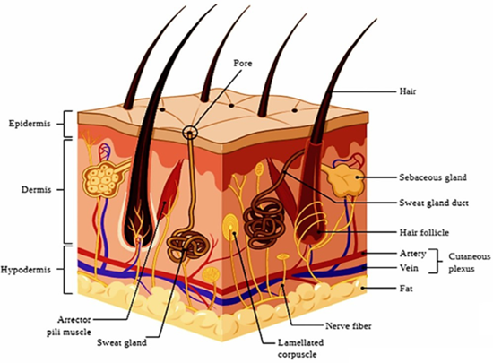
The skin is the largest organ of the human body, acting as a primary protective barrier against dehydration, physical injury, and microbial invasion. In adults, its surface area ranges from approximately 1.5 to 2.0 m². Structurally, the skin is composed of three main layers: the epidermis, dermis, and hypodermis¹². The epidermis is the outermost layer, with a thickness between 60 and 800 μm. Its uppermost sublayer, the stratum corneum (10 - 20 μm thick), serves as a critical barrier to prevent water loss and limit the entry of external substances into the body. The dermis lies beneath the epidermis and contains connective tissues, sweat glands, nerve endings, hair follicles, lymphatic vessels, and capillaries. The hypodermis, the innermost layer, is composed mainly of adipose tissue, providing cushioning and structural support to the upper layers. Beyond its barrier role, the skin also functions as a shield against ultraviolet (UV) radiation due to its melanin content¹³. It contributes to thermoregulation through mechanisms such as sweating and participates in excreting substances including xenobiotics, excess lipids, salts, urea, uric acid, ammonia, and other metabolites. Transdermal drug delivery systems (TDDS) utilize the skin as a site for drug administration. In this route, the active compound penetrates the skin and enters systemic circulation via dermal blood vessels, subsequently distributing throughout the body. TDDS offers several patient-centered advantages, such as avoidance of first-pass metabolism, reduced dosing frequency, potential for non-invasive or minimally invasive application, ease of self-administration, and elimination of the need for trained medical staff.
Micro Needles:
Microneedles (MNs) have become an important technology in the medical field for delivering a wide range of therapeutic agents from small molecules to large macromolecules, including protein-based drugs used in the treatment of numerous diseases¹³. This platform employs microscopic needle structures to breach the stratum corneum and deliver drugs into the underlying skin layers with minimal invasiveness. The primary objective of microneedles is to create micro-scale perforations in the skin that allow drug transport while avoiding contact with pain receptors or causing significant tissue injury. This design helps improve both patient comfort and treatment adherence. Microneedle devices typically consist of arrays containing one to several thousand individual needles, each measuring approximately 100 - 1500 µm in length. These needles can be integrated into a variety of delivery formats, including patches, roller devices, pen-shaped applicators, stamps, conventional syringes, or prefilled syringes. MNs have been extensively investigated for transdermal delivery of drugs such as insulin, anticancer agents, hormones, DNA, anti-obesity medications, and nanoparticle-based formulations. Vaccine delivery using dissolvable microneedles has also gained attention due to its potential to improve immunogenicity and stability. Beyond therapeutic delivery, microneedles have been explored for diagnostic purposes, such as extracting interstitial fluid (ISF) that contains biomarkers including glucose, alcohol, lactate, cortisol, cholesterol, and various proteins. The global market for microneedle technologies is expanding rapidly. In 2021, it was valued at approximately USD 2.65 billion, increasing to USD 2.83 billion in 2022. Market forecasts predict a compound annual growth rate (CAGR) of 17.8%, with revenues expected to reach USD 7.8 billion by 2027¹?.
Classification Of Microneedles:
Microneedles can be categorized into four primary types based on their drug delivery mechanism¹?:
These microneedles are manufactured from a variety of materials, including metals, glass, silicon, and biodegradable polymers. Each type has a distinct delivery strategy:
This classification system allows for tailored design and selection of microneedle types depending on the drug’s physicochemical properties, therapeutic target, and intended clinical application.
Solid microneedles (SMNs) were first proposed in 1971 as a method to pretreat the skin before drug application. They function according to the “poke and patch” principle, where the microneedles create microscopic channels in the stratum corneum, enhancing skin permeability and allowing subsequent drug diffusion from a topical formulation into the dermal layer. In this approach, the SMNs are applied to puncture the skin, after which a drug-loaded patch is placed over the treated area. The drug then migrates through the transient microchannels into the skin, either by passive diffusion or, in certain cases, through iontophoresis when an electric field is applied. The key advantage of SMNs lies in their ability to significantly increase skin permeability without causing deep tissue damage, making them suitable for pre-treatment in various transdermal applications.
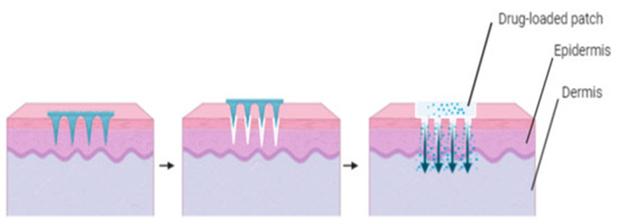
Figure 1: SMN device designed to create micro channel pores on the skin, followed by transdermal patches application.
Coated Micro Needles:
Coated microneedles (CMNs) operate using the “coat and poke” technique, in which a solid microneedle base is uniformly coated with a drug formulation. Upon insertion into the skin, the coating dissolves rapidly, releasing the active compound directly into the targeted tissue¹?. A variety of coating methods have been explored, with dip-coating being the most common. However, dip-coating requires precise control to ensure uniform deposition and accurate drug dosing. Spray-coating offers an alternative approach but can result in drug wastage due to deposition on areas of the device other than the microneedle tips. Aerosol coating methods have shown improved efficiency by directing more of the formulation onto the microneedle surfaces. Typically, a coated microneedle array consists of micron-sized sharp projections fixed onto a base substrate, with the drug and water-soluble excipients applied as a surface layer. Upon insertion, the coating dissolves almost instantly in the interstitial fluid, enabling rapid onset of therapeutic action.

Figure 2: Coated micro needle
Dissolving microneedles (DMNs) were first introduced by Miyano et al. in 2005 as an alternative to solid and coated microneedle designs. They offer multiple advantages, including simplified manufacturing, user convenience, and the ability to encapsulate relatively large drug doses within the microneedle structure¹?. DMNs function via the “poke and release” mechanism. During fabrication, the drug is incorporated directly into the microneedle matrix, typically composed of biodegradable and water-soluble polymers. Upon insertion into the skin, the microneedles dissolve in the interstitial fluid, releasing the encapsulated therapeutic agents. One of the key benefits of DMNs is the elimination of post-application sharps waste, as the microneedles dissolve completely within the skin. This feature reduces the risk of accidental needle-stick injuries and improves overall safety for both patients and healthcare providers.
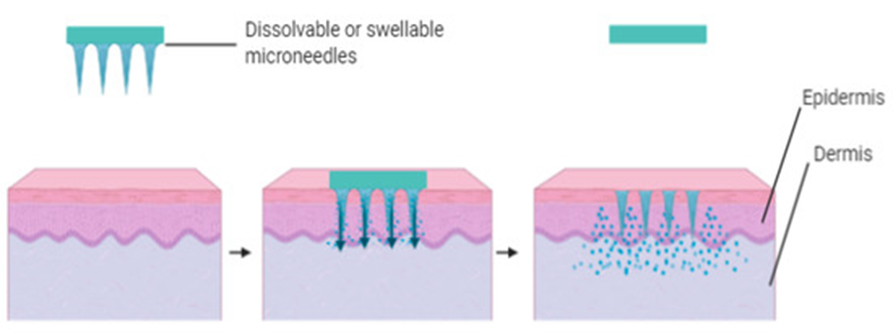
Figure 3: Illustration of poke and release using dissolving or swellable micro needles.
Hollow microneedles (HMNs) are designed to deliver liquid formulations directly from a reservoir such as a patch or syringe into the skin’s microcirculation¹?. They function similarly to miniature hypodermic needles, featuring a central lumen that allows the flow of drug solutions into the epidermis or upper dermis. These devices can administer volumes of up to 200 µL, enabling the delivery of both small-molecule drugs and large biomolecules, including proteins, oligonucleotides, and vaccines. HMNs can operate via diffusion-driven or pressure-driven flow, providing flexibility in dosing and delivery rate. Unlike dissolving or coated microneedles, HMNs can deliver relatively large quantities of drug in a single application. Their versatility makes them suitable for applications requiring precise dosing, rapid onset, or delivery of high-molecular-weight therapeutics.
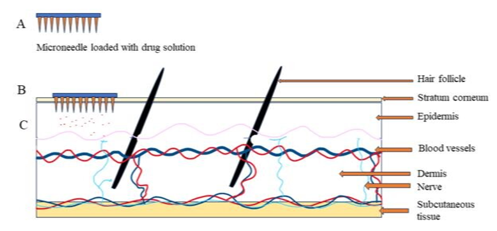
Figure 4: Illustration of poke and flow using hollow micro-needles
Mechanism Of Transdermal Micro Needles:
In microneedle-mediated drug delivery, the mechanism is based primarily on diffusion through microchannels created in the skin. The device typically contains hundreds or thousands of microneedles arranged on a small patch resembling a conventional transdermal patch¹?. When applied, these microneedles puncture the stratum corneum, bypassing its barrier properties, and deposit the therapeutic agent either on the skin surface or within the upper dermal layer. The drug then enters the systemic circulation and exerts its pharmacological effect at the intended site of action. Early approaches to microneedle delivery used solid microneedles to puncture the skin, followed by application of a medicated patch known as the “poke and patch” method. Silicon wafers were among the first materials used for microneedle fabrication. This method was also explored for non-invasive biomarker monitoring, such as glucose measurement, by extracting interstitial fluid (ISF)¹?. Subsequent developments introduced dip-coated solid microneedles, where the drug solution was applied to the needle surface and released after skin insertion. The “poke and release” approach allowed for controlled release profiles by tailoring the composition of the coating materials, often using biodegradable carbohydrates or polymers. Hollow microneedles were later developed to overcome the limitation of small payload capacity seen with dissolving or degradable microneedles. This “poke and flow” method allows liquid drug formulations to flow directly from a reservoir into the skin¹?.
Application:
The skin is a suitable site for the delivery of genes and oligonucleotides because it is well-characterized at both the molecular and cellular levels²?. Microneedle (MN) systems have been explored for the treatment of various genetic disorders, skin-related malignancies, infectious diseases, and for immunization purposes. Compared to conventional microinjection techniques, MN-mediated gene delivery offers the advantage of treating multiple cells simultaneously. The effectiveness of a transdermal delivery system depends largely on the ability of a drug molecule to penetrate the skin barrier. Large molecules, such as peptides and vaccines, have traditionally posed manufacturing and delivery challenges due to their high molecular weight²¹. MNs have demonstrated the capability to deliver such macromolecules intradermally and, in some instances, at lower doses than traditional injection methods, while achieving equivalent or improved clinical outcomes²². For example, similar hemagglutinin inhibition antibody levels were achieved with less than half the usual vaccine dose when delivered using MNs compared to conventional intramuscular injections. The efficacy of MN-based vaccination has been recognized by regulatory bodies, including approval for seasonal influenza vaccines. Moreover, MN systems have shown superior thermal stability maintaining up to 90% immunological potency after four months of storage at 40 °C²³.
CONCLUSION:
Transdermal drug delivery using microneedle (MN) technology has gained increasing attention in recent years due to its potential to improve patient access to medications while offering a less invasive alternative to traditional administration routes²?. Microneedles are available in several forms including solid, coated, dissolving, and hydrogel-based designs each with distinct advantages for specific applications. Ongoing clinical trials continue to expand the scope of MN applications by exploring their use with a variety of therapeutic agents. Their primary advantage lies in delivering drugs through a minimally invasive approach that preserves the protective barrier function of the skin, while reducing pain and discomfort compared to hypodermic injections. Notably, studies have shown that MN application induces significantly less anxiety than conventional injection techniques, a factor that may improve adherence, particularly in pediatric populations²?. From their origins in simple skin patches to their integration into advanced point-of-care systems, microneedles have transformed the field of transdermal pharmaceutical delivery. With continued research and technological innovation, MN platforms are expected to play an increasingly important role in the future of drug administration.
REFERENCES


M. Mayuri*, Nithu Kawar. J, V. Priyadharshini, R. Akshaya, Recent Development in Transdermal Microneedle Drug Delivery, Int. J. of Pharm. Sci., 2025, Vol 3, Issue 8, 1514-1523. https://doi.org/10.5281/zenodo.16870854
 10.5281/zenodo.16870854
