Department of Pharmaceutics, Krupanidhi College of Pharmacy, Bangalore
Medicinal plants have long been valued for their therapeutic potential, offering a rich source of bioactive compounds such as flavonoids, alkaloids, and phenolic acids. Despite their pharmacological benefits, most phytoconstituents face significant barriers to effective clinical use due to their hydrophilic nature, poor lipid solubility, large molecular size, and limited ability to cross biological membranes, all of which contribute to poor bioavailability. To address these limitations, phytosome technology has emerged as an advanced drug delivery system. Phytosomes are molecular complexes formed by binding plant extracts or active phytochemicals with phospholipids, most commonly phosphatidylcholine, to create a lipid-compatible vesicular structure. This modification enhances solubility, stability, intestinal absorption, and overall therapeutic efficacy of plant-based compounds. Several preparation techniques, including solvent evaporation, rotary evaporation, ether injection, mechanical dispersion, lyophilization, salting out, antisolvent precipitation, and modern supercritical fluid methods, have been developed to optimize phytosome formation. Characterization approaches such as transmission and scanning electron microscopy (TEM/SEM), dynamic light scattering (DLS), differential scanning calorimetry (DSC), Fourier transform infrared spectroscopy (FTIR), X-ray diffraction (XRD), and nuclear magnetic resonance (NMR) provide essential insights into vesicle morphology, stability, drug entrapment, and molecular interactions. Phytosome technology offers multiple advantages, including improved bioavailability, reduced dosage requirements, enhanced therapeutic stability, and applications in both medical and cosmetic formulations. However, limitations such as rapid clearance of phytoconstituents remain a challenge. Overall, Phytosomes represent a promising bridge between traditional herbal medicine and modern pharmaceutical innovation, ensuring more effective clinical utilization of plant-derived therapeutics.
Since ancient times medicinal plants and their bioactive constituents have been traditionally used for centuries in the treatment of various diseases and health conditions.(1) Herbal medicine is an important alternative to synthetic drugs because it is often more affordable and generally associated with fewer side effects. Many countries still rely on traditional systems of medicine.(2) Plants produce a wide range of secondary metabolites, such as alkaloids, phenolic acids, flavonoids and terpenoids, many of which possess therapeutic properties. However, absorption challenges often limit bioavailability of these bioactive compounds.(3) Most active constituents in herbal medicines are hydrophilic in nature. Ecause of this property, their asorption inside the body or through topical application is often limited,leading to reduced therapeutic effectiveness. In addition, their relatively large molecular size restricts passive diffusion, and their low lipid solubility hinders movement across the lipid-rich membranes of poor bioavailability, making higher doses necessary to achieve the desired therapeutic effect.(4)
The term phytosome s derived from the word “Phyto” meaning “plant” and “some” meaning “cell” and it is sometimes also refered to as a herbosome.(5)(4) This technology was developed by the Italian company indena S.P.A to enhance the bioavailability of specific phytomedicines by incorporating phospholipids with standardised plant extract. Through this process the absorption and utilization of herbal compounds are significantly improved. The method involves forming stable lipid compatible molecular complexes between phospholipids and either standardized plant extracts or water soluble phytoconstituents thereby markedly increasing absorption and bioavailability.(4) Phytosome technology provides a modern approach that improves not only the solubility of plant compounds but also their pharmacokinetic profile, leading to better absorption, distribution, and cellular uptake of secondary metabolites.(6)
BENEFITS OF PHYTOSOMES (1)
DISADVANTAGE OF PHYTOSOME
Fast removal of phytoconstituents can lower drug levels, showing the unstable of Phytosomes, which is key limitation.(1)
STRUCTURE OF PHYTOSOMES
The extract structure of Phytosomes is a lipid compatible complex formed when bioactive plant constituents (usually water soluble) are chemically bound to phospholipid (choline head) binds to the plant compound, while the non-polar tails remain lipid soluble, creating a vesicle like arrangement that improves solubility, membrane passage and bioavailability.(5)
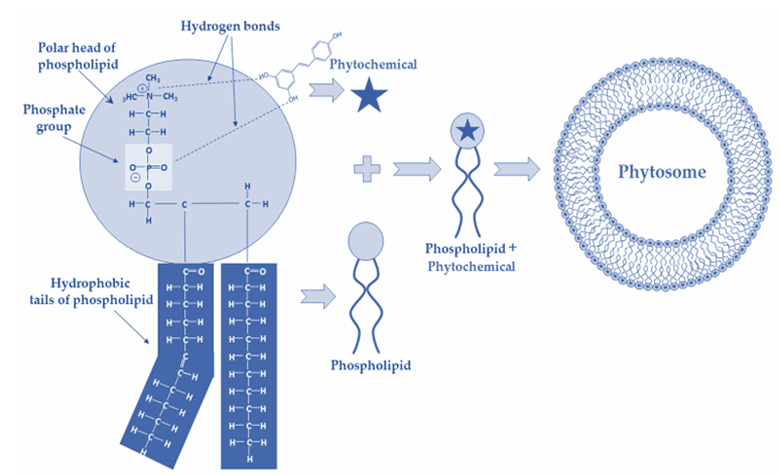
Figure 1. Schematic representation of the chemical bonds established during phytosome formation and their structure (7)
COMPONENTS OF PHYTOSOMES
Phospholipids
Phospholipids can be broadly classified based on their backbone structure into sphingomyelins and glycerophospholipids. The latter group includes phosphatidylcholine (PC), phosphatidylethanolamine (PE), phosphatidylserine (PS), phosphatidic acid (PA), phosphatidylinositol (PI), and phosphatidylglycerol (PG). Among these, PC, PE, and PS are the primary phospholipids used for forming complexes due to their characteristic structure comprising a hydrophilic head group and two hydrophobic hydrocarbon chains. Of these, phosphatidylcholine (PC) is the most widely utilized, owing to its amphipathic nature, which provides moderate solubility in both aqueous and lipid environments. Additionally, PC demonstrates excellent biocompatibility and low toxicity, reflecting its essential role in biological membranes. Importantly, PC has also been associated with therapeutic benefits, particularly in the management of liver disorders, where it exhibits notable hepatoprotective effects.(8)
Active constituents:
The active plant constituents, many of which are polyphenols with poor solubility or limited membrane permeability. For instance, hesperidin is hydrophilic and cannot cross membranes, while rutin and curcumin are lipophilic but poorly soluble in gastrointestinal fluids. Phytosomes, formed by complexing polyphenols with phospholipids, enhance their ability to cross biological barriers, improving stability, absorption, and therapeutic effectiveness while protecting them from degradation by oxidation, hydrolysis, or photolysis.(9)
Solvents
The selection of solvents plays a crucial role in phytosome preparation, influencing phospholipid solubility, complex formation, and stability. Hydrophobic solvents (e.g., dioxane, dichloromethane, ethyl acetate, n-hexane, acetone) are commonly used, while ethanol and methanol are preferred for food and pharmaceutical applications due to their low residue and eco-friendly nature, though their use is limited. A combination of protic and aprotic solvents, as well as supercritical fluids, has been shown to enhance solubility and efficiency in phytosome formulation. (10)
Table 1 Excipients used in phytosome formulation (9)
|
Chemical |
Example |
Uses |
|
Solvents |
methylene chloride, acetone |
Aprotic solvent |
|
Non-solvents |
n-hexane or aliphatic hydrocarbons |
Complex precipitation |
|
Phospholipid |
Egg phosphatidyl choline, soya phosphatidylcholine, phosphatidyl choline, dipalmitoyl phosphatidylcholine |
Cellular vesicles generating component |
|
Alcohols |
Methanol, ethanol |
solvent |
|
Buffering agents |
7 % v/v ethanol tris buffer (pH 6.5) and Saline phosphate buffer (pH 6.5) |
Hydrating medium |
METHODS OF PREPARATION (11)
Table-2 methods of Phytosomes preparation
|
Method |
Procedure |
|
Solvent Evaporation Method |
Plant extract and phospholipids are dissolved in acetone and refluxed at 50–60 °C. The mixture is concentrated, filtered, and the dried precipitate (phytosome complex) is stored in an amber glass container. |
|
Rotary Evaporation Technique |
Plant material and phospholipids are dissolved in tetrahydrofuran and stirred below 40 °C. After partial evaporation, n-hexane is added, and the resulting precipitate is collected and preserved in an amber container. |
|
Ether Injection Technique |
The drug–lipid mixture dissolved in organic solvent is slowly injected into a heated aqueous phase, resulting in vesicle formation. Depending on amphiphile concentration, structures may vary from micelles to vesicles. |
|
Mechanical Dispersion Method |
Phytoconstituents dissolved in diethyl ether are gradually added to an aqueous solution containing lipids. Removal of organic solvent under reduced pressure yields the phytophospholipid complex. Supercritical fluid (SCF) approaches (PCA, SAS, GAS) are modern alternatives. |
|
Lyophilization Technique |
Phospholipids and phytoconstituents are dissolved in suitable solvents, mixed, and stirred to form a complex. The complex is then isolated by freeze-drying (lyophilization). |
|
Salting Out Method |
Phosphatidylcholine and phytoconstituents are dissolved in aprotic solvents (e.g., acetone, dioxane) and stirred overnight. Addition of a non-solvent such as n-hexane precipitates the complex, which is then separated. |
|
Antisolvent Precipitation |
Drug and phospholipids are refluxed in dichloromethane, concentrated, and then precipitated using hexane under continuous stirring. The precipitate is filtered, dried, and sieved before storage. |
|
Supercritical Fluid (SCF) Technique |
In GAS, the drug–phospholipid solution is mixed with a supercritical antisolvent (e.g., CO?) at controlled temperature and pressure. In SEDS, both solvent and antisolvent are simultaneously introduced through a fine nozzle. These methods yield high-purity phytosome complexes with improved stability and yield. |
Characterization of Phytosomes (11)
Table 3: Characterization Of Phytosomes
|
Technique |
Description |
|
Visualization (TEM/SEM) |
Transmission electron microscopy (TEM) is employed to study vesicle size at high magnification (e.g., 1000×), while scanning electron microscopy (SEM) provides details on particle morphology and surface characteristics. Dried samples are mounted on gold-coated stubs for imaging. |
|
Particle Size & Zeta Potential |
Dynamic light scattering (DLS), supported by photon correlation spectroscopy (PCS), is used to determine vesicle size distribution and zeta potential, offering insights into dispersion stability. |
|
Transition Temperature |
Differential scanning calorimetry (DSC) measures the thermal transition of the vesicular lipid system. Samples, including drug–phospholipid complexes and controls, are heated between 0–400 °C at controlled rates in a nitrogen atmosphere. |
|
Surface Tension Measurement |
The Du Nouy ring tensiometer (ring method) assesses surface tension in aqueous drug solutions, reflecting amphiphilic behavior. |
|
Entrapment Efficiency |
Ultracentrifugation separates free drug from the phytosomal complex. The unentrapped drug concentration is quantified using UV spectroscopy to calculate entrapment percentage. |
|
Vesicle Stability |
Stability is monitored by evaluating vesicle size (via DLS) and structural integrity (via TEM) at defined time intervals. |
|
Drug Content |
The amount of incorporated drug can be quantified using validated analytical techniques, including HPLC or UV–Vis spectroscopy. |
Evaluation of Phtosomes:(9)
Ultraviolet Spectroscopy (UV): UV spectroscopy is employed to study the absorption behaviour of phytochemicals before and after complexation. Most reports indicate that phytosome formation does not significantly alter the absorption peaks of active components, suggesting that chromophoric groups remain intact.
Differential Scanning Calorimetry (DSC): DSC is used to examine thermal transitions such as melting points, peak shifts, and variations in enthalpy. Phyto phospholipid complexes usually show distinct thermograms compared to physical mixtures, indicating strong intermolecular interactions between phytoconstituents and phospholipid chains.
Fourier Transform Infrared Spectroscopy (FTIR): FTIR helps identify functional group interactions by analysing shifts in band position, intensity, and shape. Comparison of complexes with pure compounds and their mixtures often reveals new peaks or altered intensities, confirming the formation of Phyto-phospholipid complexes.
X-ray Diffraction (XRD): XRD provides information on crystallinity versus amorphous character. The disappearance of sharp crystalline peaks in complexes compared to pure phytoconstituents suggests molecular dispersion and transformation into an amorphous form, which may enhance solubility and bioavailability.
Nuclear Magnetic Resonance (NMR): ¹H and ¹³C NMR techniques allow for detailed structural analysis. Results indicate that phytosome formation is stabilized mainly by hydrogen bonding between polyphenolic groups and phospholipid head regions, rather than covalent bond formation.
CONCLUSION
Phytosome technology represents a significant advancement in enhancing the therapeutic potential of herbal medicines by overcoming the limitations of poor solubility and low bioavailability. By forming stable complexes between plant-derived bioactive compounds and phospholipids, Phytosomes improve absorption, stability, and pharmacokinetic behaviour, making them effective for both pharmaceutical and cosmetic applications. Various preparation techniques—ranging from solvent-based methods to modern supercritical fluid approaches enable the efficient development of these complexes, while advanced characterization tools such as TEM, SEM, DSC, FTIR, XRD, and NMR confirm their structural integrity and functionality. Collectively, these features establish Phytosomes as a promising drug delivery system that bridges traditional herbal medicine with modern therapeutic efficiency.
REFERENCES

Jyothi S, Nimisha Jain, Phytosomes : An Approach for Drug Delivery of Herbal Drugs, Int. J. of Pharm. Sci., 2025, Vol 3, Issue 9, 1438-1444. https://doi.org/10.5281/zenodo.17113414
 10.5281/zenodo.17113414
