View Article

Abstract

One of the body's most important defenses against infections, wounds, and irritants is inflammation. On the other hand, long-term inflammation can cause major illnesses like cancer, heart disease, and arthritis. Psidium guajava, or guava, is one of the species that are frequently used in traditional medicine to treat inflammation. Psidium guajava's roots have not gotten much scientific attention, despite the fact that its fruits and leaves have been extensively researched. This study uses the paw edema caused by carrageenan method in Wistar rats to assess the anti-inflammatory properties of the ethanolic extract of the root of Psidium that guajava.The existence of biologically active substances such as tannins, flavonoids, alkaloids, the saponins and phenolic compounds was verified by medicinal plants evaluation of the root extract. These components are known to have the ability to lessen oxidative stress and inflammation. Rats' hind paws were injected with 1?rrageenan solution to create inflammation in the experimental model. The usual medication was indomethacin (10 mg/kg), and the extract was given orally at dosages of 200 and 400 mg/kg. To evaluate the anti-inflammatory response, paw volumes were evaluated at 0, sixty, one hundred and twelve, and 180 minutes.Animals who received the ethanolic root extract showed a significant and dosage-dependent decrease in paw edema, particularly at the 400 mg/kg dose, according to the results. The extract has significant anti-inflammatory activity, as evidenced by the inhibitory impact being similar to that of the conventional medication. The results demonstrate Psidium guajava root's potential as a natural source of anti-inflammatory chemicals and validate its traditional use in inflammatory disorders. New anti-inflammatory medications made from guava root may result from additional research concentrating on the separation of active ingredients and their modes of action

Keywords

Psidium guajava, anti-inflammatory, ethanolic root extract, carrageenan paw edema, analgesic, Wistar rats

Introduction

The term "inflammatory" (derived from the Latin "inflammatory") describes how biological sensitivities of the body's tissues to detrimental stimuli include, The Latin term "Calor, dolor, rubor, tumor, and function laesa" refers to the five cardinal indicators: pain, heat, swelling, redness, and loss of function. such infections, damaged cells, or allergens. [1] The response of the immune system to dangerous stimuli, such as radiation, poisons, damaged cells, or infections, is inflammation. It works by eliminating harmful stimuli and starting the healing process. Thus, inflammation is an essential protective mechanism for good health. The danger of infection or injury during acute inflammatory reactions is frequently successfully decreased by cellular and molecular mechanisms and interactions. Restoring tissue homeostasis and resolving acute inflammation are facilitated by this mitigation strategy. Acute inflammation, however, can develop into chronic inflammation if left untreated, and this can result in a variety of chronic inflammatory diseases. Numerous pathogenic factors that result in tissue damage, Inflammation can be carried on by diseases like infection, tissue injury, and angina. Inflammation may result from non-infectious or viral causes. Tissue damage sets off a series of chemical signaling events in the body that lead to the repair of damaged tissues. These cues initiate leukocyte chemotaxis to areas of injury from the general circulation. During excitement, these leukocytes produce cytokines that lead to inflammation. [2] With the discovery that therapeutically effective NSAIDs inhibited the cyclo-oxygenase enzyme, which was also found in the gastric mucosa, the discovery of NSAIDs with a lower risk of stomach ulcers was ultimately made possible. Celecoxib and other selective COX2 inhibitors were developed as a result of the discovery that the cyclo-oxygenase found in signs of inflammation (COX2) differed from that found in the intestinal tract (COX1). Although these medications have a lower risk of causing stomach ulcers, they do relieve a number of arthritis symptoms. In order determine someone medication sensitivities and create therapy that is customized for each patient, pharmacogenomics was developed in response to the varying responsiveness to these and other therapeutic agents, as well as the fact that a pain reliever can cause an inflammatory response in certain patients with asthma. [3]The old medical term "inflammation" originally referred to traditional symptoms such as edema, erythema (redness), warmth, discomfort, and reduced function (stiffness and stiffness). According to current understanding, inflammation is a collection of evolving reactions to tissue damage that is mostly brought on by environmental agents, toxic substances, trauma, overuse, or illness. Like in many chronic disease states, some of these behaviors may encourage pathology, wound healing, or infection management. Inflammation is the second line of defense against pathogenic microorganisms. The responses that inflammation triggers are a crucial component of pathophysiology. Inflammation is a significant contributing factor to diseases that are described with the suffix -itis. The main immunological responses that produce inflammation are humoral and cell-mediated. This exercise describes the links between inflammation and cardiovascular disease and cancer, two considered the world's primary causes of death and illness. [4]

2. TYPES OF INFLAMMATION

2.1 Acute inflammation

Usually appearing within a few minutes or hours, immediate inflammation is a transient process that ends as soon as the damaging stimulus is eliminated. It involves the systematic and coordinated local mobilization of several immune-mediated, the endocrine and neurological mediators that cause acute inflammation. In a normal, healthy reaction, it is activated, eliminates the infection, initiates the recovery process, and then ceases. Acute inflammation occurs immediately upon an injury and resolves within a few days. With the aid of cytokines and chemokines, neutrophils and macrophages go to the site of inflammation. [5][6]

 2.2 Chronic inflammation

Inflammation that persists for a period of time is referred to as chronic inflammation. Unlike acute inflammatory disease, which is primarily characterized by lymphocytes, lymphocytes, macrophages, and plasma cells, chronic inflammation is characterized by neutrophils. Numerous illnesses, such as the disease, heart disease, responses, and a lung ailment known as chronic obstructive pulmonary disease, are mediated by chronic inflammation. Stress, smoking, obesity, poor nutrition, and other factors can contribute to chronic inflammation. [7]

3. Mechanism of anti-inflammatory agents

The main way that NSAIDs work is by inhibiting the cyclooxygenase (COX) enzyme. Cyclooxygenase is required to convert arachidonic acid into prostacyclin, the cytokines and thromboxane. The therapeutic benefits of NSAIDs are believed to be caused by a lack of these eicosanoids. Specifically, thromboxane promotes platelet adhesion, prostaglandins cause vasodilation, increase the hypothalamus temperature set-point, and aid in anti-nociception. [8]

4. Inflammation and herbal plant relation

The intricate process of inflammation is vital to the host's defense mechanism. Chronic conditions may result from the overproduction of certain inflammatory mediators. Different stages of the inflammatory process can be impacted by the anti-inflammatory effects of plant-based raw materials. They reduce skin flare, itching, or excessive exfoliation, stop the inflammatory response cascade from beginning, and limit the production of cytokines and eicosanoids. The majority of the herbal remedies on the market are used to treat inflammatory skin conditions based on in vitro clinical and pharmacological investigations as well as in vivo experiments. However, some of them are only used because of their many years of traditional folk medicine use. [9]

5. Process of inflammation

  • Histamine
  • cytokines
  • interleukin
  • prostaglandin [10]

6. MATERIAL AND METHODS

6.1 Preparation of extract

The root of the guava was cleaned and then cut into pieces and dried in the sunlight for several days. The root was ground into coarse powder using a mechanical grinder. 100 grams of the powdered root is placed in a thimble and kept inside the Soxhlet extractor. (95%

) ethanol is added to the round bottom flask as the solvent. The extraction process is carried out continuously for 24 hours. After the extraction is completed, the ethanol is removed using a rotary evaporator at 40? under reduced pressure. The thick extract that remains is dried is stored in an airtight container at 4? for later use.


Fig.: 2.1 Soxhlet Extraction


7. Pharmacological Evaluation

7.1 Animal study

One albino Wistar rat of each sex, weighs between 150 and 200 grams, bought on Ms. Chakraborty Enterprises in Kolkata, West Bangel, India, and kept in the Shambhunath Institute of Pharmacy, Jhalwa, Prayagraj, during the experiment. five animal per group They were kept within cages made of polypropylene that had a 12-hour cycle of light and darkness, a controlled ambient A relative humid of 55% and a temperature of 25±2°C. They were provided with an endless supply of water and a standard chow diet for the duration of the trial. The Animal Ethical Committee in the Institution (IAEC) authorized all of study protocols, which are also approved by the CPCSEA.


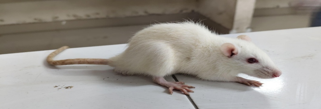
Fig.: 3.1 Albino Wistar rat


7.2 Acute Toxicity Studies

According to the OECD guidelines (423), which were obtained from CPCSEA, the Department of Social Affairs and Empowerment, and the Government of India, acute studies on toxicity must be conducted.Animals are chosen at random, labeled to enable unique identification, and housed in their cages for a minimum of five days. before the dosage to give them time to adjust to the lab environment. The test material is gavage via an intestinal tube or an appropriate intubation canula in a single dosage. The recommended amount may be administered in smaller portions over a maximum of 24 hours in the exceptional case that one dose is not feasible.Each phase involves four animals. One of 5 fixed dose levels—50 mg, one hundred mg, three hundred milligrams, 1000 mg, and 2000 milligrams per kilogram of body weight—is chosen to constitute the initial dose. The dose level that is believed to cause mortality for some of the administered animals should be the beginning dose level. The steps that need to be taken for each beginning dose are outlined in the flow charts in Annex 2.

7.3 Experimental Design

Twenty rats weighing between 150 and 190 g were used in Albino Wistar, and they were housed for an average of 4-5 weeks. Five groups of albino rats were established, with four rats in each group. The rats were then placed in separate cages.

  • Group I/Disease Control: Rats were given a single dose of carrageenan (1.0% wt./vol.) 0.10 milliliter with pure water in left hind paw (sub-plantar).
  • Group II/Standard Control: Administered 0.10 milliliter of 1.0% wt./vol. carrageenan in the left hind paw (sub-plantar) as a single dosage, and 10 mg/kg of the drug indomethacin in 0.5% CMC in distilled water orally.
  • Group III/Test Control 1: Rats received an oral dose of ethanol extract of Psidium guajava 200 mg/kg body wt. in 0.5% CMC and a single dose of carrageenan (1.0% wt./vol.) administered sub-plantar in the left hind paw (0.10 milliliter).
  • Group IV/Test Control 2: Rats received an oral dose of ethanol extract of Psidium guajava 400 mg/kg body wt. in 0.5% CMC and a single dose of carrageenan (1.0% wt./vol.) administered sub-plantar in the left hind paw (0.10 milliliter).

8. Screening Model

8.1 Anti-inflammatory activity

8.1.1 Carrageenan induced rat paw oedema model

The anti-inflammatory properties of carrageenan were evaluated in an experiment involving rat paw edema. An edema-including 0.1 ml of sub plantar injection of newly produced, every animal in every group received 1% the carrageenan in distilled water. Group 1 animals 2, 3, and 4 were given a single dosage of the vehicle thirty minutes before to carrageenan injection. Using a plethysmometer, the thickness of the paw was measured immediately before the carrageenan injection, or at “0 hour,” and then again one, two, three and four hours after the carrageenan injection and A standard test and medications administered orally. When the tibiotarsal articulation, the difference was measured as the increase in paw thickness between the "0 hour" paw thickness and at different times in relation to hours.

% Inhibition = Control mean- treated mean/ Control mean × 100

8.2 Analgesic Activity

8.2.1 Hot Plate method

The hotplate technique was also used to assess the extract's analgesic properties. Rats will be kept in a restrainer onto a hot plate that is kept at 55°C. The latency period, also known as the reaction (measured in seconds), was the amount of time it took the rats to react to thermal discomfort by jumping or licking their paws. It was recorded before the treatments were administered (0 minutes) and fifteen, thirty, forty-five, and 60 minutes later. The highest level of analgesia is necessary to avoid damaging the paw tissues. The formula below will be used to determine the minimum possible analgesia (MPA):

MPA = Reaction time for treatment – Reaction time for the saline/ 45 sec. reaction for saline × 100

9. RESULT AND DISCUSSION

9.1 PHARMACOLOGICAL EVALUATION

9.1.1 Evaluation of acute oral toxicity

Since there was not harmful effect and rat mortality up to 2000.0 mg/kg oral, the Psidium guajava root extract was deemed safe for use in further biological studies. At dosages of 1000.0, 2000.0 mg/kg, food consumption rose by 20% in just four hours, but then resumed as usual.

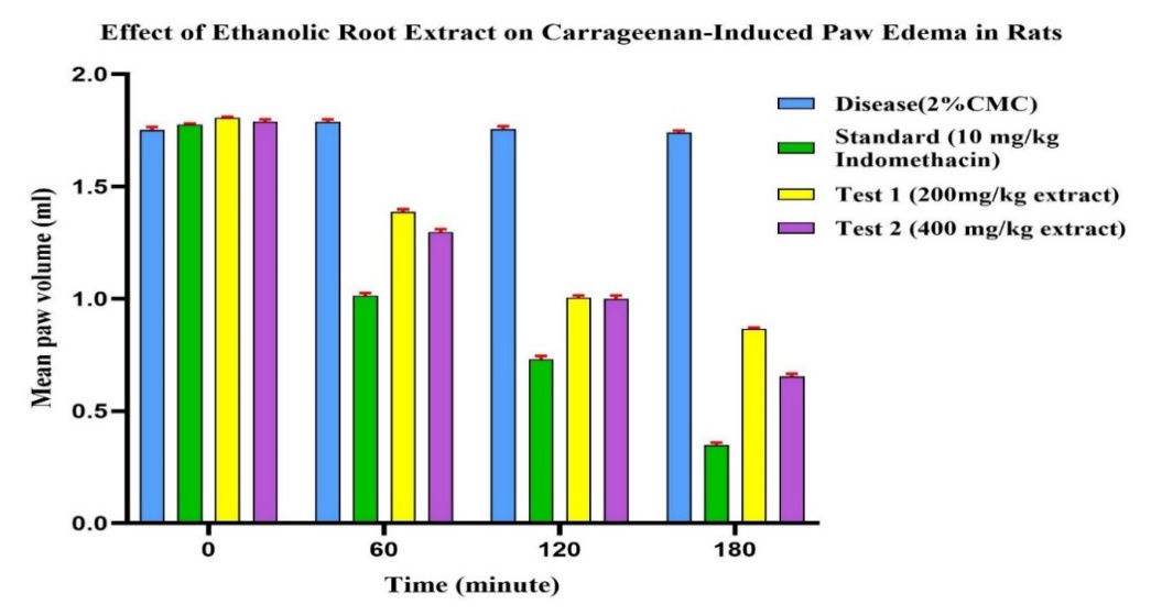
9.1.2 Anti-inflammatory activity of carrageenan


Table: 1 Effect of the Ethanol Extract On acute inflammation in rats using carrageenan


Group


Dose


Mean paw volume in ml

% inhibition

0 min

60 min

120 min

180 min

Diseased

2% CMC Solution

1.753 ± 0.012

1.788 ± 0.011

1.755 ± 0.012

1.740 ± 0.009

1.16%

Standard

10 mg/kg Indomethacin

1.775 ± 0.006

1.013 ± 0.013***

0.730 ± 0.013***

0.350 ± 0.004***

82.13%

Test 1

200 mg/kg ethanol root extract

1.805 ± 0.006

1.388 ± 0.011***

1.005 ± 0.011***

0.865 ± 0.007***

52.71%

Test 2

400 m/kg ethanol root extract

1.790 ± 0.009

1.298 ± 0.011***

1.000 ± 0.011***

0.655 ± 0.011***

65.81%


Statistical analysis

 Values are expressed as Mean ± Standard Error of Mean (SEM) for 4 rats per group (n = 4). Statistical analysis was performed using Two-Way ANOVA followed by Dunnett’s multiple comparisons test. Comparisons were made between each treatment group and the diseased control group at corresponding time points. * p < 0.05; ** p < 0.01; *** p < 0.001; **** p < 0.0001.


9.1.3 Analgesic activity of Hot plate method


Table: 2 Effect of the ethanol extract on acute Analgesic Activity in rats by using hot plate method


Group

Dose

Reaction time seen after certain time period of giving medication

0 min

30 min

60 min

120 min

Control

2% CMC Solution

10.00 ± 0.41

15.00 ± 0.41

13.00 ± 0.41

13.00 ± 0.41

Standard

10 mg/kg Indomethacin

12.00 ± 0.41

19.00 ± 0.41

15.00 ± 0.41

17.00 ± 0.41

Test 1

200 mg/kg ethanol root extract

11.00 ± 0.41

28.00 ± 0.41 ****

20.00 ± 0.41 ****

22.75 ± 0.25 ****


Test 2

400 mg/kg ethanol root extract

10.00 ± 0.41

33.00 ± 0.41 ****

21.00 ± 0.41 ****

24.00 ± 0.41 ****


Statistical analysis

Values represent Mean ± SEM (n = 4 rats per group). Statistical analysis: Two-Way Repeated Measures ANOVA followed by Tukey's multiple comparisons test. Asterisks indicate significant difference vs. Control group at the same time point: * = < 0.05 ** = p < 0.01 *** = p < 0.001 **** = p < 0.0001.


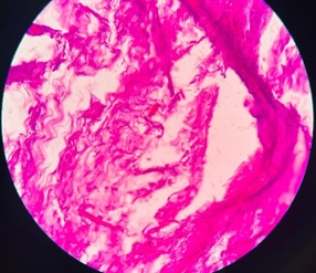
10. Histopathological Analysis


  1. Normal group                                                         (2) Diseased group


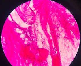
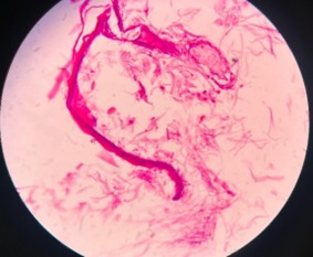
(Shows no tissue damage)                                               (Shows tissue damage)


  1. Standard group                                              (4) Test group


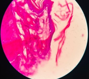
(Shows less tissue damage)                            (Shows less tissue damage than disease)


DISCUSSION

The person’s defense mechanism against damage and infection depends heavily on inflammation, but it also frequently results in chronic illnesses that are painful or hazardous and necessitate medical generation. Paw oedema brought on by carrageenan can be utilized to test for anti-inflammatory qualities in natural therapies since it is amenable to oral active anti-inflammatory medication, particularly as inflammation is at its height Carrageenan is widely used since it is antigenic model is very repeatable, and there are no discernible systemic effects. Rat paw oedema after a carrageenan injection occurs in two stages. The histamine and serotonin release are too responsible for the first hours of observation. Protease, prostaglandins, and the release of IL cause the second stage of oedema. Increased vascular permeability causes venules and arterioles to enlarge. Oedema is brought on by extravasating fluid and plasma proteins. After four hours, P. guajava root extract administered orally (100, 200 mg per kilogram of body weight) significantly reduced oedema by percentage. The release of mediators linked to inflammation may be inhibited by Psidium guajava root extract.

CONCLUSION

Extracts from Psidium guajava root considerably decreased inflammation in the current investigation. Oxidative stress may be the primary source of the inflammation that carrageenan causes since it generates inflammatory mediators. The current study shows that presence of substances derived from plants, such as tannins, alkaloids, and flavonoids, and glycosides reduces oxidative stress and inflammatory mediators (such as prostaglandin, histamine, IL, etc.). the phytoconstituents indicated above may all have the same anti-inflammatory qualities. The precise method by which phytoconstituents are influencing the aforementioned behaviours needs further research.

ACKNOWLEDGMENTS

The management and staff of the Shambhunath Institute of Pharmacy, Prayagraj, are appreciated by the authors for giving the resources and assistance needed to complete this study. Additionally, the authors would like to thank the Botanical Survey of India (BSI), Prayagraj, for his assistance for authenticating the plant material. We would especially want to thank the laboratory team for their technical support throughout the experiment.

REFERENCES

  1. Ferrero-Miliani, L., Nielsen, O. H., Andersen, P. S., & Girardin, S. (2007). Chronic inflammation: importance of NOD2 and NALP3 in interleukin-1β generation. Clinical & Experimental Immunology147(2), 227-235. https://doi.org/10.1111%2Fj.1365-2249.2006.03261.x
  2. Chen, L., Deng, H., Cui, H., Fang, J., Zuo, Z., Deng, J., ... & Zhao, L. (2017). Inflammatory responses and inflammation-associated diseases in organs. Oncotarget9(6), 7204. https://doi.org/10.18632/oncotarget.23208.
  3. Vane, J. R. (1971). Inhibition of prostaglandin synthesis as a mechanism of action for aspirin-like drugs. Nature new biology, 231, 232-235. https://doi.org/10.1038/newbio231232a0.
  4. Stone, W. L., Basit, H., Zubair, M., & Burns, B. (2024). Pathology, inflammation. In Stat Pearls [Internet]. Stat Pearls Publishing. https://www.ncbi.nlm.nih.gov/books/NBK534820/.
  5. Kumar, R., Clermont, G., Vodovotz, Y., & Chow, C. C. (2004). The dynamics of acute inflammation. Journal of theoretical biology230(2), 145-155. https://doi.org/10.1016/j.jtbi.2004.04.044.
  6. Bhaskar, K., Anbu, J., Ravichandiran, V., Venkateswarlu, V., & Rao, Y. (2009). Lipid nanoparticles for transdermal delivery of flurbiprofen: Formulation, in vitro, ex vivo and in vivo studies. Lipids in Health and Disease, 8(1), 6. https://doi.org/10.1186/1476-511X-8-6.
  7. Palombo, E. A. (2009). Traditional medicinal plant extracts and natural products with activity against oral bacteria: Potential application in the prevention and treatment of oral diseases. Evidence-Based Complementary and Alternative Medicine, 2011(1). https://doi.org/10.1093/ecam/nep067.
  8. Jaiarj, P., Khoohaswan, P., Wongkrajang, Y., Peungvicha, P., Suriyawong, P., Saraya, M. L., & Ruangsomboon, O. (1999). Anticough and antimicrobial activities of Psidium guajava Linn. leaf extract. Journal of Ethnopharmacology, 67(2), 203–212. https://doi.org/10.1016/S0378-8741(99)00022-7
  9. Ojewole, J. A. O. (2006). Anti-inflammatory and analgesic effects of Psidium guajava Linn. (Myrtaceae) leaf aqueous extract in rats and mice. Methods and Findings in Experimental and Clinical Pharmacology, 28(7), 441–446. ttps://doi.org/10.1358/mf.2006.28.7.1003578
  10. Díaz-de-Cerio, E., Verardo, V., Gómez-Caravaca, A. M., Fernández-Gutiérrez, A., & Segura-Carretero, A. (2017). Health-promoting properties of Psidium guajava L. leaves: An overview. Food & Function, 8(4), 1287–1305. https://doi.org/10.1358/mf.2006.28.7.1003578.
  11. Aiarj, P., Khoohaswan, P., Wongkrajang, Y., Peungvicha, P., Suriyawong, P., Saraya, M. L., & Ruangsomboon, O. (1999). Anticough and antimicrobial activities of Psidium guajava Linn. leaf extract. Journal of Ethnopharmacology, 67(2), 203–212. https://doi.org/10.1016/S0378-8741(99)00022-7.
  12. Costa, J. V., Silva, A. C. A., & Vieira, R. S. (2021). Psidium guajava extract modulates pro-inflammatory gene expression in human chondrocytes. Journal of Ethnopharmacology, 275, 114082. https://doi.org/10.1016/j.jep.2021.114082.
  13. Ghaderi, R., Tayarani-Najaran, Z., Ziaee, M., & Kianfar, G. (2022). The effect of hydroalcoholic extract of Psidium guajava L. on experimentally induced oral mucosal wound in rats. BMC Complementary Medicine and Therapies, 22, 201. https://doi.org/10.1016/S0378-8741(99)00022-7.
  14. Sen, S., de Araújo, J. R., & Oliveira, M. (2024). Ethyl acetate fraction of Psidium guajava leaf inhibits NF?κB pathway and leukocyte recruitment in LPS/carrageenan-induced inflammation models. Frontiers in Pharmacology, 15, 145906. https://doi.org/10.3389/fphar.2024.145906.
  15. Jayachandran, K., Phromnoi, T., & Kumar, R. (2023). Guava leaf extract suppresses pro-inflammatory cytokines and oxidative stress via NF?κB and MAPK pathway inhibition in vitro and in vivo. Bioactive Compounds from Guava Leaves, Phytomedicine, 90, 154678. https://doi.org/10.1016/j.phytomed.2023.154678.
  16. Sarwade, P. P., Gaisamudre, K. N., Kumar, S. R. S., Bongale, M. M., Otia, M., & Maurya, C. (2025, June 10). Psidium guajava: Botanical description, phytochemistry, pharmacological activities and its role in antiulcer activity. Journal for Research in Applied Sciences and Biotechnology, 4(3), 35–44. https://doi.org/10.55544/jrasb.4.3.5.
  17. Obasi, C. N., Glad, U., Prince?Nwogu, A. T., Unyinigwung, F. P., Nkwocha, E. C., & Agala, V., et al. (2025, May 15). Ameliorative effects of Psidium guajava leaf extract on ibuprofen?induced renal toxicity. Journal of Drug Delivery and Therapeutics, 15(5), 35–41. https://doi.org/10.22270/jddt.v15i5.7122.
  18. Jayachandran, K., Phromnoi, T., & Kumar, R. (2023). Guava leaf extract suppresses pro-inflammatory cytokines and oxidative stress via NF-κB and MAPK pathway inhibition in vitro and in vivo. Phytomedicine, 90, 154678. https://doi.org/10.1016/j.phytomed.2023.154678.
  19. Alam, A., Jawaid, T., Alsanad, S. M., Kamal, M., & Balaha, M. F. (2023). Composition, antibacterial efficacy, and anticancer activity of essential oil extracted from Psidium guajava (L.) leaves. Plants, 12(2), 246. https://doi.org/10.3390/plants12020246.
  20. Fredia Heppy, Mulyana, R., & Syah, N. A. (2023). The effect of Psidium guajava leaves’ extract for mild and asymptomatic coronavirus disease?19. Saudi Pharmaceutical Journal, 31(4), 592–596. https://doi.org/10.1016/j.jsps.2023.02.012.
  21. Jadaun, P., Shah, P., Harshithkumar, R., et al. (2023). Antiviral and ROS scavenging potential of Carica papaya Linn and Psidium guajava leaves extract against HIV-1 infection. BMC Complementary Medicine and Therapies, 23, 82. https://doi.org/10.1186/s12906-023-03916-x.
  22. Kuang, W., Wang, C., Xia, X., Kou, K. I., Kraithong, S., Lin, Q., Zhang, J., Liu, J., & Huang, R. (2025). Structural Characterization and Anti-Inflammatory Effects of Oligosaccharides in Psidium guajava Linn. Journal of agricultural and food chemistry73(20), 12474–12487. https://doi.org/10.1021/acs.jafc.5c02148.
  23. Chechani, B., Roat, P., Hada, S., Yadav, D. K., & Kumari, N. (2024). Psidium guajava: An Insight into Ethnomedicinal Uses, Phytochemistry, and Pharmacology. Combinatorial chemistry & high throughput screening27(1), 2–39. https://doi.org/10.2174/1386207326666230426093315.
  24. Lok B, Babu D, Tabana Y, Dahham SS, Adam MAA, Barakat K, Sandai D. The Anticancer Potential of Psidium guajava (Guava) Extracts. Life. 2023; 13(2):346. https://doi.org/10.3390/life13020346.
  25. Zhang, H., Shen, G., Lu, H., Jiang, C., Hu, W., Jiang, Q., Xiang, X., Wang, Z., & Chen, L. (2024). Psidium guajava seed oil reduces the severity of colitis induced by dextran sulfate sodium by modulating the intestinal microbiota and restoring the intestinal barrier. Foods, 13(17), 2668. https://doi.org/10.3390/foods13172668.
  26. Kareem, A. T., & Kadhim, E. J. (2024). Psidium guajava: A review on its pharmacological and phytochemical constituents. Biomedicine & Pharmacology Journal, 17(2). https://doi.org/10.13005/bpj/2924.
  27. Garrido, G. (2024). Antidiarrheal effect of Psidium guajava L. extract in acute diarrhea: A systematic review. Journal of the Science of Food and Agriculture, 104(13), 7731–7753. https://doi.org/10.1002/jsfa.13515.
  28. Costa, J. V., Silva, A. C. A., & Vieira, R. S. (2021–2023). Effect of Psidium guajava hydroalcoholic extract on TNF?α and resistin gene expression in human osteoarthritic chondrocytes. Journal of Ethnopharmacology, 275, 114082. https://doi.org/10.1016/j.jep.2021.114082.
  29. Phromnoi, T., Jayachandran, K., & Ghaderi, F. (2022). Effect of hydroalcoholic extract of Psidium guajava L. on experimentally induced oral mucosal wound in rat. BMC Complementary Medicine and Therapies, 22, 201. https://doi.org/10.1186/s12906-022-03694-3.

Reference

  1. Ferrero-Miliani, L., Nielsen, O. H., Andersen, P. S., & Girardin, S. (2007). Chronic inflammation: importance of NOD2 and NALP3 in interleukin-1β generation. Clinical & Experimental Immunology147(2), 227-235. https://doi.org/10.1111%2Fj.1365-2249.2006.03261.x
  2. Chen, L., Deng, H., Cui, H., Fang, J., Zuo, Z., Deng, J., ... & Zhao, L. (2017). Inflammatory responses and inflammation-associated diseases in organs. Oncotarget9(6), 7204. https://doi.org/10.18632/oncotarget.23208.
  3. Vane, J. R. (1971). Inhibition of prostaglandin synthesis as a mechanism of action for aspirin-like drugs. Nature new biology, 231, 232-235. https://doi.org/10.1038/newbio231232a0.
  4. Stone, W. L., Basit, H., Zubair, M., & Burns, B. (2024). Pathology, inflammation. In Stat Pearls [Internet]. Stat Pearls Publishing. https://www.ncbi.nlm.nih.gov/books/NBK534820/.
  5. Kumar, R., Clermont, G., Vodovotz, Y., & Chow, C. C. (2004). The dynamics of acute inflammation. Journal of theoretical biology230(2), 145-155. https://doi.org/10.1016/j.jtbi.2004.04.044.
  6. Bhaskar, K., Anbu, J., Ravichandiran, V., Venkateswarlu, V., & Rao, Y. (2009). Lipid nanoparticles for transdermal delivery of flurbiprofen: Formulation, in vitro, ex vivo and in vivo studies. Lipids in Health and Disease, 8(1), 6. https://doi.org/10.1186/1476-511X-8-6.
  7. Palombo, E. A. (2009). Traditional medicinal plant extracts and natural products with activity against oral bacteria: Potential application in the prevention and treatment of oral diseases. Evidence-Based Complementary and Alternative Medicine, 2011(1). https://doi.org/10.1093/ecam/nep067.
  8. Jaiarj, P., Khoohaswan, P., Wongkrajang, Y., Peungvicha, P., Suriyawong, P., Saraya, M. L., & Ruangsomboon, O. (1999). Anticough and antimicrobial activities of Psidium guajava Linn. leaf extract. Journal of Ethnopharmacology, 67(2), 203–212. https://doi.org/10.1016/S0378-8741(99)00022-7
  9. Ojewole, J. A. O. (2006). Anti-inflammatory and analgesic effects of Psidium guajava Linn. (Myrtaceae) leaf aqueous extract in rats and mice. Methods and Findings in Experimental and Clinical Pharmacology, 28(7), 441–446. ttps://doi.org/10.1358/mf.2006.28.7.1003578
  10. Díaz-de-Cerio, E., Verardo, V., Gómez-Caravaca, A. M., Fernández-Gutiérrez, A., & Segura-Carretero, A. (2017). Health-promoting properties of Psidium guajava L. leaves: An overview. Food & Function, 8(4), 1287–1305. https://doi.org/10.1358/mf.2006.28.7.1003578.
  11. Aiarj, P., Khoohaswan, P., Wongkrajang, Y., Peungvicha, P., Suriyawong, P., Saraya, M. L., & Ruangsomboon, O. (1999). Anticough and antimicrobial activities of Psidium guajava Linn. leaf extract. Journal of Ethnopharmacology, 67(2), 203–212. https://doi.org/10.1016/S0378-8741(99)00022-7.
  12. Costa, J. V., Silva, A. C. A., & Vieira, R. S. (2021). Psidium guajava extract modulates pro-inflammatory gene expression in human chondrocytes. Journal of Ethnopharmacology, 275, 114082. https://doi.org/10.1016/j.jep.2021.114082.
  13. Ghaderi, R., Tayarani-Najaran, Z., Ziaee, M., & Kianfar, G. (2022). The effect of hydroalcoholic extract of Psidium guajava L. on experimentally induced oral mucosal wound in rats. BMC Complementary Medicine and Therapies, 22, 201. https://doi.org/10.1016/S0378-8741(99)00022-7.
  14. Sen, S., de Araújo, J. R., & Oliveira, M. (2024). Ethyl acetate fraction of Psidium guajava leaf inhibits NF?κB pathway and leukocyte recruitment in LPS/carrageenan-induced inflammation models. Frontiers in Pharmacology, 15, 145906. https://doi.org/10.3389/fphar.2024.145906.
  15. Jayachandran, K., Phromnoi, T., & Kumar, R. (2023). Guava leaf extract suppresses pro-inflammatory cytokines and oxidative stress via NF?κB and MAPK pathway inhibition in vitro and in vivo. Bioactive Compounds from Guava Leaves, Phytomedicine, 90, 154678. https://doi.org/10.1016/j.phytomed.2023.154678.
  16. Sarwade, P. P., Gaisamudre, K. N., Kumar, S. R. S., Bongale, M. M., Otia, M., & Maurya, C. (2025, June 10). Psidium guajava: Botanical description, phytochemistry, pharmacological activities and its role in antiulcer activity. Journal for Research in Applied Sciences and Biotechnology, 4(3), 35–44. https://doi.org/10.55544/jrasb.4.3.5.
  17. Obasi, C. N., Glad, U., Prince?Nwogu, A. T., Unyinigwung, F. P., Nkwocha, E. C., & Agala, V., et al. (2025, May 15). Ameliorative effects of Psidium guajava leaf extract on ibuprofen?induced renal toxicity. Journal of Drug Delivery and Therapeutics, 15(5), 35–41. https://doi.org/10.22270/jddt.v15i5.7122.
  18. Jayachandran, K., Phromnoi, T., & Kumar, R. (2023). Guava leaf extract suppresses pro-inflammatory cytokines and oxidative stress via NF-κB and MAPK pathway inhibition in vitro and in vivo. Phytomedicine, 90, 154678. https://doi.org/10.1016/j.phytomed.2023.154678.
  19. Alam, A., Jawaid, T., Alsanad, S. M., Kamal, M., & Balaha, M. F. (2023). Composition, antibacterial efficacy, and anticancer activity of essential oil extracted from Psidium guajava (L.) leaves. Plants, 12(2), 246. https://doi.org/10.3390/plants12020246.
  20. Fredia Heppy, Mulyana, R., & Syah, N. A. (2023). The effect of Psidium guajava leaves’ extract for mild and asymptomatic coronavirus disease?19. Saudi Pharmaceutical Journal, 31(4), 592–596. https://doi.org/10.1016/j.jsps.2023.02.012.
  21. Jadaun, P., Shah, P., Harshithkumar, R., et al. (2023). Antiviral and ROS scavenging potential of Carica papaya Linn and Psidium guajava leaves extract against HIV-1 infection. BMC Complementary Medicine and Therapies, 23, 82. https://doi.org/10.1186/s12906-023-03916-x.
  22. Kuang, W., Wang, C., Xia, X., Kou, K. I., Kraithong, S., Lin, Q., Zhang, J., Liu, J., & Huang, R. (2025). Structural Characterization and Anti-Inflammatory Effects of Oligosaccharides in Psidium guajava Linn. Journal of agricultural and food chemistry73(20), 12474–12487. https://doi.org/10.1021/acs.jafc.5c02148.
  23. Chechani, B., Roat, P., Hada, S., Yadav, D. K., & Kumari, N. (2024). Psidium guajava: An Insight into Ethnomedicinal Uses, Phytochemistry, and Pharmacology. Combinatorial chemistry & high throughput screening27(1), 2–39. https://doi.org/10.2174/1386207326666230426093315.
  24. Lok B, Babu D, Tabana Y, Dahham SS, Adam MAA, Barakat K, Sandai D. The Anticancer Potential of Psidium guajava (Guava) Extracts. Life. 2023; 13(2):346. https://doi.org/10.3390/life13020346.
  25. Zhang, H., Shen, G., Lu, H., Jiang, C., Hu, W., Jiang, Q., Xiang, X., Wang, Z., & Chen, L. (2024). Psidium guajava seed oil reduces the severity of colitis induced by dextran sulfate sodium by modulating the intestinal microbiota and restoring the intestinal barrier. Foods, 13(17), 2668. https://doi.org/10.3390/foods13172668.
  26. Kareem, A. T., & Kadhim, E. J. (2024). Psidium guajava: A review on its pharmacological and phytochemical constituents. Biomedicine & Pharmacology Journal, 17(2). https://doi.org/10.13005/bpj/2924.
  27. Garrido, G. (2024). Antidiarrheal effect of Psidium guajava L. extract in acute diarrhea: A systematic review. Journal of the Science of Food and Agriculture, 104(13), 7731–7753. https://doi.org/10.1002/jsfa.13515.
  28. Costa, J. V., Silva, A. C. A., & Vieira, R. S. (2021–2023). Effect of Psidium guajava hydroalcoholic extract on TNF?α and resistin gene expression in human osteoarthritic chondrocytes. Journal of Ethnopharmacology, 275, 114082. https://doi.org/10.1016/j.jep.2021.114082.
  29. Phromnoi, T., Jayachandran, K., & Ghaderi, F. (2022). Effect of hydroalcoholic extract of Psidium guajava L. on experimentally induced oral mucosal wound in rat. BMC Complementary Medicine and Therapies, 22, 201. https://doi.org/10.1186/s12906-022-03694-3.

Photo
Preeti maurya
Corresponding author

Department of pharmacology, Shambhunath institute of pharmacy, jhalwa, prayagraj, Uttar Pradesh, India

Photo
Shweta Singh
Co-author

Department of pharmacology, shambhunath institute of pharmacy jhalwa prayagraj Uttar Pradesh India

Photo
Shruti Shukla
Co-author

Department of pharmacology, shambhunath institute of pharmacy jhalwa prayagraj Uttar Pradesh India

Photo
Rishita Shrivastava
Co-author

Department of pharmacology, shambhunath institute of pharmacy, jhalwa prayagraj, Uttar Pradesh, India

Preeti Maurya, Shweta Singh, Shruti Shukla, Rishita Shrivastava, Evaluation Of Anti-Inflammatory Activity of Ethanolic Root Extract of Psidium Guajava, Int. J. of Pharm. Sci., 2026, Vol 4, Issue 2, 2806-2817. https://doi.org/10.5281/zenodo.18682595

More related articles
A Review on Lidocaine...
Nagaveni Pommala , Swetha Meeniga , Saravanakumar Kasimedu , Moun...
Detection of Corona Virus: A Review on Applicatio...
Sahil Agrawal, B. Potdar, J. Shaikh, K. Pimpale, L. Kochage, N. C...
Formulation and Evaluation Study of Anti-Dandruff ...
Sivadas K. M., Lakshmi R., Zainabathul Hiba L. K., Fathimathu Naj...
Brand survey-based study of Ciprofloxacin-500 tablet marketing for Tuberculosis...
Ravi Maurya, Ishu Garg, Harish Kumar, Divyanshu Raj, Devbrath Tripathi , ...
Analytical Quality by Design Approach To RP-HPLC Method Development and Validati...
Rohan Pote, Dr. Sushil Patil, Ramdas Darade, Amol Gayake, Vikas Shinde, ...
Related Articles
Design, Synthesis and Pharmacological Evaluation of Novel Pyrimidine Derivatives...
Avinash Patil, Prashant Chavan, Amitkumar Rawal, Kiran Patil, ...
Teratogenic Effect of Different Drug at Different Stage of Pregnancy ...
Doltade Sonal, Dr. Malpani Suraj, Siral Vaishnavi, Bhakare Mahesh, Kedar Ganesh, Khandekar Janardan,...
A Formulation and Evaluation of Herbal Hair Oil ...
Seema Gaikwad, Dipmala Ghorpade, Gajanan Sanap, ...
Development And Validation of New RP HPLC Method For the Simultaneous Estimation...
P. Seetharamaiah, Nagaraju Pappula, JVLN. Seshagiri Rao, D. Gowrisankar, ...
A Review on Lidocaine...
Nagaveni Pommala , Swetha Meeniga , Saravanakumar Kasimedu , Mounika Gandham , Pranuth Atthoti , ...
More related articles
A Review on Lidocaine...
Nagaveni Pommala , Swetha Meeniga , Saravanakumar Kasimedu , Mounika Gandham , Pranuth Atthoti , ...
Detection of Corona Virus: A Review on Applications of RT PCR in Detection of C...
Sahil Agrawal, B. Potdar, J. Shaikh, K. Pimpale, L. Kochage, N. Chougule, ...
Formulation and Evaluation Study of Anti-Dandruff Herbal Shampoo...
Sivadas K. M., Lakshmi R., Zainabathul Hiba L. K., Fathimathu Naja, Remseena M., Priya Abraham, ...