View Article

Abstract

The present study aimed to evaluate the hepatoprotective potential of mangosteen (Garcinia mangostana L.) leaf extract prepared using the maceration method. Mangosteen leaves are known to be rich in bioactive compounds such as xanthones, flavonoids, and phenolic constituents, which exhibit strong antioxidant and cytoprotective properties. The hepatoprotective activity of the extract was assessed using the MTT assay on HepG2 human liver cell lines exposed to a hepatotoxic agent. Various concentrations of the extract were tested to determine their effect on cell viability. The results demonstrated a significant, concentration-dependent increase in cell viability and a marked reduction in toxin-induced cellular damage. These findings indicate that mangosteen leaf extract possesses notable hepatoprotective activity and supports its potential use as a natural therapeutic agent for the management of liver injury

Keywords

Garcinia mangostana, Hepatoprotective activity,HepG2cell line,MTT assay, Acetaminophen induced hepatotoxicity

Introduction

Mangosteen (Garcinia mangostana L.) is a tropical species known for its abundance of biologically active constituents, including xanthones, flavonoids, triterpenoids, tannins, saponins, alkaloids, and benzophenones, which collectively demonstrate potent antioxidant, anti-inflammatory, cytotoxic, and liver-protective effects. The liver is particularly susceptible to injury from toxic agents such as carbon tetrachloride (CCl?), pharmaceuticals, alcohol, and environmental contaminants, resulting in oxidative stress, membrane lipid peroxidation, enzymatic leakage, fibrosis, and progressive loss of hepatic function. Bioactive compounds present in mangosteen leaves exert hepatoprotective effects mainly by strengthening endogenous antioxidant systems, suppressing the generation of reactive oxygen species (ROS), limiting lipid peroxidation, and maintaining the structural integrity of hepatocyte membranes, thereby reducing elevated serum levels of hepatic markers including aspartate aminotransferase (AST), alanine aminotransferase (ALT), alkaline phosphatase(ALP) and bilirubin. Experimental evidence from both in vitro and in vivo models of liver injury induced by CCl?, paracetamol, ethanol, and related hepatotoxins indicates that mangosteen phytochemicals significantly lower malondialdehyde (MDA) concentrations, conserve intracellular antioxidants such as glutathione and tocopherols, and support restoration of normal liver histoarchitecture. Thus, the use of mangosteen leaves represents a promising plant-based strategy for liver protection, combining pharmacological efficacy with sustainable utilization of botanical resources.

MATERIALS AND METHODS

3.1 Plant Material Collection and Authentication

Healthy leaves of Garcinia mangostana L. were collected from local cultivation areas in southern India during the post-monsoon season. The plant was authenticated by a qualified taxonomist, and a reference specimen was preserved for documentation. The collected leaves were rinsed with distilled water to remove surface contaminants and shade-dried at ambient temperature until a constant weight was obtained. The dried material was ground into a coarse powder and stored in airtight containers for further experimental use.

3.2 Preparation of Aqueous Leaf Extract

The dried leaf powder (50 g) was soaked in ethanol (500 mL) and subjected to cold maceration for 72 hours with intermittent agitation to enhance extraction efficiency. The mixture was filtered through muslin cloth followed by Whatman No. 1 filter paper. The resulting filtrate was concentrated using a rotary vacuum evaporator at controlled temperature and further dried to obtain a solid aqueous extract. The extract was preserved at 4 °C until required for analysis.

3.3 Preliminary Phytochemical Analysis

The aqueous extract was qualitatively screened for major secondary metabolites using standard phytochemical procedures. Tests were conducted to identify the presence of phenolic compounds, flavonoids, tannins, alkaloids, saponins, and xanthone derivatives.

3.4 In Vitro Hepatoprotective Activity

3.4.1 Cell Line and Culture Conditions

Human liver carcinoma cells (HepG2) were obtained from a certified cell repository and maintained in Dulbecco’s Modified Eagle Medium supplemented with fetal bovine serum (10%) and antibiotic solution. The cells were cultured in a humidified incubator at 37 °C with 5% carbon dioxide.

3.4.2 Cell Seeding and Induction of Hepatotoxicity

Exponentially growing HepG2 cells were trypsinized and seeded into 96-well plates at a density of 5 × 10³ cells per well. After 24 hours of incubation, hepatotoxicity was induced by treating the cells with acetaminophen (20 µM).

3.4.3 Treatment with Aqueous Leaf Extract

Following toxicity induction, the cells were treated with different concentrations of the aqueous leaf extract and incubated for an additional 24 hours. Untreated cells served as the normal control, while acetaminophen-treated cells served as the toxic control.

3.4.4 MTT Cell Viability Assay

After treatment, the culture medium was removed and MTT reagent (5 mg/mL) was added to each well. The plates were incubated for 4 hours to allow formation of formazan crystals. Dimethyl sulfoxide was then added to dissolve the crystals, and absorbance was measured at 570 nm using a microplate reader.

3.5 Evaluation of Hepatoprotective Activity

Cell viability was calculated and compared between control, toxic, and extract-treated groups. An increase in cell viability in the treated groups compared to the toxic control was considered indicative of hepatoprotective activity.


RESULTAND DISCUSSION

4.1 EXTRACTION

The powdered mangosteen (garcinia mangostana) leaf extract were subjected to maceration extraction using ethanol as solvent. The extract was concentrated to semi solid mass.


Parameter

Observation

Weight of dried leaf powder

40g

Solvent used

Ethanol(95%)

Extraction method

maceration

Temperature

Room temperature

duration

72 hours

Extract obtained

5.5g

Percentage yield

13.75%


extraction details

Percentage yield = weight of extract ×100

weight of sample

A 13.75% yield indicates good extraction efficiency. Ethanol, being a polar solvent effectively extracts phenolic compounds, xanthones and flavonoids present in mangosteen leaves. Higher extract yield suggest a higher concentration of bioactive compounds responsible for antioxidant and hepatoprotective activity.

4.2 PHYTOCHEMICAL SCREENING


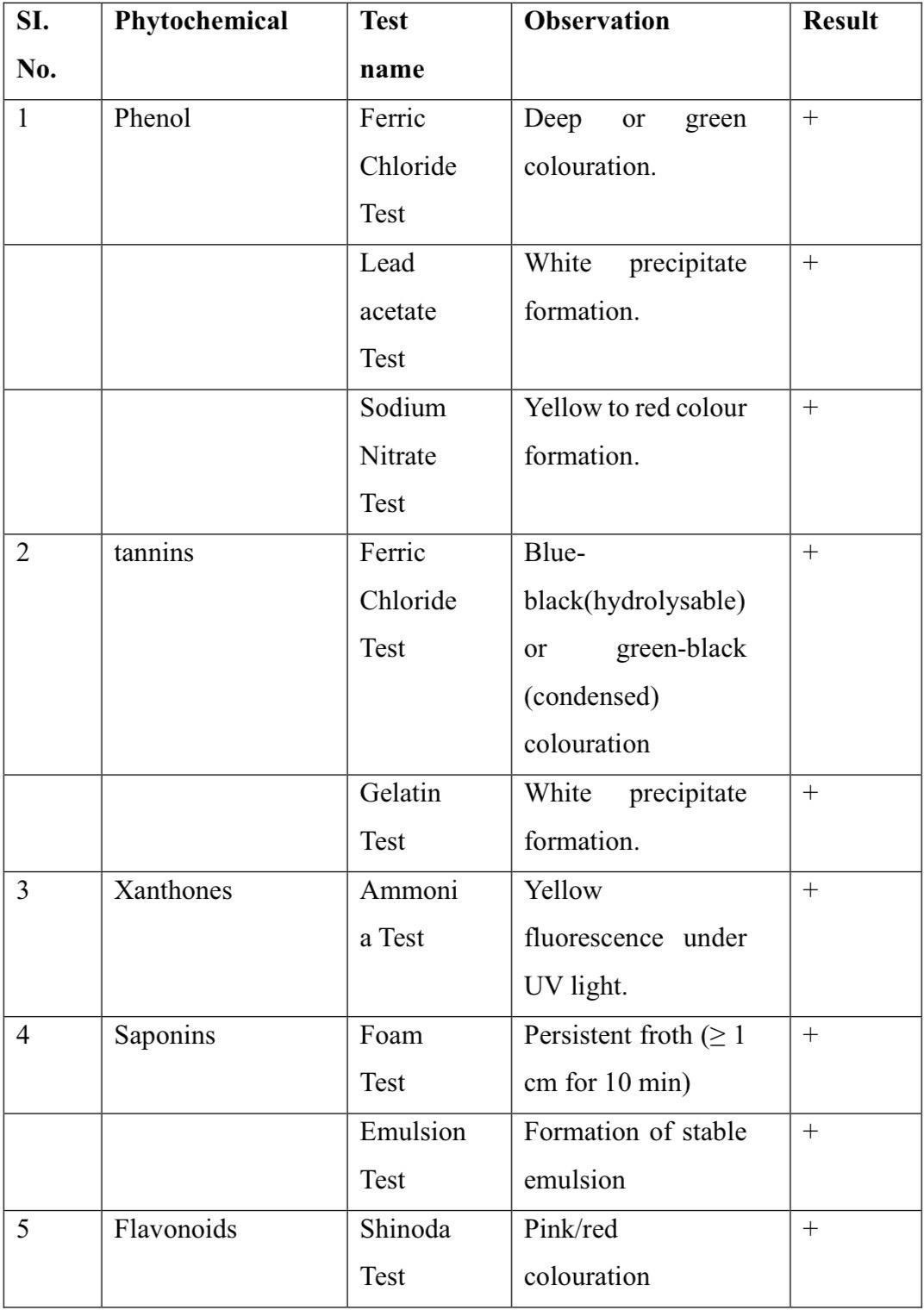
Preliminary phytochemical screening was carried out on the mangosteen leaf to identify the major classes of secondary metabolites present in the leaf. The phytochemical screening clearly show that mangosteen leaf extract contains a wide range of bioactive compounds. The presence of flavonoids, phenols, tannins,xanthones and saponins suggests strong potential for antioxidant, anti-inflammatory, antimicrobial and hepatoprotective activity.
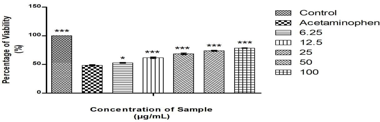
4.3 MTT ASSAY

The cytotoxic activity of the extract was evaluated using the MTT assay. The percentage of cell viability decreased with increasing concentrations of the extract ,showing a dosedependent effect.


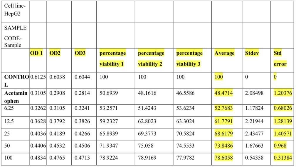
The study showed that acetaminophen greatly reduced the viability of HEPG2 liver cells, confirming its toxic effect. The sample treatment improved cell survival at all tested concentrations. As the dose increased, with the protective effect also increased, with the highest recovery seen at 100 g/mL. This shows that the sample can reduce the harmful impact caused by acetaminophen. Therefore, the sample demonstrates strong potential as a hepatoprotective agent.


CONCLUSION

This study demonstrates that mangosteen (Garcinia mangostana L.) leaf extract prepared through the maceration method. The extract, enriched with xanthones, flavonoids, and phenolic compounds, showed notable antioxidant and cell-protective effects. In HepG2 liver cells exposed to a toxic agent, the extract significantly increased cell viability, indicating its ability to reduce toxin-induced cellular damage. A clear dose-dependent response was observed, where higher concentrations of the extract provided greater protective effects. Overall, these findings highlight the promising role of mangosteen leaf extract as a natural agent for supporting and maintaining liver health.

REFERENCE

  1. Vinnakota S, Jayasree N. A new insight into the morphology of the human liver: a cadaveric study. ISRN Anat. 2013;2013:689564.
  2. Peeters G, Debbaut C, Laleman W, Monbaliu D, Vander Elst I, Detrez JR, et al. A multilevel framework to reconstruct anatomical 3D models of the hepatic vasculature in rat livers. J Anat.2017;230(3):417-83.
  3. MacParland SA, Liu JC, Ma XZ, Innes BT, Bartczak AM, Gage BK, et al. Single cell RNA sequencing of human liver reveals distinct intrahepatic macrophages population. Nat commun.2018;9(1):1-21.
  4. Shankar S, Rammohan A, Rela M, Srinivasan P. Surgical anatomy of segment four of liver and its implications in hepato-biliary surgery and liver transplantation. J Liver Transplant.2022;6(100076):100076.
  5. Chen H, Li T, Cai M, Huang Z, Gao J, Ding H, et al. study on gene expression in the liver at various developmental stages of human embryos. Front Cell Dev Biol. 2024;12:1515524.
  6. Friedman SL. Liver fibrosis-from bench to bedside. J Hepatol. 2021;75(3):795- 812.
  7. Asrani SK, Devarbhavi H, Eaton J, Kamath PS. Burden of liver diseases in theworld. J Hepatol. 2029;70(1):151-71.
  8. Ozturk, N. B., Herdan, E., Saner, F. H., & Gurakar, A. (2023). A comprehensive review of the diagnosis and management of acute liver failure. Journal of Clinical Medicine, 12(23), 7451.
  9. Liu Y, Wu Y, He S. Clinical value of microRNA-130a as marker of acute liver failure and its involvement in disease development. Hum Immunol. 2024;85(6);111-173.
  10. Manikat, R., Ahmed, A., & Kim, D. (2024). Current epidemiology of chronic liver disease. Gastroenterol Rep (Oxf), 12.
  11. Heidelbaugh JJ, Bruderly M. Cirrhosis and chronic liver failure: part. Diagnosis and evaluation. Am Fam Physician. 2006;74(5):756-62.
  12. Sumadewi Embryology, K. T. (2023). anatomy and physiology of the liver Indian. J. Clin. Anat. Physiol, 10(3), 138–144.
  13. Wang, L., Shao, Z., Wang, X., Lu, W., & Sun, H. (2025). Xenobiotic-induced liver injury: Molecular mechanisms and disease progression. Ecotoxicology and Environmental Safety, 303(118854), 118854.
  14. Messina,       A.,            &            Duclos-Vallée,            J.-C.     (2023). Molecular        mechanisms     of hepatotoxicity. International Journal of Molecular Sciences, 24(4), 3791.
  15. Roth, R. A., & Ganey, P. E. (2010). Intrinsic versus idiosyncratic drug-induced hepatotoxicity-two villains or one? J Pharmacol Exp Ther, 332(3), 692–697.
  16. Delgado-Montemayora, C., Cordero-Pérez, P., Salazar-Aranda, R., & Waksman Minsky, N. (2015). Models of hepatoprotective activity assessment. Medicina Universitaria,17(69), 222–228.
  17. Guduguntla, S. (2013). Hepatoprotective activity: a review. Pharma Tutor, 1.
  18. Arige, S. S., Arige, S. D., & Rao, A. (2017). A review on hepatoprotective activity. Int J Curr Res, 9(06), 51876–51881.
  19. Kumar, A., Rai, N., Kumar, N., Gautam, P., & Kumar, J. S. (2013). Mechanisms involved in hepatoprotection of different herbal products: a review. Int J Res Pharm Sci,4(2), 112-117.
  20. Pedraza-Chaverri J, Cárdenas-Rodríguez N, Orozco-Ibarra M, Pérez-Rojas JM. Medicinal properties of mangosteen (Garcinia mangostana). Food Chem Toxicol. 2008;46(10):3227–3239.
  21. Gutierrez-Orozco F, Failla ML. Biological activities and bioavailability of mangosteen xanthones: a critical review of the current evidence. Nutrient. 2013 Aug 13;5(8):3164.
  22. Chin, Y.; Kinghorn, A.D. Structural characterization, biological effects, and synthetic studies on xanthones from mangosteen (Garcinia mangostana), a popular botanical dietary supplement. Mini Rev. Org. Chem. 2008, 5, 355–364.
  23. Assemian ICCA, Bouyahya A, Dakka N, Bakri Y. Garcinia mangostana leaf extract from Ivory Coast possess remarkable antioxidant, anti-inflammatory, and cytotoxicological properties. Biomed Pharmacol J. 2019;12(2):517-526.24. Walker, E.B. HPLC analysis of selected xanthones in mangosteen fruit. J. Sep. Sci.2007, 30, 1229–1234.
  24. Erikania S, Juniatmoko R, Susilowati AA, Wardani TS Hi Wahid RA. Antioxidant and anticancer activity of mangosteen (Garcinia mangostana L.) leaf extract against human hepatocellular carcinoma (HepG-2) cells. Res J Pharm Technol.
  25. 2025;18(7):3017-3023.
  26. Andani R, Fajrina A, Asra R, Eriadi A. Antibacterial activity test of mangosteen plants (Garcinia mangostana L): A review. Journal Pharmaceutics and Pharmacology Research.2023;5(2):45-52.
  27. Aizat WM, Ahmad-Hashim FH, Syed Jaafar SN. Valorization of mangosteen,“The Queen of Fruits,” and new advances in postharvest and in food and engineering applications: A review. Food Research International. 2022;152:110921
  28. AizatWM, Ahad-Hashim FH, Syed Jaafar SN. Valorization of mangosteen, “The Queen of Fruits”, and new advances in postharvest and in food and engineering applications: A review. Food Research International. 2022;152:110921.
  29. Sun M, Gao AX, Ledesma-Amaro R. Microbial conversion of ethanol to high-value product: progress and challenges. Bio techno Biofuels Bioprod. 2024;17:83.
  30. R Suhartali, F Apriyani, Khusnul, D P Virgianti, M Fathurohman :Antimicrobial Activity Test of mangosteen Leaves Ethanol Extract (Garcinia mangostana Linn) Against Pseudomonas aeruginosa Bacteria” Journal of Physics: Conference series:1- 2.
  31. Ravichandran Veerasamy, et al, “Biosynthesis of silver non-particles usingmangosteen leaf extract and evaluation of their antimicrobial activities” 2011;113120.
  32. Rusli, R.K., et al. “Optimization of solvent and extraction time on secondary metabolite content of mangosteen leaf (Garcinia mangostana L) as a feed additivescandidate on poultry” JAVARVOL. 11;139-145.
  33. Alsultan, Q. M.N., Sijam, K., Rashid, T.S., & Ahamad, K.B. “GC-MS Analysis and Antibacterial Activity of Mangosteen Leaf Extract against Plant Pathogenic Bacteria” American Journal of Plant Sciences,2016;1013-1020.
  34. Johan Van Meerloo, et al, “Cell sensitivity assays: the MTT assay” 2011;237- 240.
  35. Rosa Supino, “Invitro toxicity testing protocols-MTT assay”1995;137-149.
  36. Laia Tolosa, et al. “General cytotoxicity assessment by means of the MTT assay” 2014;333-348.
  37. Jerard C, Michael BP, Chenicheri S, Vijayakumar N, Ramachandran R. Rosmarinic Acid-Rich Fraction from Menthaarvensis Synchronizes Bcl/Bax Expression and Induces Go/Gl Arrest in Hepatocarcinoma Cells. ProcNatlAcadSci, India, Sect B Biol Sci.2020 Sep;90(3):515-22.
  38. Mosmann T. Rapid colorimetric assay for cellular growth and survival: Application to proliferation and cytotoxicity assay. Journal of Immunological Methods. 1983 Dec;65(1-2):55-63.

Reference

  1. Vinnakota S, Jayasree N. A new insight into the morphology of the human liver: a cadaveric study. ISRN Anat. 2013;2013:689564.
  2. Peeters G, Debbaut C, Laleman W, Monbaliu D, Vander Elst I, Detrez JR, et al. A multilevel framework to reconstruct anatomical 3D models of the hepatic vasculature in rat livers. J Anat.2017;230(3):417-83.
  3. MacParland SA, Liu JC, Ma XZ, Innes BT, Bartczak AM, Gage BK, et al. Single cell RNA sequencing of human liver reveals distinct intrahepatic macrophages population. Nat commun.2018;9(1):1-21.
  4. Shankar S, Rammohan A, Rela M, Srinivasan P. Surgical anatomy of segment four of liver and its implications in hepato-biliary surgery and liver transplantation. J Liver Transplant.2022;6(100076):100076.
  5. Chen H, Li T, Cai M, Huang Z, Gao J, Ding H, et al. study on gene expression in the liver at various developmental stages of human embryos. Front Cell Dev Biol. 2024;12:1515524.
  6. Friedman SL. Liver fibrosis-from bench to bedside. J Hepatol. 2021;75(3):795- 812.
  7. Asrani SK, Devarbhavi H, Eaton J, Kamath PS. Burden of liver diseases in theworld. J Hepatol. 2029;70(1):151-71.
  8. Ozturk, N. B., Herdan, E., Saner, F. H., & Gurakar, A. (2023). A comprehensive review of the diagnosis and management of acute liver failure. Journal of Clinical Medicine, 12(23), 7451.
  9. Liu Y, Wu Y, He S. Clinical value of microRNA-130a as marker of acute liver failure and its involvement in disease development. Hum Immunol. 2024;85(6);111-173.
  10. Manikat, R., Ahmed, A., & Kim, D. (2024). Current epidemiology of chronic liver disease. Gastroenterol Rep (Oxf), 12.
  11. Heidelbaugh JJ, Bruderly M. Cirrhosis and chronic liver failure: part. Diagnosis and evaluation. Am Fam Physician. 2006;74(5):756-62.
  12. Sumadewi Embryology, K. T. (2023). anatomy and physiology of the liver Indian. J. Clin. Anat. Physiol, 10(3), 138–144.
  13. Wang, L., Shao, Z., Wang, X., Lu, W., & Sun, H. (2025). Xenobiotic-induced liver injury: Molecular mechanisms and disease progression. Ecotoxicology and Environmental Safety, 303(118854), 118854.
  14. Messina,       A.,            &            Duclos-Vallée,            J.-C.     (2023). Molecular        mechanisms     of hepatotoxicity. International Journal of Molecular Sciences, 24(4), 3791.
  15. Roth, R. A., & Ganey, P. E. (2010). Intrinsic versus idiosyncratic drug-induced hepatotoxicity-two villains or one? J Pharmacol Exp Ther, 332(3), 692–697.
  16. Delgado-Montemayora, C., Cordero-Pérez, P., Salazar-Aranda, R., & Waksman Minsky, N. (2015). Models of hepatoprotective activity assessment. Medicina Universitaria,17(69), 222–228.
  17. Guduguntla, S. (2013). Hepatoprotective activity: a review. Pharma Tutor, 1.
  18. Arige, S. S., Arige, S. D., & Rao, A. (2017). A review on hepatoprotective activity. Int J Curr Res, 9(06), 51876–51881.
  19. Kumar, A., Rai, N., Kumar, N., Gautam, P., & Kumar, J. S. (2013). Mechanisms involved in hepatoprotection of different herbal products: a review. Int J Res Pharm Sci,4(2), 112-117.
  20. Pedraza-Chaverri J, Cárdenas-Rodríguez N, Orozco-Ibarra M, Pérez-Rojas JM. Medicinal properties of mangosteen (Garcinia mangostana). Food Chem Toxicol. 2008;46(10):3227–3239.
  21. Gutierrez-Orozco F, Failla ML. Biological activities and bioavailability of mangosteen xanthones: a critical review of the current evidence. Nutrient. 2013 Aug 13;5(8):3164.
  22. Chin, Y.; Kinghorn, A.D. Structural characterization, biological effects, and synthetic studies on xanthones from mangosteen (Garcinia mangostana), a popular botanical dietary supplement. Mini Rev. Org. Chem. 2008, 5, 355–364.
  23. Assemian ICCA, Bouyahya A, Dakka N, Bakri Y. Garcinia mangostana leaf extract from Ivory Coast possess remarkable antioxidant, anti-inflammatory, and cytotoxicological properties. Biomed Pharmacol J. 2019;12(2):517-526.24. Walker, E.B. HPLC analysis of selected xanthones in mangosteen fruit. J. Sep. Sci.2007, 30, 1229–1234.
  24. Erikania S, Juniatmoko R, Susilowati AA, Wardani TS Hi Wahid RA. Antioxidant and anticancer activity of mangosteen (Garcinia mangostana L.) leaf extract against human hepatocellular carcinoma (HepG-2) cells. Res J Pharm Technol.
  25. 2025;18(7):3017-3023.
  26. Andani R, Fajrina A, Asra R, Eriadi A. Antibacterial activity test of mangosteen plants (Garcinia mangostana L): A review. Journal Pharmaceutics and Pharmacology Research.2023;5(2):45-52.
  27. Aizat WM, Ahmad-Hashim FH, Syed Jaafar SN. Valorization of mangosteen,“The Queen of Fruits,” and new advances in postharvest and in food and engineering applications: A review. Food Research International. 2022;152:110921
  28. AizatWM, Ahad-Hashim FH, Syed Jaafar SN. Valorization of mangosteen, “The Queen of Fruits”, and new advances in postharvest and in food and engineering applications: A review. Food Research International. 2022;152:110921.
  29. Sun M, Gao AX, Ledesma-Amaro R. Microbial conversion of ethanol to high-value product: progress and challenges. Bio techno Biofuels Bioprod. 2024;17:83.
  30. R Suhartali, F Apriyani, Khusnul, D P Virgianti, M Fathurohman :Antimicrobial Activity Test of mangosteen Leaves Ethanol Extract (Garcinia mangostana Linn) Against Pseudomonas aeruginosa Bacteria” Journal of Physics: Conference series:1- 2.
  31. Ravichandran Veerasamy, et al, “Biosynthesis of silver non-particles usingmangosteen leaf extract and evaluation of their antimicrobial activities” 2011;113120.
  32. Rusli, R.K., et al. “Optimization of solvent and extraction time on secondary metabolite content of mangosteen leaf (Garcinia mangostana L) as a feed additivescandidate on poultry” JAVARVOL. 11;139-145.
  33. Alsultan, Q. M.N., Sijam, K., Rashid, T.S., & Ahamad, K.B. “GC-MS Analysis and Antibacterial Activity of Mangosteen Leaf Extract against Plant Pathogenic Bacteria” American Journal of Plant Sciences,2016;1013-1020.
  34. Johan Van Meerloo, et al, “Cell sensitivity assays: the MTT assay” 2011;237- 240.
  35. Rosa Supino, “Invitro toxicity testing protocols-MTT assay”1995;137-149.
  36. Laia Tolosa, et al. “General cytotoxicity assessment by means of the MTT assay” 2014;333-348.
  37. Jerard C, Michael BP, Chenicheri S, Vijayakumar N, Ramachandran R. Rosmarinic Acid-Rich Fraction from Menthaarvensis Synchronizes Bcl/Bax Expression and Induces Go/Gl Arrest in Hepatocarcinoma Cells. ProcNatlAcadSci, India, Sect B Biol Sci.2020 Sep;90(3):515-22.
  38. Mosmann T. Rapid colorimetric assay for cellular growth and survival: Application to proliferation and cytotoxicity assay. Journal of Immunological Methods. 1983 Dec;65(1-2):55-63.

Photo
Noora Latheef
Corresponding author

Mount Zion College of Pharmaceutical Sciences and Research, Adoor

Photo
Amina Rasheed
Co-author

Mount Zion College of Pharmaceutical Sciences and Research, Adoor

Photo
Arya A
Co-author

Mount Zion College of Pharmaceutical Sciences and Research, Adoor

Photo
Shibina S
Co-author

Mount Zion College of Pharmaceutical Sciences and Research, Adoor

Photo
Archana SV
Co-author

Mount Zion College of Pharmaceutical Sciences and Research, Adoor

Photo
Rakhi A. R.
Co-author

Assistant Professor, Mount Zion College of Pharmaceutical Sciences and Research, Adoor

Noora Latheef*, Amina Rasheed, Arya A., Shibina S, Archana S. V., Rakhi A. R., Assessment of the hepatoprotective potential of Garcinia mangostana Leaf extract, Int. J. of Pharm. Sci., 2026, Vol 4, Issue 2, 48-54. https://doi.org/10.5281/zenodo.18453685

More related articles
Jackfruit Seed as a Source of Natural Bioactives: ...
Sonali Somase , Amol Gayke, Shivani Sangale, Sakshi Jadhav, ...
A Review on Promising Approach of Transferosomes f...
Kalugade Sanika, Deshmukh Akshada, ...
Metachromatic Leukodystrophy: A Comprehensive Review of Pathogenesis, Diagnosis,...
Sachitanand Biradar , Nikhil Chapule, Govind Soni, Vishnu Bharti, Harshvardhan Shinde, ...
Related Articles
The Role of Vitamin-D Receptor in Alzheimer’s Disease: A Molecular and Clinica...
Vrushali Murari, Arun Pachauri, Kailash Singh Bisht, Anupama Sinha, Lokesh Paranjape, Omkar Sutar, ...
Thiourea Based Antidiabetic Candidates: Integrating InVitro, InVivo, and In Sili...
Raja Waleed Sajjad, Hammad Nasir, Ahmad Nawaz, Saba Manzoor, Raja Ahmed, ...
Jackfruit Seed as a Source of Natural Bioactives: A Review on Its Therapeutic Po...
Sonali Somase , Amol Gayke, Shivani Sangale, Sakshi Jadhav, ...