Abstract
Varicose vein is one type of venous insufficiency that presents with any dilated, elongated, or tortuous veins caused by permanent loss of its valvular efficiency. Destruction of venous valves in the axial veins results in venous hypertension, reflux, and total dilatation, causing varicosities and transudation of fluid into subcutaneous tissue. Varicose Veins can be explained as a disorder of the veins (especially of legs) wherein they get affected due to the backward flow and turbulence in the circulation of the blood. The veins get perverted, become enlarged due to a condition called edema. The disease also shows many associated symptoms which worsens the condition of the varicose veins. The present article deals with brief introduction regarding the etiopathogenesis of varicose veins, related causes and symptoms, the current surgical and non- surgical treatments.
Keywords
Varicose vein. Valvular insufficiency, Thrombophlebitis.
Introduction
Varicose veins are very common problem with broadly varying estimates of prevalence and it cause disability and impairment in the quality of life. It is easily recognized by their twisted, bulging, superficial appearance on the lower extremities, but they also can be found in rectum (hemorrhoids), and esophagus (esophageal varices) etc.Varicose veins are very common: 40% of men and 32% of women aged 18-64 years have this condition.
Definition Varicose vein is one type of venous insufficiency which falls under the broad heading superficial venous disease. In Western populations, the incidence of varicose veins varies with the definition applied. Most investigators favor the definition of Arnoldi, who said that varicosities are “any dilated, elongated, or tortuous veins, irrespective of size” The normal flow of blood occurs from the superficial veins to the deep veins and from the legs up to the heart. Both the pathway for the flow of blood consists of a single-way venous valve. The ineffectiveness of the systems causes difficulty in the flow of blood and hence, leads to backward flow of blood, pooling of blood and in turn venous hypertension. Prior to the venous hypertension, there is increase in the hydrostatic pressure of the calves which then results in failure of the pump mechanism due to the perforating veins.
Pathophysiology
Venous hypertension, venous valvular incompetence, structural changes in the vein wall, inflammation, and alterations in shear stress are the major pathophysiological mechanisms resulting in varicose veins. Venous hypertension is caused by reflux attributable to venous valvular incompetence, venous outflow obstruction, or calf-muscle pump failure. Venous reflux may occur in either or both the superficial or deep venous system and results in venous hypertension below the area of venous valvular incompetence.

FIG:-1 Diagrammatic Representation of Normal Veins and Varicose Veins.
Thrombophlebitis
Superficial thrombophlebitis (“phlebitis”) can complicate varicose veins. The risk of deep vein thrombosis is remote, but in a case series it occurred very occasionally if phlebitis extended above the knee.4 Veins may sometimes remain permanently occluded. Treatment of the varicose veins may be appropriate if phlebitis is recurrent or severe, or if the veins also cause other symptoms. Note that thrombophlebitis is not caused by infection, and treatment with antibiotics is unnecessary: drug treatment should be limited to antiinflammatory analgesics.
Bleeding, skin changes, and ulcers
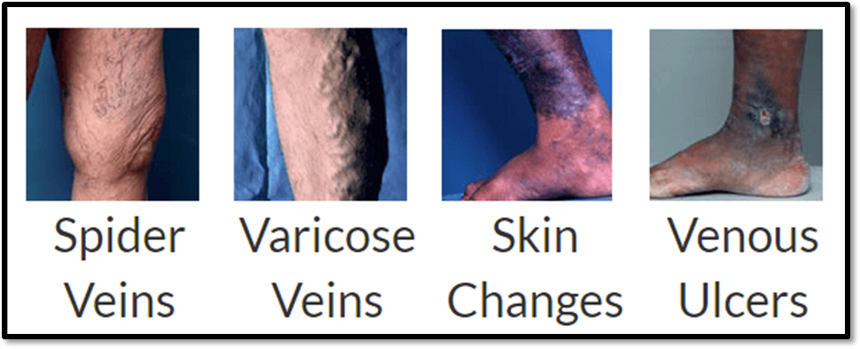
These are the complications of varicose veins that mandate consideration of treatment. They are all associated with high venous pressure in the upright position, as a result of incompetent venous valves. Bleeding is uncommon and usually occurs from a prominent vein on the leg or foot with thin, dark, unhealthy skin overlying it. “Skin changes” range from eczema, through brown discoloration, to florid lipodermatosclerosis with induration of the subcutaneous tissues.
Leg swelling
This is an uncommon symptom of varicose veins other causes are much commoner. Unilateral swelling of a leg with big varicose veins is the most typical presentation.

FIG:2 Skin changes (lipodermatosclerosis) caused by venous hypertension. Recognition of skin damage is fundamental in examination of varicose veins
Clinical Presentation.
Venous varicosities can be categorized according to the CEAP classification
Clinical Classification
C0: no visible or palpable signs of venous disease C1: telangiectasies or reticular veins
C2: varicose veins C3: edema
C4a: pigmentation or eczema
C4b: lipodermatosclerosis or atrophieblanche C5: healed venous ulcer
C6: active venous ulcer
S: symptomatic, including ache, pain, tightness, skinirritation, heaviness, and muscle cramps, and othercomplaints attributable to venous dysfunction
A: asymptomatic
Etiologic Classification
Ec: congenital
Ep: primary
Es: secondary (postthrombotic) En: no venous cause identified
Anatomic Classification
As: superficial veins Ap: perforator veins Ad: deep veins
An: no venous location identified
Pathophysiologic Classification
Basic CEAP Pr: reflux
Po: obstruction
Pr,o: reflux and obstruction
Pn: no venous pathophysiology identifiable.
Risk Factors:
Age As person get older, the tissues of vein walls lose elasticity and as causing the valve system to fail. Evans CJ et al.(1999) done cross sectional survey on 1566 participants concluded that approximately one third of men and women aged 18-64 years had trunk varices. Gender Women have a higher incidence of varicose vein disease due to female hormones and their effect on the vein walls. Brand FN et al. (1988) examined 3,822 adults, concluded that incidence of varicose veins is higher among women than men, and who had lower levels of physical activity and higher systolic blood pressure and higher smoking rates .
Heredity If parents and grandparents had the problem, it will increased risk of varicose veins. Lee AJ et al. (2003) conducted study which conclude that, self-reported evidence suggested a familial susceptibility . CornuThenard et al (1994) conducted a case control study on 134 families demonstrated a prominent role of heredity in the development of varicose veins . Kohno K et al (2016) reviewed the data and concluded that genetic factors make a strong contribution to the familial transmission of varicose vein from parents to offspring . Prolonged Standing Occupations that involve prolonged standing cause increased volume and pressure of blood in the lower limbs due to the effects of gravity. Kohno K et al. (2014) concluded that that exposure to both prolonged standing at work and overweight exacerbate varicose vein development. Hormonal Changes These occur during puberty, pregnancy, multiparous, and menopause, post- menopausal, hormone replacement and other medicines containing estrogen and progesterone may contribute to the forming of varicose veins. Lesiak M et al (2012) critically examined the data and conclude that Caesarean section, pregnancy, family factors are associated with inheritance of the formation of varicose changes and venous insufficiency. M. Dindelli et al.(1993) conducted survey on 611 women it concluded that to be secondiparae or more was associated with an increased risk of developing venous disease in pregnancy. Women who developed venous disease in pregnancy reported more frequently a family history of varicose disease than those who did not
Obesity Being overweight can put extra pressure on veins; this can lead to varicose veins. Seidell JC et al (1986) conducted retrospective cohort study it is concluded that incidence of registered morbidity in the overweight group was higher for varicose veins for women. Alcohol and Smoking Alcohol/ smoking also increases the risk of varicose veins. Ahti TM et al. (2010) conducted cross sectional study on 4903 participants, It is concluded that alcohol is likely to increase the risk of varicose veins in women and Smokers had a higher incidence of varicose veins compared with non-smokers in both genders . Musil D et al. (2016) conducted retrospective study 70 years and?on 641 patients concluded that age obesity were strongly associated with an occurrence of venous thromboembolism.
Diagnosis:
Clinical Examination
The clinical evaluation of a patient with varicose veins begins with the physical examination to determine the type, location, extent, and possibly the cause of the venous disease. Varicose v
eins should be examined in the standing position and inspected for erythema, tenderness, or induration that may suggest superficial vein thrombosis. The clinical examination should identify any signs of more advanced chronic venous disease such as edema, hyperpigmentation, lipodermatosclerosis, atrophie blanche, or ulceration. A complete pulse examination should be performed.
History taking
Detailed physical examination in sufficient light
A positive tap test and negative Perthes test.
Angiogram
Doppler test - an ultrasound scan to check the direction of blood flow in the veins and checks for blood clots in the veins.
Color duplex ultrasound scan
Tourniquet tests (such as the Trendelenberg test)
Venography
Ambulatory venous pressure measurements
Therapies for Varicose Veins:
Physical Therapy:
Exercise and Yogasans increase the muscle strength, stimulate the flow of blood and enhance the circulation. This relieves pain and other complications and thus promotes healthy veins. Sarvangasana, Halsana, Pawan Muktasana are some the vitalizing and effective yogasans for reducing the complications resulting from Varicose veins. In addition to this, the simple everyday activities such as walking, cycling, swimming, etc. help toning the muscles. The elevation of the legs using pillows or any other props overnight or for a few hours in the day time is recommended as it helps in better flow of blood. Massage therapy in which the tension is applied onto the muscles in the upward direction of the legs using oils such as citrus oils, olive oil, mustard oil, castor oil etc. also results in good circulation and proper drainage of blood .
Compression therapy:
The therapy uses the special type of compression stockings which constricts the dilated veins by creating pressure on surface of the calves. Therefore, there is decrease in the passage of the veins which in turn results in increased blood movement towards the heart.
Non-surgical Treatment:
Sclerotherapy:
Spider veins or angioectasis is treated using this technique. The technique involves use of sclerosing agents such as sodium salicylate, polidacanol, chroamted glycine which is injected using small needles. The treatment is accompanied with compression stockings to be worn after the sclerotherapy so as to constrict the treated vessels. Side effects to this treatment include scars at the site of injection, neovascularization (formation of petite veins which may take a couple of months to year to disappear), swelling and small ulcers (in severe cases)
Ultrasound guided foam sclerotherapy:
The method involves the damaging of the endothelial layer of the vein so as to create a blockage and scar formation in the dilated veins. The sclerosing agent here is in the form of foam as it provides larger surface area on the wall of the veins. The side effects to thistreatment were bubble embolism and thrombophlebitis.
Endothermal Ablation:
The treatment involves use of energy from radiofrequency and lasers to fasten the affected veins. These treatments ensure a rapid recovery. It includes two of the following methods:
Radiofrequency ablation of the Varicose Veins:
The affected veins are heated by using the bipolar generator and inducing radiofrequency catheter into it along with sheath able electrodes. This method is carried out at the temperature of 85±3 °C
Endovenous Ablation:
The method involves the closure of the vein by placing the catheter through the saphenous vein at the saphenofemoral junction (under the knee) and passing the laser fiber through it. This method is 98% successful method to cure the venous insufficiency. Complications observes were stiffness in the limb, pain and bruising
Surgical Treatments:
Vein Stripping: This is a surgical technique in which the affected veins are treated by insertion of special wires made of any suitable material by providing a tear onto the saphenous vein so as to “strip” the veins. The leg is operated by giving general anesthesia and known as bilateral surgery. Bleeding, bruising, infections may be observed as side effects.
Ambulatory Phlebectomy: The method in which the superficial veins are removed by performing incisions in the skin. The procedure is performed on the out patients by the dermatologist. The compression socks are continued to be worn after the surgery for some period of time. Temporary swelling and inflammation may be observed.
CONCLUSION:
Patients suffering from varicose veins usually have to undergo through various complex treatments, surgical or non-surgical, that involves number of intricate processes and other complications. Although, these methods are highly recommended by the physicians, they have certain drawbacks. The symptoms of the disease may reoccur in some cases if proper care is not takenThe agenda of this article is to knock down the clinical presentation that can be more beneficial to know the cause and other existing treatments of varicose veins. There is a vast scope for research in this field so as to bring up these various therapy in the form of various dosage forms and assigned doses.
REFERENCES
- Beebe-Dimmer, Pfeifer J, Engle J and Schottenfeld: The Epidemiology of Chronic Venous Insufficiency and Varicose Veins. Annals of Epidemiology. 2005; 15(3), 175-184.
- Sadat U and Gaunt M: Current management of varicose veins. British Journal of Hospital Medicine. 2008; 69(4), 214-217.
- McGuckin M, Waterman R, Brooks J, Cherry G, Porten L, Hurley S, Kerstein MD. Validation of venous leg ulcer guidelines in the United States and United Kingdom. Am J Surg. 2002;183:132–137.
- Shepherd AC, Gohel MS, Lim CS, Davies AH. A study to compare disease-specific quality of life with clinical anatomical and hemodynamic assessments in patients with varicose veins. J Vasc Surg. 2011;53:374–382.
- Gourgou S, Dedieu F, Sancho-Garnier H. Lower limb venous insufficiency and tobacco smoking: a case-control study. Am J Epidemiol. 2002;155:1007–1015.
- Raju S, Neglén P. Clinical practice. Chronic venous insufficiency and varicose veins. N Engl J Med. 2009;360:2319–2327.
- Eklöf B, Rutherford RB, Bergan JJ, Carpentier PH, Gloviczki P, Kistner RL, Meissner MH, Moneta GL, Myers K, Padberg FT, Perrin M, Ruckley CV, Smith PC, Wakefield TW; American Venous Forum International Ad Hoc Committee for Revision of the CEAP Classification. Revision of the CEAP classification for chronic venous disorders: consensus statement. J Vasc Surg. 2004;40:1248–1252.
- Jones RH, Carek PJ. Management of Varicose Veins. Am Fam Physician 2008;78(11):1289- 1294.
- M-C Nogaro, D J Pournaras, C Prasannan, A Chaudhuri. Varicose vein. BMJ 2012;344:e667 doi: http://dx.doi.org/10.1136/bmj.e667.
- Franz A, Wann Hansson C. Patients’ experiences of living with varicose veins and management of the disease in daily life. J clinNurs 2016;25(5-6):733-41
- Cardia G, Catalano G, Rosafio I, Granatiero M, De Fazio M. Recurrent varicose veins of the legs. Analysis of a social problem; GChir 2012;33(11-12): 450-4.
- National Clinical guidelines centre. Varicose veins in legs: The diagnosis and management of varicose veins; Commissioned by the National Institute for Health and Care Excellence. July 2013.
- Ebrahimi H, Amanpour F, BolbolHaghigi N. Prevalence and risk factors of varicose veins among female hairdressers: a cross sectional study in northeast of Iran. J Res Health Sci 2015;15(2):119-23.
- Statistics by country for varicose veins. Available from: http://www.curesearch.com/v/varicoseveins/stats.html. Accessed 24 may 2017.
- Eklof B et al. Revision of the CEAP classification for chronic venous disorders: consensus statement. JVascSurg 2004;40(6):1248-52.
- Evans CJ, Fowkes FG, Ruckley CV, Lee AJ. Prevalence of varicose veins and chronic venous insufficiency in men and women in the general population: Edinburgh Vein Study. J. Epidemiol Community Health 1999;53(3):149–153.
- Brand FN, Dannenberg AL, Abbott RD, Kannel WB. The epidemiology of varicose veins: the Framingham Study.Am. J. Prev. Med 1988;4(2):96–101.
- Lee AJ, Evans CJ, Allan PL, Ruckley CV, Fowles FS. Lifestyle factors and the risk of varicose veins: Edinburgh Vein Study. J ClinEpidemiol 2003;56 (2):171-9.
- CornuThenard A, Boivin P, Baud JM, De Vincenzi I, CarpentiorPH.Importance of the familial factor in varicose disease. Clinical study of 134 families. J DermatolSurgOncol 1994;20(5):318-26.
- Kohno K et al. Familial Transmission of HospitalTreated Varicose Veins in Adoptees: A Swedish Family Study. J Am collSurgDermatol 2016;223(3): 452-60.
- Kohno K et al. Standing posture at work and overweight exacerbate varicose veins: Shimane CoHRE Study. J Dermatol 2014;41(11):964-8.
- Tuchsen F, Krause N, Hannerz H, Burr H, Kristensen. Standing posture at work and overweight exacerbate varicose veins: Shimane CoHRE Study. Scand J Work Environ Health 2000;26(5):414-20..


 Sahil F. Patel*
 Rahul V. Takawale


 10.5281/zenodo.14619397