Department of Pharmacology, The Oxford College of Pharmacy
Urolithiasis, the formation of calculi in the urinary tract, was identified as a frequent urological disorder influenced by environmental, genetic, and dietary factors. Nephrolithiasis is the formation of calculi in the kidney. Allopathic treatment was available, but it often came with side effects and a risk of recurrence, sparking curiosity in alternative methods. In addition to discussing the pathophysiology of urolithiasis, this review looked at medicinal herbs that have historically been used in treating it. Herbs with Anti-urolithiatic properties included Phyllanthus niruri, Aerva lanata, Cucumis melo, Asparagus racemosus, Scoparia dulcis, Bryophyllum pinnatum, Moringa oleifera, and Melia azedarach. They were said to have diuretic, antioxidant, and crystal-formation and aggregation-inhibiting actions. Flavonoids, alkaloids, phenolics, terpenoids, and saponins are among the bioactive substances present in these plants that have been shown to enhance kidney health and reduce the production of stones. Overall, the study indicated that herbal remedies had the potential to serve as complementary or alternative strategies in the prevention and management of urolithiasis.
Urolithiasis is a multifaceted and complex urological disorder defined by the development of calculi in the kidney, bladder, and urethra. The term urolithiasis is derived from the Greek words ouron(urine), oros(flow), and lithos(stones). Urolithiasis is defined as the formation of calculi in any part of the urinary tract (like the bladder, ureter), whereas nephrolithiasis is defined as the formation of stones in the kidney. Renal calculi are most prevalent in Western countries compared to the eastern hemisphere, with an incidence rate of 1-5% in Asia, 5-9% in Europe, 12% in Canada, and 13-15% in the USA1. Renal calculi are calcium oxalate stones which is developed by the combination of calcium with oxalate in urine, leading to the formation of a crystalline structure2. It is considered the third most frequently occurring disorder of the urinary tract, after other urinary tract infections and benign prostatic hyperplasia. Nephrolithiasis is most commonly found in men compared to women in the age group between 20 and 40 in both sexes3. Risk factors include climate, dietary habits, genetics, and ethnicity4. Even though the etiology of urolithiasis remained unclear, it can be ascertained by correlation between data from laboratory and clinical sources 5. Renal calculi are formed due to the accumulation of crystals of phosphate, oxalate, uric acid, ammonium phosphate, apatite, and struvite. Approximately 75% of renal calculi are hard deposits of calcium, in which 50% is pure calcium oxalate crystal, 5% calcium phosphate crystal, 45% is a combination of both compounds. A 24-hour urine collection test helps to identify risk factors contributing to kidney stones. Some of them are:
Renal calculi can be potently treated with herbs and herbal medication, as the clinically demonstrated effects of these medications, such as immunomodulation, adaptogenicity, and antimutagenicity, have captivated public attention.
Types of Renal Calculi:
The following are types of renal calculi:
Pathophysiology of Urolithiasis:
A biological process that involves physicochemical alteration and supersaturation of urine leads to kidney stone development. A solution that contains more dissolved material than could be dissolved by the solvent at a given temperature or pressure is referred to as a supersaturated solution, which results in precipitation of crystals in the urine 8. Further, the order of events that stimulate the stone formation inside the kidney includes nucleation, growth, aggregation, and adhesion of crystals within the kidney 9,10. The development of a nucleus from the supersaturated urine initiates the stone formation; this process is called nucleation 11,12. Crystal nucleation occurs in the kidney by free particle mechanism, where the ions or atoms present in supersaturated urine combine and form a microscopic cluster that is precipitated 13,8. These clusters present in urine adhere together to form a small solid mass stone, known as crystal growth 14. The process by which small solid masses stick together to form a large solid mass is called crystal aggregation. The attachment of developed crystals with the lining of epithelial cells in the renal tubules is referred to as crystal retention or crystal-cell interaction15,16.
Urinary supersaturation

Oxidative stress

Cell injury and cell membrane rupture

Nucleation

Crystal growth

Crystal aggregation

Crystal – cell interaction

Crystal retention or adhesion

Stone formation
Fig 01: Schematic representation of Pathophysiology of urolithiasis
Natural remedies overview:
The tiny stones do not need extensive treatments as they can be eliminated by consuming a specific amount of water. Consuming a large quantity (4-5 litres/day) of water aids in disappearance of stones from the body via urine17. Specific dietary changes can significantly assist in preventing kidney stones like reducing salt consumption and foods that are high in oxalate.
Herbal remedies:
It is estimated that 80% of the global population depends on traditional medicine for the treatment of their disease18. Medicinal herbs have history of application and are considered to be safe than the synthetic drugs19. “Pashanbheda” is ayurvedic term used to refer the plant which is having the ability to dissolve or disintegrate the stones. Some natural herbs used in urolithiasis treatment,
Phyllanthus niruri L. is generally known as ‘Bhumi amla’ or ‘Stone breaker’, belonging to the Euphorbiaceae family. It has been utilized as a remedy in Ayurveda for centuries. In Charaka Samhita, it is stated to be advantageous in treating different health issues. The extract of this plant is said to be beneficial in hepatitis and AIDS, and it also possesses anti-viral, anti-fungal, anti-inflammatory, anti-oxidant, hepatoprotective, hypoglycaemic, hypotensive, and analgesic properties. The active ingredient in Phyllanthus niruri L. is niruriside, it also contains numerous bioactive compounds like lignans, phyllanthin, flavonoids, hypophyllanthin, tannins, alkaloids, glycosides, triterpenes, phenylpropanoids, steroids, phyltetralin, and ricinolic acid 20. Biochemical compounds discovered in this plant are shown to exhibit an inhibiting effect on stones. The triterpenes have been observed to suppress the cytotoxicity caused by calcium oxalate and also lower the excretion of constituents that form kidney stones, along with the indicators of crystal deposition in the kidneys21. The root of this plant is also said to facilitate the removal of stones in individuals with kidney stones, along with the normalisation of calcium levels in patients with hypercalciuria20. The aqueous extract of Phyllanthus niruri L. was observed to have a strong inhibitory effect on the development of matrix calculus and decreases the quantity of stone; it might affect the initial phases (crystal growth and aggregation) of stone formation and could signify a different method of treatment or prevention for urolithiasis22.
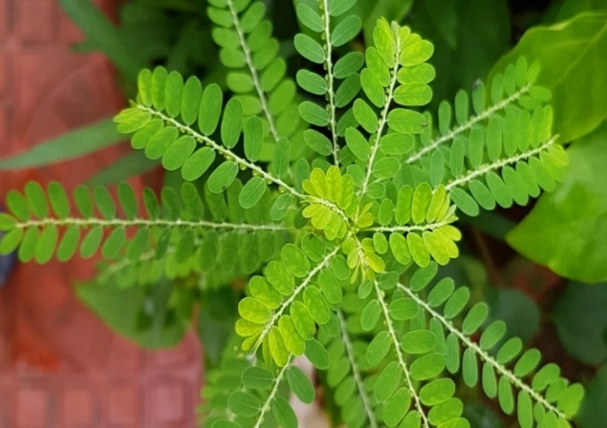
Fig 02: Phyllanthus niruri (L.)
As a member of Amaranthaceae family, Aerva lanata (L) is also called as Pashanabheda, Bhui and Gorakshaganjaa, which has several therapeutic applications, such as diuretic and anti-urolithiatic activity, anti-microbial activity, anti-fertility activity, anti-parasitic, anti-asthmatic, and anti-diabetic activity. In the Indian traditional medical system, ayurvedic practitioner uses the whole Aerva lanata plant as an anti-urolithiatic medication. It has several phytochemical components that have been identified to be responsible for variety of therapeutic activities. It includes alkaloids (like ervolanine, aervolanine, ervoside, ervine, methylervine), flavonoids (like quercetin, kaempferol, isorhamnetin, flavanone glucoside persinol, persinosides A and B), terpenoids (lupeol, betulin, beta-Sitosterol), phenolic compounds, tannins, and saponins. Roots of this plant are used to treat headaches as well as a demulcent. Quercetin and betulin were isolated from the plant and mainly show anti-urolithiatic activity by inhibiting oxalate oxidase enzyme activity (causes stone formation), leading to decreased production of oxalates23,24.
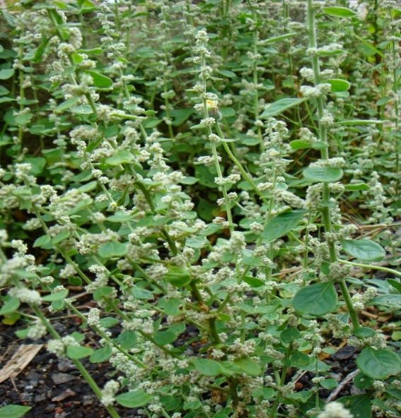
Fig 03: Aerva lanata (L.)
The eudicot diploid plant species known as muskmelon (Cucumis melo L.) is a member of Cucurbitaceae family, which is a species of Cucumis and is produced all over the world for its nutritional and commercial benefits. There are many varieties of active ingredients present in different parts of melon, the peel and seeds of melon contains phenolic acids like hydroxybenzoic acid (gallic acid, isovanillic acid, 3 and 4 hydroxybenzoic acid), methoxybenzoic acid (vanillic acid) and hydroxycinnamic acid (coumaric acid, chlorogenic acid), flavones and flavanones (luteolin-7-glycoside, naringenin), phenolic alcohols(tyrosol), phenylethanoids, benzenacetic acid derivatives, aromatic aldehydes(vanillin), vitamin E and secoiridoides. The pulp of melon contains tetraterpenoids (beta carotene), vitamin C (ascorbic acid). The medicinal activity exhibited by melons is anti-oxidant activity, analgesic activity, anti-inflammatory activity, anti-ulcer activity, anti-hypothyroidism, anti-diabetic, and anti-bacterial activity. Elevated levels of uric acid, creatine and blood urea nitrogen (BUN) are the important markers of renal injury brought by stones, the anti-urolithiatic activity of methanol and chloroform extract of melons peel and pulp shows significant reduction in renal calculi by decreasing the levels of indicators of the kidney (flavonoids and phenolic compound from extract prevent crystal formation, oxidative stress and inflammation)25,26.
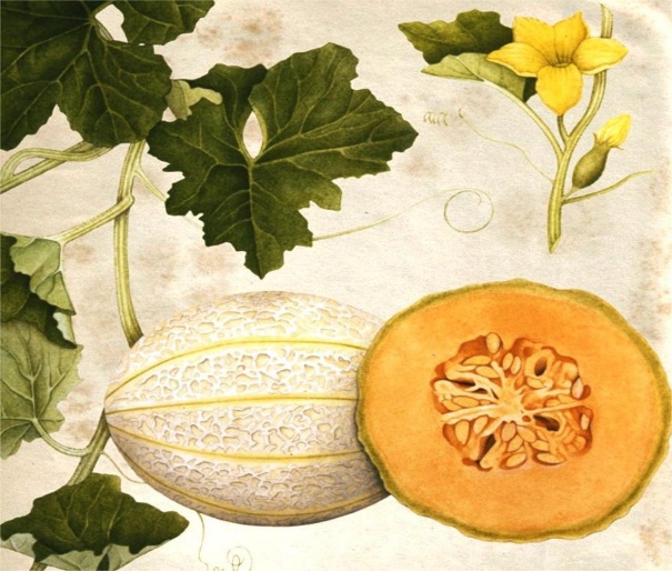
Fig 04: Cucumis melo (L.)
Shatavari, Satavari, and Asvel are the synonyms of the therapeutic plant Asparagus racemosus belonging to the family Asparagaceae, which has abundant phytochemical, nutritional and medical properties (especially roots). It consist of alkaloids (Asparagamine A, polycyclic alkaloids), flavonoids (quercetin, rutin, kaempferol, cynidin-3-glucoronide), steroidal saponins (Shatavarins, asparanin, adscendin), immunoside or oligospirostanoside (Racemoside, shatavaroside), sterols (Diosgenin, sitosterol), phenanthrene and furan derivatives, carbohydrates and polysaccharides, trace elements like zinc, iron, magnesium, potassium, calcium present mainly in roots as well as essential oil, tannins and quinones (anthraquinone). It exhibits multiple pharmacological actions like anti-depressant, anti-tussive, neuroprotective, anti-bacterial, anti-oxidant, anti-diabetic, anti-ulcer, nootropic, immunomodulatory, anti-inflammatory, anti-cancer, anti-urolithiatic and hepatoprotective action. The aqueous root extract of Asparagus racemosus shows anti-urolithiatic activity by inhibiting steps involved in kidney stone formation (step shown in Fig. 01). It also changes the calcium oxalate crystal structure from monohydrate to dihydrate, which is unstable and less likely to adhere to the walls of the kidney or renal tubular cells and is easily eliminated in urine. The phytochemicals from the extract form a soluble complex by binding with calcium ions and other compounds that form stones, causing less free and insoluble calcium availability for stone formation27,28.
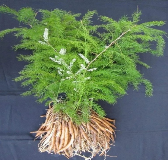
Fig 05: Asparagus racemosus
The plant Scoparia dulcis belongs to the Scrophulariaceae family, commonly called sweet broom weed, Mithipatti, Ghodatulsi. It exhibits various pharmacological and medicinal properties with numerous phytochemical constituents. Some constituents are flavonoids (luteolin, apigenin, cirsimarin, vitexin), steroids and triterpenes (betulinic acids, glutinol, stigmasterol), terpenoids and diterpenes (scopadulcic acid A and B, scopadulin, scoparic acid), phenols, polyphenols, amino acids, catechol amines, tannins, and coumarins. The pharmacological activities of this plant are anti-microbial, anti-fungal, anti-diabetic, nephroprotective, analgesic, anti-inflammatory, anti-pyretic, anti-ulcer, anti-urolithiatic, anti-sickling, anti-allergenic, and anti-hyperlipidemic as well as sedative and hypnotics. Serum levels of creatine, uric acid, and marker enzymes like ACP, ALP, AST, and ALT rise as a result of kidney stone development, leading to renal injury. These parameters nearly returned to normal after receiving the ethanolic leaf extract of Scoparia dulcis treatment. It indicates that kidney function had been restored and that stone development is decreased. The extract opposes the ethylene glycol-induced oxidative damage and hyperoxaluria, it may reduce renal tissue damage and stop the formation of calcium oxalate crystals, maintain kidney function, and normalize biochemical indicators to act as an anti-urolithiatic agent29,30,31.
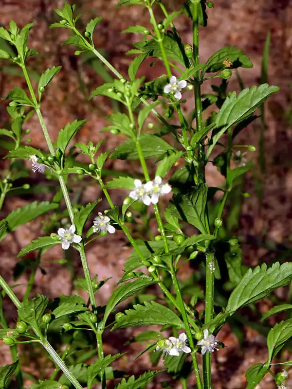
Fig 06: Scoparia dulcis
Bryophyllum pinnatum (Lam.), belonging to the Crassulaceae family, kalanchoe pinnata, pers and Bryophyllum calycinum salisb are synonyms. The common names of this plant are parnabija, Canterbury, love plant, air plant, cathedral bells, and zakham-e-hyat. In traditional medicine, Bryophyllum pinnata is widely used to treat a variety of illnesses. It has significant pharmacological activity due to various phytochemicals or active medicinal ingredients present in the plant abundantly. The phytochemicals are alkaloids, flavonoids(kaempferol, rutin, quercetin, luteolin and glycosylated flavones), phenolic acids and phenylpropanoids(syringic acid, caffeic acid, ferulic acid, p-coumaric acid, phosphoenolpyruvate and hydroxycinnamic acid), triterpenoids(alpha and beta amyrin, pseudo taraxasterol, taraxerol, friedelin, glutinol, bryophollenone and bryophollone), steroids and cardiac glycosides(bufadienolides, beta sitosterol, stigmasterol, campesterol, bryophyllin A,B,C, bryotoxins, clerosterol, ergosta type sterols, digoxin and digitoxin), amino acids and proteins(glycine, cysteine, tyrosine, phenylalanine, glutamic acid, methionine), carbohydrates and sugars(lactose, sucrose, glucose, raffinose, fructose, galactose), minerals(sodium, calcium, potassium, magnesium, iron, copper, zinc, iodine), tannins, phenanthrene derivatives, vitamins(vit.C, vit.B1,2,3,6), lipids and fatty acids(palmitic acid, stearic acid, arachidic acid, behenic acid), organic acids(isocitric acid, succinic acid, citric acid, oxalic acid, malic acid, oxaloacetate) and small traces of toxic compound hydrogen cyanide. The therapeutic activity possessed by this plant is anti-oxidant, anti-inflammatory, diuretic, anti-lithic, anti-tumor, cytotoxic-anticancer, anti-ulcer, anti-oxidant, anti-microbial, immunomodulatory, anti-viral, nephroprotective, astringent, cardio protective, anti-coagulant, and hypocholesterolemic activities. The hydroalcoholic extract of this plant shows anti-urolithiatic activity by reducing the levels of stone-forming ions, enhancing urine production to remove stones, defending renal tissue from oxidative stress or damage, and encourages stone dissolution or disintegration, also prevents stone reccurance32,33.
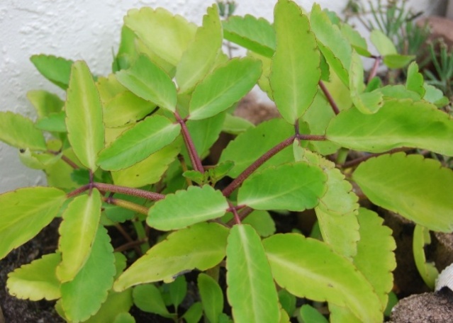
Fig 07: Bryophyllum pinnatum (Lam.)
Moringa oleifera, also known as drumstick, subhanjana, morigkai, saguna, belonging to the family Moringaceae, grows widely in India and tropical regions. The leaves, bark, roots, and flower parts of this plant have various beneficial therapeutic uses. The leaves of this plant are enriched in potassium, calcium, beta carotene, and other vital elements, which is used to treat malnutrition in infants and nursing women. The roots are traditionally utilized to treat paralysis and helminthiasis. Bark is used to treat toothaches, ulcers, and hypertension. Moringa oleifera contains major phytochemicals like flavonoids(rutin, kaempferol, quercetin, myricetin, isorhamnetin, procyanidins), glucosinolates and their derivatives like glucomoringin, niazirin, niazirinin, niazimin A and B, phenolic compounds(coumaric acid, ferulic acid, caffeic acid, gallic acid, ellagic acid, sinapic acid, syringic acid), fatty acids like oleic acid, palmitic acid, arachidic acid, phytosterols like beta-sitosterol, stigmasterol, vitamins like beta-carotene, alpha-tocopherol, and carbohydrates like mannose. The pharmacological activity shown by this plant is anti-inflammatory, anti-microbial, anti-cancer, anti-oxidant, anti-urolithiatic, diuretic, anti-fertility, hepatoprotective, anti-ulcer, anti-pyretic, anti-diabetic, cardiovascular, analgesic, anti-allergic, wound healing, anti-helminthic, local anaesthetic, and CNS activity. The aqueous extract of the bark of this plant shows a significant decrease in the stone weight, inhibits nucleation, and urinary crystal growth. The diuresis activity causes a decrease in supersaturation of urine, increases urine volume, and dilutes salts, which causes stones34,35,36.
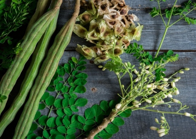
Fig 08: Moringa oleifera
Melia azedarach Linn is a tiny shrub closely related to the neem plant, belonging to Meliaceae family. It is commonly called as chinaberry, Indian lilac, padric, cinnamumo, bakain, This plant possess various phytochemicals like terpenoids, diterpenes like phytol, squalene, azedarachtin A and b, flavonoids like quercetin, rutin, kaempferol, isoquercetin, tetra-nor-triterpenoids or limonoids like meliacarpin, meliartenin, melianol, meliacin, steroids like stigmasterol, beta-sitosterol, campesterol, phenolic compounds like vanillin, vanillic acid, benzoic acid, hydroxyl-3-methoxycinnamaldehyde, alkaloids or beta-carboline alkaloids like 4,8-dimethoxy-1-vinyl-beta-carboline, fatty acids like palmitic acid, pentadecanoic acid, hexadecenoic acid, anthraquinones compounds like 1,3,5,8-tetrahydroxy-2-methylanthraquinones and vitamins A and E. The medicinal properties of this plant are anti-oxidant, anti-fertility, hepatoprotective, anti-pyretic, anti-bacterial, anti-viral, anti-nephrolithiasis, anti-ulcer, anti-malarial, anti-protozoal, anti-helminthic, cytotoxic activity, wound healing capacity, and diuretic activity. The aqueous leaf extract of the plants shows anti-urolithiasis by reducing elevated levels of oxalates, inhibiting crystal nucleation and aggregation, maintaining serum creatine level, uric acid, and blood urea nitrogen (BUN) levels, flushing out small stones by enhanced urine output37,38.
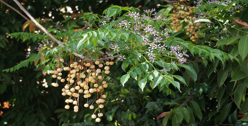
Fig 09: Melia azedarach Linn
Table 01: List of plants with active components and their role as anti-urolithiatic activity
|
Plant |
Scientific name |
Family |
Part of the plant and extract used |
Anti-urolithiasis effect |
|
Bhumi amla |
Phyllanthus niruri (L.) |
Euphorbiaceae
|
Aqueous extract of the whole plant |
Inhibition of the development of matrix calculus or inhibits the initial stages of stone formation. |
|
Gorakshaganjaa or Bhui |
Aerva lanata (L.) |
Amaranthaceae |
Hydroalcoholic extract of dried plant |
Inhibition of oxalate oxidase enzyme activity leads to a decrease in the production of oxalate crystals. |
|
Muskmelon |
Cucumis melo (L.) |
Cucurbitaceae |
Methanolic extract of peel and chloroform extract of pulp |
Reduction in elevated levels of serum creatine, BUN, and uric acid, as well as preventing crystal formation and oxidative stress caused by stones. |
|
Shatavari or satavari |
Asparagus racemosus |
Asparagaceae |
Aqueous root extract |
It converts calcium oxalate crystal structure from monohydrate to dihydrate, which is unstable and less likely to adhere to the walls of the kidney. It also forms a soluble complex with calcium ions and makes less available free ca+2 for stone development. |
|
Ghodatulsi, or sweet broom weed, or mithipatti |
Scoparia dulcis |
Scrophulariaceae |
Ethanolic leaf extract |
The biomarkers of kidney stones are reduced, decreasing stone formation, reducing oxidative stress, and hyperoxaluria levels. |
|
Kalanchoe pinnata or parnabija |
Bryophyllum pinnatum (Lam.) |
Crassulaceae |
Hydroalcoholic leaf extract |
It reduces levels of stone-forming ions and enhances urine production to flush out stones. |
|
Drumstick or morigkai |
Moringa oleifera |
Moringaceae |
Aqueous extract of the plant's bark |
Inhibition of nucleation and urinary crystal growth by decreasing the supersaturation of urine. |
|
Indian lilac or chinaberry |
Melia azedarach Linn |
Meliaceae |
Aqueous leaf extract |
It reduces hyperoxaluria and inhibits crystal nucleation and aggregation. Enhanced urine output causes flushing of stones. |
CONCLUSION:
Urolithiasis remains a major health concern due to its recurrent nature and high frequency. The use of herbal remedies, which were common in older medical systems, may be advantageous as safer and more helpful alternatives for kidney stone treatment. The plants examined in this study demonstrated a variety of methods of action that preserved renal tissue, slowed the formation of stones, and made it easier to remove them. To ensure the safety and therapeutic effectiveness of herbal preparations, however, clinical validation and standardization were still necessary. A viable method for enhancing long-term results in urolithiasis patients seemed to be in combination with researched herbal remedies and traditional therapies.
REFERENCES

A. Sravanthi, Meghna, Mohamed Ashwaq S., Mohammed Sameer H., Mouneshwar V., Nature's Remedy: The Role of Herbs in Urolithiasis Treatment, Int. J. of Pharm. Sci., 2025, Vol 3, Issue 11, 2757-2768. https://doi.org/10.5281/zenodo.17645822
 10.5281/zenodo.17645822
