View Article

Abstract

In terms of public health, central nervous system (CNS) illnesses play a critical role. One of the most frequent CNS illnesses is epilepsy. Epilepsy is a chronic condition that causes significant morbidity and death. Many individuals worldwide suffer from epilepsy, and the reasons of this condition are yet unknown. Epilepsy affects roughly 50 million individuals worldwide. Antiepileptic medications are often used in the treatment of epilepsy, however the problem with these treatments is the development of drug resistance, and antiepileptic drugs may be taken orally or intravenously. These therapies, however, are not always successful. Drugs used to treat epilepsy, on the other hand, must be supplied properly and securely in order to preserve the brain. As a result, novel delivery methods are required to administer medications at doses found to have great therapeutic effectiveness in epilepsy while minimising negative effects. In light of this knowledge, new therapeutic options are required. Dendrimers are one such therapeutic option which can help to active site specific and more successful results. Hence, in present article we have summarized the epilepsy condition, dendrimers and its use in epilepsy in brief.

Keywords

CNS, epilepsy, dendrimers, Novel delivery, Carbamazepine

Introduction

Epilepsy is a neurological disorder that produces seizures in various areas of the brain due to aberrant electrical activity. This condition has neurological, cognitive, psychological, and social consequences, and it affects about 50 million individuals worldwide (1). Estimates of worldwide prevalence and incidence of epilepsy differ by nation. While it is more prevalent in low-income nations than in high-income countries, there is a considerable rise in the number of patients in infancy compared to old age (2). The fatality rate, however, is minimal. Unintentional injury and suicide were shown to be among the fatalities caused by epilepsy (3). These findings indicate that the patients' psychological well-being is impacted by the disease's prognosis.

Seizures, according to the World Health Organisation (WHO), are the result of excessive electrical discharges in a group of brain cells. These discharges may occur in many areas of the brain. Seizures triggered by these discharges may occur anywhere from once per year to many times per day. According to the International League Against Epilepsy (ILAE) categorization, epilepsy may be defined as any of the following conditions:

  1. At least 2 times to provoke the occurrence of seizure in 1 day
  2. Seizure occurring in the next 10 years and their probability (having at least a 60% chance of seizures)
  3. Diagnosis of epilepsy syndrome (4,5)
  1. Pathology of Epilepsy

Extreme and unexpected electrical activity in the brain is the underlying mechanism of epileptic seizures (6). This abnormal state in the brain may be detected in a variety of brain areas, including the temporal, frontal, parietal, and occipital-lobe (7,8). Although the aetiology of epilepsy is not fully understood, various causes are suspected. In addition to brain injury, stroke, brain tumours, brain infections, or birth abnormalities (6), genetic changes are considered to have a significant influence in the disease's incidence (9). Epilepsy research based on genetic variables is continuously continuing. For example, based on the findings of sample isolation from patients with infantile-onset seizures, it is believed that the condition is caused by a mutation in the PCDH19 gene (10). Cerebrovascular problems, cortical development anomalies, and metabolic diseases are also linked to epilepsy pathophysiology (11).

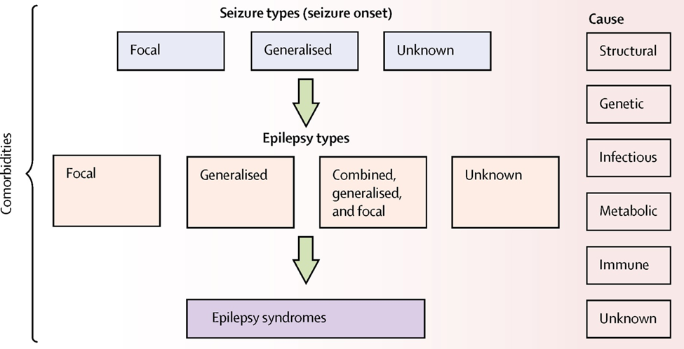
Fig.1:- showing the general pathophysiology of epileptic symptoms.

  1. Diagnosis of Epilepsy

Anamnesis should be used to determine if the individual experienced an epileptic seizure, the description of the attack, and the mix of symptoms. Electroencephalography (EEG), long-term video electroencephalography (EEG), high-resolution MRI, and neurophysiological testing are all required for a proper diagnosis. EEG is now widely used in the non-invasive diagnosis of epilepsy. Aside from anamnesis, EEG is the most significant supplementary diagnostic procedure, and it should be conducted on any patient suspected of having a seizure. EEG and video EEG give information regarding deep brain activities and disease processes (12). Existing approaches for detecting seizures rely on simulations to extract features from EEG recordings. Stimulants are used during EEG to analyse epileptic results (13). As a result, flash stimuli in the EEG utilised for diagnosis are among the major elements that cause seizures. PET and SPECT scans, functional MRI, MR spectroscopy, magnetoencephalography, and the Wada test are all options (14). The magnetic resonance imaging (MR) approach is utilised to determine seizure onset locations (15). Nuclear Medicine Examinations Nuclear medicine imaging technologies such as SPECT and PET are utilised to locate the epileptic focal prior to surgery. Tc99m HMPAO and Tc-99m ECD are the radiopharmaceuticals utilised in SPECT. F-18 FDG is the most often used radioactive substance for PET imaging, and F-18 flumazenil (FMZ) also displays the seizure region, although in a more restricted area than FDG (16). Neuroimaging may be used to identify structural abnormalities that may be causing epilepsy. The chosen imaging examination is magnetic resonance imaging (MRI). The MRI approach may be employed in individuals who have a focused onset based on their history, symptoms on examination, or EEG (17).

  1. Treatment of epilepsy

It is critical to assess the seizure type, frequency, and risk of recurrence in those who have epilepsy. Treatments attempt to eliminate or lessen the incidence of seizures while improving the patient's quality of life (18,19). Antiepileptic medications are often utilised in therapy. One of the primary issues in therapy is drug resistance (19). However, antibiotic resistance reduces the chance of illness recovery. Alternative therapeutic options, in addition to medication therapy, are being explored. Epilepsy surgery is one of these techniques. Surgery may be an option for controlling focal seizures altogether. Individuals who do not react to drugs may have three kinds of surgery: vagus nerve stimulation, anterior thalamic stimulation, and stimulation responsive to the closed-loop (18). Vagus nerve stimulation (20), deep brain stimulation (21), responsive neurostimulation (22), and the ketogenic diet (23) are some other therapy options. However, the medications used to treat epilepsy are just supportive, and the disease's healing impact is minimal. Although different ways have been shown to reduce the incidence of seizures, these strategies need to be improved. In at-risk individuals, antiepileptogenic medicines are used to prevent epilepsy before the first seizure occurs.-modifying novel medicines are also required to manage continued severe seizures linked with worsening underlying illness (24-27).

  1. Novel treatment : dendrimers

Dendrimers are nanosized macromolecules with a hyper-branched globular shape that are often employed for drug delivery. Dendrimers, in contrast to typical polymer nanovehicles, are monodisperse and have well-defined chemical structures. Furthermore, the unique structure of dendrimers allows them to load medicinal medicines through covalent conjugation or electrostatic adsorption (28). Dendrimers are the size of a swarm of biological structures. For example, generation 5 (G5) polyamidoamine (PAMAM) dendrimers (5.5 nm in diameter) are about the same size and shape as haemoglobin (29). Dendrimers are made up of a central core (a single atom or group of atoms), generations of repeating building units linked to the core, and functional groups on the surface. Dendrimers' physiochemical and biological properties are therefore governed by three structural components: core, building blocks, and functional groups. To produce a dendrimer, two main synthetic approaches are used: divergent and convergent. Dendrimers are built from the core to the shell, generation by generation, in the divergent method, while compounds are built from the peripheral to the centre in the convergent approach.

Surface groups may have positive, negative, or neutral charges, which is useful in identifying potential dendrimers as drug delivery carriers (30). Dendrimers ended with positively charged functional groups often induce anionic cell membrane destabilisation and even cell lysis, resulting in poor biocompatibility, while neutral or anionic charged dendrimers display reduced hemolysis (31). To address this problem, surface functionalization such as PEGylation may affect zeta-potential, blood retention, and even distribution in vivo. Gene therapy is a promising method for many illnesses; nevertheless, it still faces certain challenges, such as the difficulty of free gene molecules to penetrate target cells in vivo (32). To produce stable dendriplexes, the cationic dendrimers electrostatically attach the negatively charged polynucleotides. After cellular absorption, dendriplexes are released through a "proton sponge" function in endosomes, promoting gene expression in cells (33,34). Dendrimers, as gene delivery carriers, may shield gene molecules from biodegradation, allow nucleic acids to enter cells more easily, and keep gene molecules biologically active.

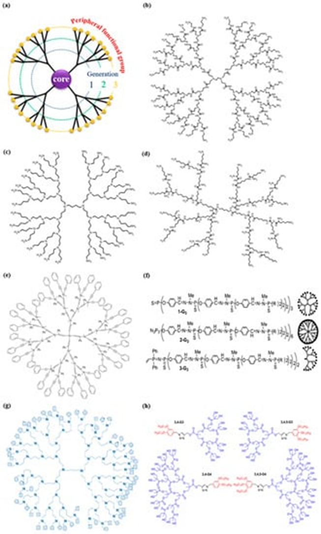
Fig.2:- Commonly used dendritic macromolecules in drug delivery: (a) A schematic diagram revealing the three components of a dendrimer molecule and chemical structures of (b) polyamidoamine (PAMAM), (c) polypropyleneimine (PPI), (d) poly-l-lysine (PLL) dendrimers, and (e) carbosilane dendrimers; (f) examples of cores of phosphorus (PPH) dendrimers, (g) glycodendrimers, and (h) Janus dendrimers. Fig. b–h is reproduced with permission from References (35-39).

  1. Dendrimers and its derivatives
    1. Dendrimers made from PAMAM

The term "polyamidoamine dendrimers" (PAMAM) is traditionally used to describe dendrimers that have (i) a core (the earliest core was an ethylenediamine core), (ii) branches made up of amide groups emanating from a branching point that forms the walls of cavities, and (iii) amine functional groups on the periphery (Figure 1b). Concurrently, there is a wide range of PAMAM dendrimers with various surface groups (40). PAMAM dendrimers, one of the most thoroughly researched dendrimers, include interior cavities and peripheral functional groups that may be changed to encapsulate agents or other cargos for biomedical purposes. Dendrimers, as dendrimer creator Donald Tomalia has suggested, might be used as nano-agents against tumours, germs, and viruses (41). However, one worry with the usage of PAMAM, particularly those with positively charged groups, is their in vitro cytotoxicity, which is concentration and generation dependent. PAMAM dendrimers of G5 or lower generation are deemed harmless (42). Surface modification using PEG was used to address this limitation. PAMAM has the most uses in the area of drug delivery, antibacterial, antiviral, and antioxidant agents for diagnostics, and medication targeting when compared to other dendrimers.

    1. PPI Dendrimers

The earliest known dendrimer commercially utilised for medication delivery is polypropyleneimine (PPI). It is made up of diaminobutane and propyleneimine repeat units (Figure 1c). Because diaminobutane (DAB) is a component of PPI, DAB-dendrimer is the second most often used acronym for polypropyleneimine (40). PPI's amino terminal groups give appropriate water solubility. PPI dendrimers may thereby increase the water solubility of hydrophobic substances entrapped in the PPI's hydrophobic inner cavities. The positively charged surface of PPI, on the other hand, often destabilises cell membranes and promotes cell lysis. Another issue is that PPI has a lower drug loading capacity than PAMAM (43). PPI/drug complexes are less stable than PPI alone. PEGylation and acetylation of surface groups have been selected as surface group modifications. Acetylation is chosen due to its great effectiveness and penetration. Furthermore, PEG chain steric hindrance may alter the interaction of surface functional groups with medicinal molecules (44).

    1. PLL Dendrimers

Poly-l-lysine (PLL) dendrimers (also known as dendri-grafted poly-l-lysine or DGL) are dendrimers made up of lysine residues (Figure 1d). These dendrimers have higher biocompatibility, lower cytotoxicity, easier enzymatic breakdown, and subsequent excretion of low molecular weight metabolites (45). Furthermore, the ability to add stimuli responsiveness when needed by including certain amino acid sequences of PLL dendrimers is highly intriguing (46). Gene delivery has made considerable use of PLL dendrimers and its variants. When compared to PLL dendrimers, the higher generation of PLL demonstrates superior gene transfection, while the typical linear poly(lysine) exhibits poorer gene transfection efficacy and increased cytotoxicity.

    1. Carbosilane Dendrimers

A variety of carbosilane dendritic macromolecules with hydrophobic scaffolds and excellent thermal stability have also been created using extensive silicon chemistry (47). The use of silicon chemistry to synthesise dendrimers is prevalent due to the fact that nucleophilic molecules may easily access electrophilic silicon (Si+) (48). Notably, the advantage of carbosilane dendrimers is connected with the low polarity and high energy of the C-Si bond, which confers strong hydrophobicity (49). Despite having a hydrophobic core structure, carbosilane dendrimers may be transformed into hydrophilic molecules by surface functionalization with polar moieties. Reactive groups such as Si-H, Si-Cl, Si-CH=CH2, and Si-CH2CH=CH2 may aid in the introduction of many additional fascinating inorganic, organic, and organometallic substituents, leading to an increase in pharmaceutic applications (40).

    1. PPH Dendrimers

Phosphorus (PPH) dendrimers, which are synthesised specifically for drug delivery systems (DDSs), are also of particular interest in the cationic dendrimer family. PPH dendrimers, or dendrimers with phosphorus atoms at each branching point, have a particular position in the chemistry because of their comparatively simple production and the rich chemical variety, which is due in most instances to the presence of reactive end groups (aldehydes or P(S)Cl2 groups) (37). Such dendrimers may have a hydrophilic surface and a hydrophobic backbone, allowing them to interact with cell membranes and be internalised (50,51). Recent studies have demonstrated that PPH dendrimers have enormous promise in biological applications. Such dendritic materials have been shown to impact amyloid peptide and Tau protein aggregation in neurodegenerative disorders (52). Furthermore, the ability of PPH dendrimers to transport anticancer siRNAs to target cells (53) and gene therapy for HIV infection (54) has been revealed.

    1. Janus Dendrimers

"Janus" dendrimers, also known as bow-tie dendrimers, diblock dendrimers, co-dendrimers, and "surface-block" dendrimers, are a relatively novel family of amphiphilic dendrimers for pharmaceutical applications. They are characterised by two dendrimeric wedges with distinct terminal groups. The name "Janus" alludes to the ancient God of gates and doorways, who is usually represented with a two-faced head looking in opposing directions (55). Janus dendrimers' broken symmetry allows for the formation of sophisticated self-assembled materials and the presentation of new sets of characteristics that are now unthinkable for homogeneous or symmetrical dendrimers. As a result, Janus dendrimers, whose characteristics are closely related to the two separate peripheral surface groups, are very attractive. Because of their biofunctionality and thermal characteristics, these Janus dendrimers are gaining attention in self-assembling and medicinal applications (56).

    1. Linear-Dendritic Block Copolymers

With the creation of dendrimers, a new amphiphilic block copolymer, linear-dendritic block copolymers (LDBC), has emerged. They are made up of linear chains that are covalently bonded to the dendrimer. The majority of LDBCs participate in the conversion of hydrophobic dendrons to hydrophilic linear chains (57). The amphiphilic copolymers may self-assemble in aqueous solution to create a stable core-shell structure with distinct properties from typical micelles (e.g., reduced critical micelle concentration (CMC) and longer blood stability) (58). In terms of architecture, copolymers can be split into six prime groups: AB diblock linear-dendritic copolymers which contain the linear A block and dendritic B block, ABA triblock linear-dendritic copolymers which employ B as the linear block and A as the dendritic block, linear-hyperbranched polymers, side chain functional or dendronized linear-dendritic copolymers, multi-arm star copolymers, and linear-dendrimer-grafts polymers (58).

    1. Dendrimers of Other Types

Aside from the aforementioned LDBC dendrimers, which are members of the hybrid dendrimer family, a plethora of additional dendrimers with well-defined nanoscopic size and an abundance of functional terminal groups are now accessible. Core shell (tecto) dendrimers (a polymeric architecture with highly ordered structure achieved through controlled covalent attachment of dendrimer building blocks) (59), peptide dendrimers (60), glycodendrimers (61), PAMAM-organosilicon (PAMAMOS) dendrimers (consists of hydrophilic and nucleophilic PAMAM interiors and hydrophobic organosilicon (OS) exteriors) (62), and others are among these.

  1. Recent advancement
    1. Carbamazepine dendrimers

Carbamazepine (CBZ) is an antiepileptic medication that may potentially be used to treat neurodegenerative illnesses such as Alzheimer's. However, owing to its poor solubility, unfavourable pharmacokinetic profiles, and many adverse effects, its usage has been restricted. PAMAM dendrimers, ethylenediamine core, generation 4.0 (amine terminal groups) and 4.5 (carboxylate terminal groups) (DG4.0 and DG4.5) polymers may enhance drug solubility through complexation. As a result, the goal of this study was to produce and characterise complexes between CBZ and dendrimers. Both DG4.0 and DG4.5 facilitated the integration of 20 molecules of CBZ into their hydrophobic pockets per dendrimer. The complexes DG4.0-CBZ and DG4.5-CBZ were shown to be stable for 90 days at 37 °C and resistant to lyophilization, allowing for regulated drug release. The nanotoxicity of the complexes was further examined ex vivo (human red blood cells), in vitro (N2a cell line), and in vivo (zebrafish). In the ex vivo model, no hemolytic impact was seen. In terms of in vitro toxicity, the DG4.5-CBZ complexes dramatically decreased the free drug's toxicity. Furthermore, DG4.5-CBZ had neither neurotoxicity or cardiotoxicity in zebrafish larvae. Finally, a stable and biocompatible drug delivery system capable of complexing the CBZ based on the DG4.5 has been designed. This accomplishment illustrates the benefits of utilising negatively charged dendrimers in nanomedicine.(63)

    1. Naloxonazine complex dendrimers

The goal of this work was to see whether the host dendrimer DAB-Am-16 could be used as a drug carrier to shorten the time it took for the encapsulated naloxonaxine to form an irreversible covalent link with the 1-opioid receptor (resulting in a pharmacologically selective action). The tail-flick test was used to assess the efficiency of the dendrimer-naloxonazine nanocomplex (DNC) in antinociception generated by convulsions induced by intraperitoneal (IP) injection of pentylenetetrazole. We discovered that animals' tail-flick latencies increased after convulsions. Furthermore, acute DNC pre-treatment (10 minutes), but not naloxonazine alone, reduced post-ictal analgesia compared to control pre-treatment. However, whereas naloxonazine administration 24 hours before PTZ reduced post-ictal antinociception, DNC failed to counteract tonic-clonic seizure-induced analgesia. Furthermore, whether provided 10 minutes or 24 hours before PTZ, naloxonazine, DAB-Am-16 dendrimer, or DNC had no effect on seizure severity according to Racine's index.(64)

    1. Click chemistry-based dendrimers

Neurological illnesses have a high death rate and place a significant cost on society. Because of the difficulty to transport effective medications across the blood-brain barrier, therapeutics for neurological illnesses are often ineffective. Previous studies have identified generation 4 hydroxyl-terminating polyamidoamine (PAMAM) dendrimers as a viable drug delivery platform due to its excellent functionality, biocompatibility, and adaptability. The development of click chemistry and CuAAC (copper-catalyzed azide-alkyne cycloaddition) reactions allows for the simple and regulated conjugation of medicines and biocompatible small molecules onto dendrimers. CuAAC click reactions are high-yielding and broad in scope, producing regiospecific products and only easily removed byproducts. It has been identified as an appropriate method for synthesising promising dendrimer nanomedicine with a high degree of functionality and customisation for the possible treatment of neurological illnesses. Building on this promise, we developed a CuAAC click reaction-based technique for attaching medicines of interest to G4 PAMAM-OH dendrimers. We specifically investigated the use of click chemistry to synthesise (1) dendrimer-PBA (4-phenylbutyric acid) conjugate for the potential treatment of ALD (adrenoleukodystrophy) and (2) dendrimer-VPA (valproic acid) conjugate for the potential treatment of epilepsy. The synthesis and characterization of dendrimer-drug conjugates will be covered in this section.(65)

    1. Dendrimers as bio imaging

Dendrimer science is a newer discipline of nanotechnology that focuses on non-linear macromolecules known as dendrimers. These hyperbranched polymers have monodispersity, a well-defined structure, and, depending on the kind of dendrimer, excellent biocompatibility. Because a dendrimer may encapsulate or surface conjugate guest molecules, it can be used as a delivery agent in life sciences. Dendritic polymers have been widely researched as traditional fluorophore carriers for bioimaging based on fluorescence. The complexation of organic dyes with these macromolecules increases their solubility and cellular absorption. Furthermore, it aids in overcoming other limitations in their use for photobleaching, such as their lack of specificity or cytotoxicity. Dendrimer's protective characteristics are particularly useful for usage with quantum dots, which have a high optical potential but include heavy metals that may harm biological items. Fluorescent probes based on dendrimers have been extensively used in in vitro and in vivo bioimaging. Non-traditional intrinsic fluorescence (NTIF) of dendrimers is an intriguing phenomena. Some of them exhibit pH and oxygen-dependent fluorescence in the absence of conjugation with other particles. Although NTIF is not completely understood, efforts have been made to use it for bioimaging. This phenomenon has enormous promise since it allows dendrimers to function as both a delivery and diagnostic tool.(64)

    1. Dendrimers fluorescent imaging

The discovery of fluorescent proteins and synthetic compounds whose fluorescence characteristics are modulated by the environment allows for the minimum disturbance of physiological and pathological processes in biological systems. There are several tiny organic dyes available that are commonly employed to evaluate physiologically relevant characteristics. Unfortunately, their use is hampered by a variety of constraints resulting from their employment in the biological environment. As a result, we offer a new dendrimer-based design that leads to multifunctional sensing components that potentially solve several of these issues. There are reports of use in vitro, in live cells, and in vivo. In specifically, we image extracellular pH in the brain for the first time in a mouse epilepsy model. The authors feel that the suggested design may be a helpful and new tool in fluorescence imaging that can be used in combination with a wide variety of sensing dyes and experimental settings.(65)

CONCLUSION

Finally, the involvement of dendrimers in anti-epileptic medicines offers tremendous promise and possibility for enhancing epilepsy treatment techniques. Dendrimers, with their distinct structural traits and customizable qualities, provide a multimodal approach to improving anti-epileptic medication administration, bioavailability, and therapeutic effectiveness.

Dendrimers address medication solubility, stability, and targeting concerns by their capacity to encapsulate, protect, and regulate drug release. Their adaptable surface functions allow for particular interactions with biological systems, allowing for tailored medication administration to epileptic foci while minimising off-target effects. Furthermore, dendrimer-based therapeutic formulations have the potential to reduce concerns with dosage frequency and adverse responses, enhancing patient compliance and overall quality of life.

While the promise of dendrimers in anti-epileptic medication delivery is obvious, further study is needed to optimise formulations, understand pharmacokinetics, and prove long-term safety and effectiveness. Furthermore, the development of dendrimer-based epilepsy medicines should entail multidisciplinary cooperation among pharmaceutical scientists, chemists, neurologists, and other relevant professionals.

Dendrimers are anticipated to play an increasingly important role in revolutionising the landscape of anti-epileptic medication therapy in the future years as our knowledge of dendrimer technology increases and their applications grow. As more targeted and efficient drug delivery systems are developed, the possibility of providing more personalised and effective therapeutic interventions for epilepsy patients becomes a tantalising possibility, offering hope for improved seizure management and, eventually, improving their overall quality of life.

REFERENCES

    1. Devinsky O, Vezzani A, O’Brien TJ, Jette N, Scheffer IE, de Curtis M, Perucca P. Epilepsy. Nat Rev Dis Primers. 2018 Mar 22;4:18024. doi: 10.1038/nrdp.2018.24.
    2. Fiest KM, Sauro KM, Wiebe S, Patten SB, Kwon CS, Dykeman J, Pringsheim T, Lorenzetti DL, Jette N. Prevalence and incidence of epilepsy: A systematic review and meta-analysis of international studies. Neurology. 2017 Jan 17;88(3):296-303. doi: 10.1212/WNL.0000000000003509.
    3. Thurman DJ, Logroscino G, Beghi E, Hauser WA, Hesdorffer DC, Newton CR, Scorza FA, Sander JW, Tomson T, Epidemiology Commission of the International League Against Epilepsy. The burden of premature mortality of epilepsy in high-income countries: A systematic review from the Mortality Task Force of the International League Against Epilepsy. Epilepsia. 2017 Jan;58(1):17-26. doi: 10.1111/epi.13604.
  1. Beghi E. The Epidemiology of Epilepsy. Neuroepidemiology. 2020;54(2):185-191. doi: 10.1159/000503831.
  2. Fisher RS, Acevedo C, Arzimanoglou A, Bogacz A, Cross JH, Elger CE, Engel J Jr, Forsgren L, French JA, Glynn M, Hesdorffer DC, Lee BI, Mathern GW, Moshe SL, Perucca E, Scheffer IE, Tomson T, Watanabe M, Wiebe S. ILAE official report: a practical clinical definition of epilepsy. Epilepsia. 2014 Apr;55(4):475-82. doi: 10.1111/epi.12550.
  3. Aronica E, Muhlebner A. Neuropathology of epilepsy. Handb Clin Neurol. 2017;145:193-216. doi: 10.1016/B978-0-12-802395-2.00015-8.
  4. Kwan P, Schachter SC, Brodie MJ. Drug-resistant epilepsy. N Engl J Med. 2011 Sep 8;365(10):919-26. doi: 10.1056/NEJMra1004418.
  5. Fisher RS, van Emde Boas W, Blume W, Elger C, Genton P, Lee P, Engel J Jr; ILAE Commission on Classification and Terminology. Epileptic seizures and epilepsy: definitions proposed by the International League Against Epilepsy (ILAE) and the International Bureau for Epilepsy (IBE). Epilepsia. 2005 Apr;46(4):470-2. doi: 10.1111/j.0013-9580.2005.66104.x.
  6. Pandolfo M. Genetics of epilepsy. Semin Neurol. 2011 Oct;31(5):506-18. doi: 10.1055/s-0031-1299789.
  7. Hynes K, Tarpey P, Dibbens LM, Bayly MA, Berkovic SF, Smith R, Raisi ZA, Turner SJ, Brown NJ, Desai TD, Haan E, Turner G, Christodoulou J, Leonard H, Gill D, Stratton MR, Gecz J, Scheffer IE. Epilepsy and mental retardation limited to females with PCDH19 mutations can present de novo or in single generation families. J Med Genet. 2010 Mar;47(3):211-6. doi: 10.1136/jmg.2009.068817.
  8. Reif PS, Strzelczyk A, Rosenow F. The history of invasive EEG evaluation in epilepsy patients. Seizure. 2016 Sep;41:191-195. doi: 10.1016/j.seizure.2016.04.006.
  9. Ullah I, Hussain M, Qazi E, Aboalsamh H. An automated system for epilepsy detection using EEG brain signals based on deep learning approach. Expert Syst Appl. 2018;107:61-71.
  10. Ergun E, Salanc? B, Erbas B. SPECT and PET in epilepsy, Nuclear Medicine Department, Hacettepe University Faculty of Medicine. Turk Klin J Nucl Med Spec Top. 2017;3(2):123-31.
  11. Ryvlin P, Rheims S. Epilepsy surgery: eligibility criteria and presurgical evaluation. Dialogues Clin Neurosci. 2008;10(1):91-103. doi: 10.31887/DCNS.2008.10.1/pryvlinv.
  12. Yalnizoglu D, Hirfanoglu T, Serdaroglu A, Turanli G, Topcu M. Intractable epilepsy in childhood: presurgical evaluation and treatment. Epilepsi: J Turk Epilepsi Soc. 2012;18(1):7-14.
  13. Kinay D. Neuroimaging in epilepsy surgical applications. Bakirkoy Prof. Dr. Mazhar Osman Mental and Neurological Diseases Training and Research Hospital 1. Neurology Clinic. Epilepsi. 2012;18(1):18-29. doi: 10.5505/epilepsi.2012.36035.
  14. Erdinc O. Principles of therapy in the epilepsies. Nobel Tip Kitabevleri, Istanbul.
  15. Rowland LP, editor. Merritt's neurology. Gunes Tip Kitabevleri; 2008. ISBN:9789752771819.
  16. Kwon CS, Ripa V, Al-Awar O, Panov F, Ghatan S, Jette N. Epilepsy and Neuromodulation- Randomized Controlled Trials. Brain Sci. 2018;8(4):69. doi: 10.3390/brainsci8040069.
  17. Tellez-Zenteno JF, Hernandez-Ronquillo L, Buckley S, Zahagun R, Rizvi S. A validation of the new definition of drug-resistant epilepsy by the International League Against Epilepsy. Epilepsia. 2014;55(6):829-834. doi: 10.1111/epi.12633.
  18. Edwards CA, Kouzani A, Lee KH, Ross EK. Neurostimulation Devices for the Treatment of Neurologic Disorders. Mayo Clin Proc. 2017;92(9):1427-1444. doi: 10.1016/j.mayocp.2017.05.005.
  19. Skarpaas TL, Jarosiewicz B, Morrell MJ. Brain-responsive neurostimulation for epilepsy (RNS System). Epilepsy Res. 2019;153:68-70. doi: 10.1016/j.eplepsyres.2019.02.003.
  20. Liu H, Yang Y, Wang Y, Tang H, Zhang F, Zhang Y, Zhao Y. Ketogenic diet for treatment of intractable epilepsy in adults: A meta-analysis of observational studies. Epilepsia Open. 2018;3(1):9-17. doi: 10.1002/epi4.12098.
  21. Schmidt D, Schachter SC. Drug treatment of epilepsy in adults. BMJ. 2014;348:g254. doi: 10.1136/bmj.g254.
  22. Du X, Shi B, Liang J, Bi J, Dai S, Qiao SZ. Developing Functionalized Dendrimer-Like Silica Nanoparticles with Hierarchical Pores as Advanced Delivery Nanocarriers. Adv Mater. 2013;25:5981–5985. doi: 10.1002/adma.201300344.
  23. Esfand R, Tomalia DA. Poly(amidoamine) (PAMAM) dendrimers: from biomimicry to drug delivery and biomedical applications. Drug Discov Today. 2001;6:427–436.
  24. Kesharwani P, Jain K, Jain NK. Dendrimer as nanocarrier for drug delivery. Prog Polym Sci. 2014;39:268–307.
  25. Malik N, Wiwattanapatapee R, Klopsch R, Lorenz K, Frey H, Weener JW, Meijer EW, Paulus W, Duncan R. Dendrimers: relationship between structure and biocompatibility in vitro, and preliminary studies on the biodistribution of 125I-labelled polyamidoamine dendrimers in vivo. J Control Release. 2000;65:133–148.
  26. Vannucci L, Lai M, Chiuppesi F, Ceccherini-Nelli L, Pistello M. Viral vectors: a look back and ahead on gene transfer technology. New Microbiol. 2013;36:1–22.
  27. Perumal OP, Inapagolla R, Kannan S, Kannan RM. The effect of surface functionality on cellular trafficking of dendrimers. Biomaterials. 2008;29:3469–3476.
  28. Sonawane ND, Szoka FC Jr, Verkman AS. Chloride accumulation and swelling in endosomes enhances DNA transfer by polyamine-DNA polyplexes. J Biol Chem. 2003;278:44826–44831.
  29. Hsu HJ, Bugno J, Lee SR, Hong S. Dendrimer-based nanocarriers: a versatile platform for drug delivery. Wiley Interdiscip Rev Nanomed Nanobiotechnol. 2017;9.
  30. Kim C, Hong JH. Carbosilane and carbosiloxane dendrimers. Molecules. 2009;14:3719–3730.
  31. Caminade AM, Majoral JP. Nanomaterials based on phosphorus dendrimers. Acc Chem Res. 2004;37:341–348.
  32. Klementieva O, Benseny-Cases N, Gella A, Appelhans D, Voit B, Cladera J. Dense shell glycodendrimers as potential nontoxic anti-amyloidogenic agents in Alzheimer's disease: amyloid-dendrimer aggregates morphology and cell toxicity. Biomacromolecules. 2011;12:3903–3909.
  33. Selin M, Nummelin S, Deleu J, Ropponen J, Viitala T, Lahtinen M, Koivisto J, Hirvonen J, Peltonen L, Kostiainen MA, et al. High-generation amphiphilic Janus-dendrimers as stabilizing agents for drug suspensions. Biomacromolecules. 2018;19:3983–3993.
  34. Pedziwiatr-Werbicka E, Milowska K, Dzmitruk V, Ionov M, Shcharbin D, Bryszewska M. Dendrimers and hyperbranched structures for biomedical applications. Eur Polym J. 2019;119:61–73.
  35. Tomalia DA, Christensen JB, Boas U. Dendrimers, dendrons, and dendritic polymers: discovery, applications, and the future. Cambridge University Press; 2012.
  36. Beezer AE, King ASH, Martin IK, Mitchel JC, Twyman LJ, Wain CF. Dendrimers as potential drug carriers: encapsulation of acidic hydrophobes within water-soluble PAMAM derivatives. Tetrahedron. 2003;34:3873–3880.
  37. Richter-Egger DL, Tesfai A, Tucker SA. Spectroscopic investigations of poly(propyleneimine) dendrimers using the solvatochromic probe phenol blue and comparisons to poly(amidoamine) dendrimers. Anal Chem. 2001;73:5743–5751.
  38. Fant K, Esbjorner EK, Jenkins A, Grossel MC, Lincoln P, Norden B. Effects of PEGylation and acetylation of PAMAM dendrimers on DNA binding, cytotoxicity, and in vitro transfection efficiency. Mol Pharm. 2010;7:1734–1746.
  39. Ryan GM, Kaminskas LM, Bulitta JB, McIntosh MP, Owen DJ, Porter CJ. PEGylated polylysine dendrimers increase lymphatic exposure to doxorubicin when compared to PEGylated liposomal and solution formulations of doxorubicin. J Control Release. 2013;172:128–136.
  40. Byrne M, Victory D, Hibbitts A, Lanigan M, Heise A, Cryan SA. Molecular weight and architectural dependence of well-defined star-shaped poly(lysine) as a gene delivery vector. Biomater Sci. 2013;1:1223.
  41. Uchida H, Kabe Y, Yoshino K, Kawamata A, Tsumuraya T, Masamune S. General strategy for the systematic synthesis of oligosiloxanes. Silicone dendrimers. J Am Chem Soc. 1990;112:7077–7079.
  42. Zhou LL, Roovers J. Synthesis of novel carbosilane dendritic macromolecules. Macromolecules. 1993;26:963–968.
  43. Sepúlveda-Crespo D, Gómez R, De La Mata FJ, Jimenez JL, Muñoz-Fernández MA. Polyanionic carbosilane dendrimer-conjugated antiviral drugs as efficient microbicides: Recent trends and developments in HIV treatment/therapy. Nanomed Nanotechnol Biol Med. 2015;11:1481–1498.
  44. Pourianazar NT, Mutlu P, Gündüz U. Bioapplications of poly(amidoamine) (PAMAM) dendrimers in nanomedicine. J Nanoparticle Res. 2014;16:2342.
  45. Caminade A-M, Majoral J-P. Positively charged phosphorus dendrimers. An overview of their properties. New J Chem. 2013;37:3358.
  46. Wasiak T, Ionov M, Nieznanski K, et al. Phosphorus dendrimers affect Alzheimer's (Aβ1–28) peptide and MAP-Tau protein aggregation. Mol Pharm. 2012;9:458–469.
  47. Dzmitruk V, Szulc A, Shcharbin D, et al. Anticancer siRNA cocktails as a novel tool to treat cancer cells. Part (B). Efficiency of pharmacological action. Int J Pharm. 2015;485:288–294.
  48. Briz V, Serramia M, Madrid R, Hameau A, Caminade A-M, Majoral J, Munoz-Fernandez M. Validation of a generation 4 phosphorus-containing polycationic dendrimer for gene delivery against HIV-1. Curr Med Chem. 2012;19:5044–5051.
  49. Caminade A-M, Laurent R, Delavaux-Nicot B, Majoral J-P. “Janus” dendrimers: syntheses and properties. New J Chem. 2012;36:217–226.
  50. Tuuttila T, Lahtinen M, Huuskonen J, Rissanen K. Synthesis and thermal behavior of Janus dendrimers, part 2. Thermochim Acta. 2010;497:109–116.
  51. Kalva N, Aswal VK, Ambade AV. Effect of the branching pattern of hydrophobic dendrons on the core structure of linear-dendritic copolymer micelles. Macromol Chem Phys. 2014;215:1456–1465.
  52. Fan X, Zhao Y, Xu W, Li L. Linear–dendritic block copolymer for drug and gene delivery. Mater Sci Eng C. 2016;62:943–959.
  53. Schilrreff P, Mundiña-Weilenmann C, Romero EL, Morilla MJ. Selective cytotoxicity of PAMAM G5 core–PAMAM G2.5 shell tecto-dendrimers on melanoma cells. Int J Nanomed. 2012;7:4121–4133.
  54. Sadler K, Tam JP. Peptide dendrimers: applications and synthesis. J Biotechnol. 2002;90:195–229.
  55. Boas U, Söntjens SHM, Jensen KJ, Christensen JB, Meijer EW. New dendrimer-peptide host-guest complexes: towards dendrimers as peptide carriers. ChemBioChem. 2002;3:433–439.
  56. Agashe HB, Babbar AK, Jain S, et al. Investigations on biodistribution of technetium-99m-labeled carbohydrate-coated poly(propylene imine) dendrimers. Nanomed Nanotechnol Biol Med. 2007;3:120–127.
  57. Trzepi?ski P, Klajnert-Maculewicz B. Dendrimers for fluorescence-based bioimaging. J Chem Technol Biotechnol. 2017;92:1157-66.
  58. Igartúa DE, Martinez CS, Temprana CF, et al. PAMAM dendrimers as a carbamazepine delivery system for neurodegenerative diseases: A biophysical and nanotoxicological characterization. Int J Pharm. 2018;544:191-202.
  59. Leiro V, Duque Santos S, Lopes CD, Paula Pêgo A. Dendrimers as powerful building blocks in central nervous system disease: headed for successful nanomedicine. Adv Funct Mater. 2018;28:1700313.
  60. Lallana E, Fernandez-Trillo F, Sousa-Herves A, Riguera R, Fernandez-Megia E. Click chemistry with polymers, dendrimers, and hydrogels for drug delivery. Pharm Res. 2012;29:902-21.
  61. Liu K, Xu Z, Yin M. Perylenediimide-cored dendrimers and their bioimaging and gene delivery applications. Prog Polym Sci. 2015;46:25-54.
  62. Trzepi?ski P, Klajnert-Maculewicz B. Dendrimers for fluorescence-based bioimaging. J Chem Technol Biotechnol. 2017;92:1157-66.

Reference

    1. Devinsky O, Vezzani A, O’Brien TJ, Jette N, Scheffer IE, de Curtis M, Perucca P. Epilepsy. Nat Rev Dis Primers. 2018 Mar 22;4:18024. doi: 10.1038/nrdp.2018.24.
    2. Fiest KM, Sauro KM, Wiebe S, Patten SB, Kwon CS, Dykeman J, Pringsheim T, Lorenzetti DL, Jette N. Prevalence and incidence of epilepsy: A systematic review and meta-analysis of international studies. Neurology. 2017 Jan 17;88(3):296-303. doi: 10.1212/WNL.0000000000003509.
    3. Thurman DJ, Logroscino G, Beghi E, Hauser WA, Hesdorffer DC, Newton CR, Scorza FA, Sander JW, Tomson T, Epidemiology Commission of the International League Against Epilepsy. The burden of premature mortality of epilepsy in high-income countries: A systematic review from the Mortality Task Force of the International League Against Epilepsy. Epilepsia. 2017 Jan;58(1):17-26. doi: 10.1111/epi.13604.
  1. Beghi E. The Epidemiology of Epilepsy. Neuroepidemiology. 2020;54(2):185-191. doi: 10.1159/000503831.
  2. Fisher RS, Acevedo C, Arzimanoglou A, Bogacz A, Cross JH, Elger CE, Engel J Jr, Forsgren L, French JA, Glynn M, Hesdorffer DC, Lee BI, Mathern GW, Moshe SL, Perucca E, Scheffer IE, Tomson T, Watanabe M, Wiebe S. ILAE official report: a practical clinical definition of epilepsy. Epilepsia. 2014 Apr;55(4):475-82. doi: 10.1111/epi.12550.
  3. Aronica E, Muhlebner A. Neuropathology of epilepsy. Handb Clin Neurol. 2017;145:193-216. doi: 10.1016/B978-0-12-802395-2.00015-8.
  4. Kwan P, Schachter SC, Brodie MJ. Drug-resistant epilepsy. N Engl J Med. 2011 Sep 8;365(10):919-26. doi: 10.1056/NEJMra1004418.
  5. Fisher RS, van Emde Boas W, Blume W, Elger C, Genton P, Lee P, Engel J Jr; ILAE Commission on Classification and Terminology. Epileptic seizures and epilepsy: definitions proposed by the International League Against Epilepsy (ILAE) and the International Bureau for Epilepsy (IBE). Epilepsia. 2005 Apr;46(4):470-2. doi: 10.1111/j.0013-9580.2005.66104.x.
  6. Pandolfo M. Genetics of epilepsy. Semin Neurol. 2011 Oct;31(5):506-18. doi: 10.1055/s-0031-1299789.
  7. Hynes K, Tarpey P, Dibbens LM, Bayly MA, Berkovic SF, Smith R, Raisi ZA, Turner SJ, Brown NJ, Desai TD, Haan E, Turner G, Christodoulou J, Leonard H, Gill D, Stratton MR, Gecz J, Scheffer IE. Epilepsy and mental retardation limited to females with PCDH19 mutations can present de novo or in single generation families. J Med Genet. 2010 Mar;47(3):211-6. doi: 10.1136/jmg.2009.068817.
  8. Reif PS, Strzelczyk A, Rosenow F. The history of invasive EEG evaluation in epilepsy patients. Seizure. 2016 Sep;41:191-195. doi: 10.1016/j.seizure.2016.04.006.
  9. Ullah I, Hussain M, Qazi E, Aboalsamh H. An automated system for epilepsy detection using EEG brain signals based on deep learning approach. Expert Syst Appl. 2018;107:61-71.
  10. Ergun E, Salanc? B, Erbas B. SPECT and PET in epilepsy, Nuclear Medicine Department, Hacettepe University Faculty of Medicine. Turk Klin J Nucl Med Spec Top. 2017;3(2):123-31.
  11. Ryvlin P, Rheims S. Epilepsy surgery: eligibility criteria and presurgical evaluation. Dialogues Clin Neurosci. 2008;10(1):91-103. doi: 10.31887/DCNS.2008.10.1/pryvlinv.
  12. Yalnizoglu D, Hirfanoglu T, Serdaroglu A, Turanli G, Topcu M. Intractable epilepsy in childhood: presurgical evaluation and treatment. Epilepsi: J Turk Epilepsi Soc. 2012;18(1):7-14.
  13. Kinay D. Neuroimaging in epilepsy surgical applications. Bakirkoy Prof. Dr. Mazhar Osman Mental and Neurological Diseases Training and Research Hospital 1. Neurology Clinic. Epilepsi. 2012;18(1):18-29. doi: 10.5505/epilepsi.2012.36035.
  14. Erdinc O. Principles of therapy in the epilepsies. Nobel Tip Kitabevleri, Istanbul.
  15. Rowland LP, editor. Merritt's neurology. Gunes Tip Kitabevleri; 2008. ISBN:9789752771819.
  16. Kwon CS, Ripa V, Al-Awar O, Panov F, Ghatan S, Jette N. Epilepsy and Neuromodulation- Randomized Controlled Trials. Brain Sci. 2018;8(4):69. doi: 10.3390/brainsci8040069.
  17. Tellez-Zenteno JF, Hernandez-Ronquillo L, Buckley S, Zahagun R, Rizvi S. A validation of the new definition of drug-resistant epilepsy by the International League Against Epilepsy. Epilepsia. 2014;55(6):829-834. doi: 10.1111/epi.12633.
  18. Edwards CA, Kouzani A, Lee KH, Ross EK. Neurostimulation Devices for the Treatment of Neurologic Disorders. Mayo Clin Proc. 2017;92(9):1427-1444. doi: 10.1016/j.mayocp.2017.05.005.
  19. Skarpaas TL, Jarosiewicz B, Morrell MJ. Brain-responsive neurostimulation for epilepsy (RNS System). Epilepsy Res. 2019;153:68-70. doi: 10.1016/j.eplepsyres.2019.02.003.
  20. Liu H, Yang Y, Wang Y, Tang H, Zhang F, Zhang Y, Zhao Y. Ketogenic diet for treatment of intractable epilepsy in adults: A meta-analysis of observational studies. Epilepsia Open. 2018;3(1):9-17. doi: 10.1002/epi4.12098.
  21. Schmidt D, Schachter SC. Drug treatment of epilepsy in adults. BMJ. 2014;348:g254. doi: 10.1136/bmj.g254.
  22. Du X, Shi B, Liang J, Bi J, Dai S, Qiao SZ. Developing Functionalized Dendrimer-Like Silica Nanoparticles with Hierarchical Pores as Advanced Delivery Nanocarriers. Adv Mater. 2013;25:5981–5985. doi: 10.1002/adma.201300344.
  23. Esfand R, Tomalia DA. Poly(amidoamine) (PAMAM) dendrimers: from biomimicry to drug delivery and biomedical applications. Drug Discov Today. 2001;6:427–436.
  24. Kesharwani P, Jain K, Jain NK. Dendrimer as nanocarrier for drug delivery. Prog Polym Sci. 2014;39:268–307.
  25. Malik N, Wiwattanapatapee R, Klopsch R, Lorenz K, Frey H, Weener JW, Meijer EW, Paulus W, Duncan R. Dendrimers: relationship between structure and biocompatibility in vitro, and preliminary studies on the biodistribution of 125I-labelled polyamidoamine dendrimers in vivo. J Control Release. 2000;65:133–148.
  26. Vannucci L, Lai M, Chiuppesi F, Ceccherini-Nelli L, Pistello M. Viral vectors: a look back and ahead on gene transfer technology. New Microbiol. 2013;36:1–22.
  27. Perumal OP, Inapagolla R, Kannan S, Kannan RM. The effect of surface functionality on cellular trafficking of dendrimers. Biomaterials. 2008;29:3469–3476.
  28. Sonawane ND, Szoka FC Jr, Verkman AS. Chloride accumulation and swelling in endosomes enhances DNA transfer by polyamine-DNA polyplexes. J Biol Chem. 2003;278:44826–44831.
  29. Hsu HJ, Bugno J, Lee SR, Hong S. Dendrimer-based nanocarriers: a versatile platform for drug delivery. Wiley Interdiscip Rev Nanomed Nanobiotechnol. 2017;9.
  30. Kim C, Hong JH. Carbosilane and carbosiloxane dendrimers. Molecules. 2009;14:3719–3730.
  31. Caminade AM, Majoral JP. Nanomaterials based on phosphorus dendrimers. Acc Chem Res. 2004;37:341–348.
  32. Klementieva O, Benseny-Cases N, Gella A, Appelhans D, Voit B, Cladera J. Dense shell glycodendrimers as potential nontoxic anti-amyloidogenic agents in Alzheimer's disease: amyloid-dendrimer aggregates morphology and cell toxicity. Biomacromolecules. 2011;12:3903–3909.
  33. Selin M, Nummelin S, Deleu J, Ropponen J, Viitala T, Lahtinen M, Koivisto J, Hirvonen J, Peltonen L, Kostiainen MA, et al. High-generation amphiphilic Janus-dendrimers as stabilizing agents for drug suspensions. Biomacromolecules. 2018;19:3983–3993.
  34. Pedziwiatr-Werbicka E, Milowska K, Dzmitruk V, Ionov M, Shcharbin D, Bryszewska M. Dendrimers and hyperbranched structures for biomedical applications. Eur Polym J. 2019;119:61–73.
  35. Tomalia DA, Christensen JB, Boas U. Dendrimers, dendrons, and dendritic polymers: discovery, applications, and the future. Cambridge University Press; 2012.
  36. Beezer AE, King ASH, Martin IK, Mitchel JC, Twyman LJ, Wain CF. Dendrimers as potential drug carriers: encapsulation of acidic hydrophobes within water-soluble PAMAM derivatives. Tetrahedron. 2003;34:3873–3880.
  37. Richter-Egger DL, Tesfai A, Tucker SA. Spectroscopic investigations of poly(propyleneimine) dendrimers using the solvatochromic probe phenol blue and comparisons to poly(amidoamine) dendrimers. Anal Chem. 2001;73:5743–5751.
  38. Fant K, Esbjorner EK, Jenkins A, Grossel MC, Lincoln P, Norden B. Effects of PEGylation and acetylation of PAMAM dendrimers on DNA binding, cytotoxicity, and in vitro transfection efficiency. Mol Pharm. 2010;7:1734–1746.
  39. Ryan GM, Kaminskas LM, Bulitta JB, McIntosh MP, Owen DJ, Porter CJ. PEGylated polylysine dendrimers increase lymphatic exposure to doxorubicin when compared to PEGylated liposomal and solution formulations of doxorubicin. J Control Release. 2013;172:128–136.
  40. Byrne M, Victory D, Hibbitts A, Lanigan M, Heise A, Cryan SA. Molecular weight and architectural dependence of well-defined star-shaped poly(lysine) as a gene delivery vector. Biomater Sci. 2013;1:1223.
  41. Uchida H, Kabe Y, Yoshino K, Kawamata A, Tsumuraya T, Masamune S. General strategy for the systematic synthesis of oligosiloxanes. Silicone dendrimers. J Am Chem Soc. 1990;112:7077–7079.
  42. Zhou LL, Roovers J. Synthesis of novel carbosilane dendritic macromolecules. Macromolecules. 1993;26:963–968.
  43. Sepúlveda-Crespo D, Gómez R, De La Mata FJ, Jimenez JL, Muñoz-Fernández MA. Polyanionic carbosilane dendrimer-conjugated antiviral drugs as efficient microbicides: Recent trends and developments in HIV treatment/therapy. Nanomed Nanotechnol Biol Med. 2015;11:1481–1498.
  44. Pourianazar NT, Mutlu P, Gündüz U. Bioapplications of poly(amidoamine) (PAMAM) dendrimers in nanomedicine. J Nanoparticle Res. 2014;16:2342.
  45. Caminade A-M, Majoral J-P. Positively charged phosphorus dendrimers. An overview of their properties. New J Chem. 2013;37:3358.
  46. Wasiak T, Ionov M, Nieznanski K, et al. Phosphorus dendrimers affect Alzheimer's (Aβ1–28) peptide and MAP-Tau protein aggregation. Mol Pharm. 2012;9:458–469.
  47. Dzmitruk V, Szulc A, Shcharbin D, et al. Anticancer siRNA cocktails as a novel tool to treat cancer cells. Part (B). Efficiency of pharmacological action. Int J Pharm. 2015;485:288–294.
  48. Briz V, Serramia M, Madrid R, Hameau A, Caminade A-M, Majoral J, Munoz-Fernandez M. Validation of a generation 4 phosphorus-containing polycationic dendrimer for gene delivery against HIV-1. Curr Med Chem. 2012;19:5044–5051.
  49. Caminade A-M, Laurent R, Delavaux-Nicot B, Majoral J-P. “Janus” dendrimers: syntheses and properties. New J Chem. 2012;36:217–226.
  50. Tuuttila T, Lahtinen M, Huuskonen J, Rissanen K. Synthesis and thermal behavior of Janus dendrimers, part 2. Thermochim Acta. 2010;497:109–116.
  51. Kalva N, Aswal VK, Ambade AV. Effect of the branching pattern of hydrophobic dendrons on the core structure of linear-dendritic copolymer micelles. Macromol Chem Phys. 2014;215:1456–1465.
  52. Fan X, Zhao Y, Xu W, Li L. Linear–dendritic block copolymer for drug and gene delivery. Mater Sci Eng C. 2016;62:943–959.
  53. Schilrreff P, Mundiña-Weilenmann C, Romero EL, Morilla MJ. Selective cytotoxicity of PAMAM G5 core–PAMAM G2.5 shell tecto-dendrimers on melanoma cells. Int J Nanomed. 2012;7:4121–4133.
  54. Sadler K, Tam JP. Peptide dendrimers: applications and synthesis. J Biotechnol. 2002;90:195–229.
  55. Boas U, Söntjens SHM, Jensen KJ, Christensen JB, Meijer EW. New dendrimer-peptide host-guest complexes: towards dendrimers as peptide carriers. ChemBioChem. 2002;3:433–439.
  56. Agashe HB, Babbar AK, Jain S, et al. Investigations on biodistribution of technetium-99m-labeled carbohydrate-coated poly(propylene imine) dendrimers. Nanomed Nanotechnol Biol Med. 2007;3:120–127.
  57. Trzepi?ski P, Klajnert-Maculewicz B. Dendrimers for fluorescence-based bioimaging. J Chem Technol Biotechnol. 2017;92:1157-66.
  58. Igartúa DE, Martinez CS, Temprana CF, et al. PAMAM dendrimers as a carbamazepine delivery system for neurodegenerative diseases: A biophysical and nanotoxicological characterization. Int J Pharm. 2018;544:191-202.
  59. Leiro V, Duque Santos S, Lopes CD, Paula Pêgo A. Dendrimers as powerful building blocks in central nervous system disease: headed for successful nanomedicine. Adv Funct Mater. 2018;28:1700313.
  60. Lallana E, Fernandez-Trillo F, Sousa-Herves A, Riguera R, Fernandez-Megia E. Click chemistry with polymers, dendrimers, and hydrogels for drug delivery. Pharm Res. 2012;29:902-21.
  61. Liu K, Xu Z, Yin M. Perylenediimide-cored dendrimers and their bioimaging and gene delivery applications. Prog Polym Sci. 2015;46:25-54.
  62. Trzepi?ski P, Klajnert-Maculewicz B. Dendrimers for fluorescence-based bioimaging. J Chem Technol Biotechnol. 2017;92:1157-66.

Photo
Ankita Tripathi
Corresponding author

Swami Vivekanand Subharti University, Meerut

Photo
Dr. Neeraj Jain
Co-author

Amity University, Lucknow Campus

Photo
Dr. Neelam Jain
Co-author

Sakshi College of Pharmacy Kanpur

Photo
Ankeeta
Co-author

Chaudhary Bansi Lal University, Bhiwani

Photo
Prakhar Srivastava
Co-author

NIPER Ahmedabad

Photo
Pragati Mishra
Co-author

Maharishi University Lucknow

Photo
Suraj Neupane
Co-author

Rapti Academy of Health Sciences Nepal

Photo
Palak Chaurasia
Co-author

Bennett University Noida

Photo
Pawni Chaurasia
Co-author

Bennett University Noida

Dr. Neeraj Jain, Dr. Neelam Jain, Ankeeta, Prakhar Srivastava, Pragati Mishra, Suraj Neupane, Palak Chaurasia, Pawni Chaurasia, Ankita Tripathi, Nanodendrimers in Antiepileptic Drug Delivery: Current Progress and Future Perspectives, Int. J. of Pharm. Sci., 2026, Vol 4, Issue 3, 3086-3098. https://doi.org/10.5281/zenodo.19218755

More related articles
Formulation And Evaluation Of Herbal Face Gel Usin...
Savri A. Jadhav, Sadhana Dnyaneshwar Gachande, Dinesh Sanjay Jadh...
Niosomes-Based Controlled Drug Delivery for Oral M...
Laura SL, Vignesh R, Gopinath E, Suresh K, ...
Formulation And Evaluation of Matrix Type Rosuvast...
Aakash Saini , Sunita Rani, Deepak Saini, ...
Formulation and Evaluation of an Alcohol Based Herbal Hand Sanitizer...
Avhad Sanket, Kshirsagar Diksha, Warule Pooja, Shaikh Gulamgaus Faiyaz, Waghmare Onkar, ...
Comprehensive Study of Recent Development in Novel Drug Delivery System...
Suraj Ambale, Prashant Ware, Aniket Gayakwad, Pranav Adamapure, Sanika Charapale, Siddhi Sangare, ...
Acalabrutinib induced headache...
S. Sai Krishna, Karthik J., Vignesh A., ...
Related Articles
A Community Based Knowledge, Attitude and Practice Study on Pradhan Mantri Bhart...
Ranjan K., Dhanush G. S., Swasthik G., Nandan Naidu S. A., Charan H. M., ...
A Review on Personalized Medicine...
Chanchal Ray, Rita Soyam, ...
Identification and Quantification of Flavonoids in Musa Paradisiaca by Using Col...
V. Shirisha, K. Neha Subadra Devi, K. Gayathri Susmitha, Y. Ravi Teja, CH. Shainy, M. Bapi Raju, ...
Screening Of Anti Diuretic Effect Of Ailanthus Excelsa Leaf Extract Against Furo...
CH. Manasa, J.Divya, CH. Bharath Reddy , D. Swarna Kumari , A. Nimilitha , ...
Formulation And Evaluation Of Herbal Face Gel Using Cucumber Fruit Extract ...
Savri A. Jadhav, Sadhana Dnyaneshwar Gachande, Dinesh Sanjay Jadhav, Mayur Sakharam Gangurde, ...
More related articles
Formulation And Evaluation Of Herbal Face Gel Using Cucumber Fruit Extract ...
Savri A. Jadhav, Sadhana Dnyaneshwar Gachande, Dinesh Sanjay Jadhav, Mayur Sakharam Gangurde, ...
Formulation And Evaluation Of Herbal Face Gel Using Cucumber Fruit Extract ...
Savri A. Jadhav, Sadhana Dnyaneshwar Gachande, Dinesh Sanjay Jadhav, Mayur Sakharam Gangurde, ...