We use cookies to make sure that our website works properly, as well as some ‘optional’ cookies to personalise content and advertising, provide social media features and analyse how people use our site. Further information can be found in our Cookies policy
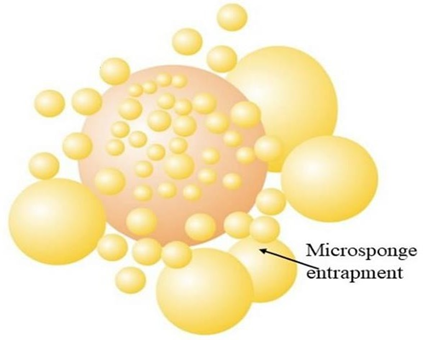
Microsponge is polymeric delivery system and a drug carrier which have tiny microscopic structure with a larger porous surface. These are micro-sized particle with average diameter of 5-30 micron. The pores of a microsponge are used for entrapment of drug. These are newer technologies which have been used to control the release of medicament from formulation and also it precises the drug in targeted delivery. Microsponges have several advantages like improved stability, extended release, improved bioavailability and précised drug targeting makes it a better carrier for drug than the conventional nanocarrier like liposome. Microsponges are formulated by various approaches in which quasi-emulsion solvent diffusion method. In this two phases (1) Internal organic phase (2) External aqueous phase are prepared separately. The organic phase contains drug and solvent (ethanol) with polymer (1-5% w/v) and outer phase contains water with surfactant (PVA) (0.1-1%w/v). The internal phase is added drop-wise to external phase with high speed continuous stirring that formulate porous microsponge after evaporation. Due to higher ranges of temperature tolerance up-to 130oC and Ph stability from 1-11, micrsponge are suitable opportunity for preparing stable formulation of wide range of drug categories. Microsponges were widely used in various drug deliveries like oral, drug delivery, topical drug delivery with uses in diseases like acne, diabetic wound healing, psoriasis, fungal infection, cancer etc.
Microsponges are spherical, porous, microscopic particles. These are polymeric delivery system and a drug carrier which have tiny microscopic structure with a larger porous surface. These are micro-sized particle with average diameter of 5-30 microns 1. Microsponge have unique compression and also have a unique dissolution due to its spongy structure 2.Microsponge can deliver drug at minimal amount of dose and work efficiently in enhancing stability, modifying drug release and reducing side effects 3. These have flexibility to entrap a wide range of active ingredients which are mostly used for prolonged topical administration for extended release of drug 4. Recently, in oral drug delivery microsponge shows increase rate of solublisation of poorly water-soluble drug 5. As the pores are very small containing about 2,50,000 pores in a typical 25 micrometer microsphere. 6. A 25 micrometer sponge may have internal pore structure equal to 10ft length, making it almost 1ml/gram for drug retention 7.
Figure 01: - Structure of microsponge
History Of Microsponge:-
The technology of microponge was developed by won in 1987 and the original patent was assigned to Advanced Polymer Systems Inc.8. The company worked on many variations of this technique and the technique was applied to various cosmetics as well as OTC and prescription drug 9and also in some FDA-approved products such as- Retin A Micro® (0.1/0.04% tretinoin) and carac (0.5% FU) 10.
Characteristics Of Microsponge11, 12, 13: -
Micrsponge are stable over wide pH range from ph 1-11 and can withstand temperature up to 130oC.
These have higher drug payload efficiency that it shows about 50-60% drug entrapment efficiency.
These are compatible with most ingredient and vehicles.
Self- sterilizing due to small pore size any bacteria can’t pass through it.
Microsponge can absorb oil up to 6 times of their weight.
Figure 02: - Characteristics Of Microsponge
Advantages Of Microsponge14, 15, 16, 17: -
Microsponges have better controlled release of API or drug from the sponge than of microcapsules.
These are non-allergic, non-irritating, non-toxic and non-mutagenic.
These have better chemical stability.
Microsponges can be easily prepared as compared to other nanoparticles like liposome.
In comparison to ointments these have ability to absorb skin secretion, so this causes less greasiness, lesser sticky and then reduces shine to skin.
Liquids can be transferred into free flowing powder so it becomes easier to handle.
These helps in improving the elegance of formulation.
Figure 03:- Advantages Of Microsponges
Mechanism Of Drug Release From Microsponge:-
In microsponge the active ingredients are free to move in and out from the peptides but the release of drug is triggered by some external factors: -
The external factors that triggers drug release are-
Pressure
Temperature
Ph
solubility18, 19
Figure 04: - Mechanism Of Drug Release from Microsponge
Pressure: - The release from microsponge is triggered by rubbing microsponge through the skin 20, 21.
Temperature: - When the formulation is applied to skin by changing the temperature of skin we can regulate drug release from the formulation 22.
Ph- triggered: - By modifying the ph of the coating material of the microspong system the drug release can also be regulated 23.
Solubility: - Solubility and partition coefficient of ingredients, active material and the chemical composition of the microsponge plays a crucial role in drug release from microsponge drug delivery system 24, 25.
Dichloromethane, ethyl cellulose, and drug are mixed together to form internal phase and the internal phase is stirred on magnetic stirrer for 15 minute.
In a separate container external phase is prepared by mixing surfactant and plasticizer in water.
Internal phase is added drop-wise to external phase for emulsification followed by continuous stirring for 1 hour.
Thus dichloromethane was eliminated and suspension is filtered and then dried for 24 hour at 40o C to obtain microsponge.
Liquid-liquid suspension polymerization 31,32:-
Non-polar active ingredientsare dissolved with monomer in a suitable solvent.
The solution is then dispersed in aqueous phase with surfactant and suspending agents helps in the formation of suspension.
Formation of ladder is occurred by cross-linking between chained monomers.
The ladder then folds to form spherical particles and agglomeration of spheres causes formation of microspheres.
The internal aqueous phase containing emulsifying agent is disseminated in organic polymeric solution.
The w/o emulsion then disseminated in exterior phase containing PVA to formulate double emulsion
This approach is suitable for both water and oil soluble drugs.
Figure 08:-Water in oil emulsion solvent diffusion method
Oil in oil emulsion solvent diffusion 34:-
The interior phase is madeup of volatile organic fluid. The volatile solvent is mainly dichloromethane.
The external phase is prepared by polymer span 80 and poly actide-glycolic acid.
The internal phase is added dropwise to external phase with continuous stirring.
Addition of porogen35: -
Porogens like hydrogen peroxide or sodium bicarbonate is disseminated in polymeric solution to form uniform dispersion.
The dispersion is than re-dispersed in aqueous phase containing PVA.
Than hydrogen peroxide is added that causes creation of linked pores.
Figure 09: - addition of porogen method of preparation
Lyophilisation 36:-
The microsponges are lyophilized after incubating in chitosan hydrochloride solution.
By lyophilisation rapid removal of solvent occurs. But due to rapid solvent elimination cracking, shrinkage may occur.
Vibrating orifice aerosol generator (VOAG) 37:-
VOAG was first reported for the preparation of lipid bilayered mesoporous silica particles.
Tetra ethyl ortho silicate, ethanol, water and dil. HCl were refluxed to prepare stock solution for formation of care.
The stock solution is diluted with solvent containing surfactant and stirred to form mono dispersed droplets. The droplets were encapsulated in liposome.
Evaluaion Paramaeters Of Microsponge:-
Particle size determination
Morphology and surface topography
Determination of loading efficiency and production field
Drug entrapment
Particle size determination38-
For particle size distribution optical microscope is used. Diffractrometry can also be used to determine the particle size of microsponges.
The texture and stability of a formulation are affected by particle size.
Morphology39–
For determination of morphology or surface morphology scanning electron microscopy is used.
The sample was mounted directly on to the SEM sample holder using double sided sticked tape. The images were recorded at different resolutions.
Production yield40,41-
The production yield of microsponges can be calculated using formula.
x=Actual yieldTheoratical yield×100
Drug content 42–
To measure drug content in microsponge 100 mg of precisely weighted microsponge is mixed in 100 ml of 6.8 ph phosphate buffer. The mixture is filtered through 0.45 µm membrane filter andsample is analyzed at suitable wavelength using UV.
Drugcontent=Actual amount of drugWeighed amount of sponge×100
Entrapment efficiency 43,44-
Drug entrapment efficiency is assessed through solvent extraction method.
10 mg of precisely weighed microsponge is dissolved in 5ml of methanol using magnetic stirrer for 20 min.
20 ml of freshly prepared PBS is added and heated at 45-50oC .Till the formation of clear solution. Methanol is evaporated and cooled at 25oC and filtered.
The drug concentration is measured by UV.
DEE=Actual drug content of spongeTheoratical drug content of sponge×100
Application Of Microsponge in Different
Pharmaceutical Formulation:-
Figure10:- Application of microsponge
Microsponge for oral drug delivery:-
Microsponge helps to maintain the drug in protected condition and protected release under regulated condition in lower GIT 45.
Microsponge increases absorption of medicament through small and large intestine by extending transit time 46.
There produced mechanically robust tablets of ketoprofen by quasi-emulsion solvent diffusion method of sponge formation. That prove plastic deformation of micosponge enhances compressibility 47.
Fluorbiprofen shows notable augmentation in drug release during 8th hour due to inclusion of enzyme in colon results in colon-targeting 48.
Microsponge for topical delivery: -
Fluocinolone acetonide, a corticosteroid is employed in dermatology to eliminate skin irritation and improve inflammatory condition 49.
By incorporating benzoyl peroxide in micrsponge its percutaneous absorption lowers which causes lessen in skin-irritation 50, 51, 52.
Microsponge can be formulated in creams, gels, powders, lotions.
Zinc pyrithione, selenium sulphide when formulated in microsponge to be use in anti-dandruff shampoo shows reduction in unpleasant odour with lowered irritation with extended safety and efficacy 54.
Microsponge in cosmetics: -
Microsponge helps in dismantling uniformity and improves covering power so colorful cosmetics can be prepared with microsponge 55.
Microsponge for bone and tissue engineering: -
Two aqueous dispersion tricalcium phosphate granules and powdered calcium hydroxyapetite and prepolymerised polymethyl metha acrylate with methyl methaacrylate monomer liquid.
A collagen sheet encapsulating basic fibroblast growth factor (bFGF) when injected subcutaneously causes increase in blood rate 56, 57, 58.
Uses Of Microsponge For Various Diseases
For psoriasis- Momentasonefurote is used in microsponge by emulsion solvent diffusion method shows initial rapid drug release effect with 29-30% drug release during first hour to 78-95 % drug release after 8 hour 59,60,61,62.
For skin infection- Mupirocin microsponge incorporated in emulgel base could provide sustained drug release till 24 hours. In draize patch test these formulation are stable and safe use. This causes slow release and shows prolonged therapeutic effect 63.
For diabetic wound healing-Nevibolol- loaded microspnge shows double effect of vasodilation with controlled and sustained release so promote faster healing. In in-vitro study 80% drug release shows within 8 hour and rapid and significant wound healing64.
For fungal infection- Voriconazole prepared using quasi-solvent diffusion method shows high encapsulation efficiency than coventional fluconazole gel and shows greater zone of inhibition65.
For acne- Benzoyl peroxide microsponge had a high drug loading capacity, sustained drug release and improved stability over conventional formulations.The microsponge gel shows greater reduction in acne lesions and skin irritation 66, 67.
Atopic dermatitis- Nargenin 1% microsponge gel have 82% entrapment and shows faster healing, reduced earflap thickness, lower WBC counts as compared to plain gel. The main benefit is three-fold greater drug deposition onto skin68.
Hyperpigmentation-glabrid microsponge using ethyl cellulose then formulated in gel causes reduction in melanin density 69.
For skin cancer- 5- fluorouracil (5-FU) shows better surface area and pore volume and have improved texture and it shows 5.5 times increasein skin deposition and lessen skin irritation 70, 71.
For Herpes-acyclovir loaded microsponge emulgel causes improved drug penetration than commercial formulation 72.
For arithritis- diclofenac used in microsponge shows prolonged release for arithritis on topical application 73, 74.
CONCLUSION
The microsponge delivery system is a unique technology for the controlled release of macroporous beads, loaded with an active agent, offering a potential reduction in side effects while maintaining their therapeutic efficacy. The microsponge delivery system is a novel, unique and emerging technology for controlled and prolonged release of drug.These are valuable drug matrix substance with several beneficial advantages like having good physical, chemical and thermal stability and allow greater flexibility in dosage from manufacturing and drug entrapment. Microsponges drug delivery system is very emerging pharmaceutical application for controlled release system, it reduces irritancy, reduces toxicity and compatible with most of ingredient and vehicles. Synthesized through techniques like quasi-emulsion solvent difusion, they found use in dermatological and oral drug delivery. Now a days it can also used for tissue engineering and controlled oral delivery using bio-erodible polymers, especially for colon specific delivery. These were mainly used in cosmetics, but due to its versatility it is source of interest in drugs ofvarious field like NSAIDs, cancer, arthritis, acne, Diabetic wound etc. Due to its stability over wide range of temperature and ph range it is suitable area of research for various sensitive drugs and its ability to control drug and it can also be controlled drug delivery and biopharmaceutical drug delivery. Due to its larger pore area it is main choice for enhance drug entrapment and control release rate of drug from sphere. Microsponges represent a promising frontier in drug delivery, with potential across pharmaceutical and cosmetic domains. MDS holds a promising future in various pharmaceutical applications in the coming years as they have unique properties like extended release, reduced irritancy, small size, self sterilize and compatible with most of vehicles and ingredients, so flexible to develop novel product forms.
REFERENCES
Tomar M and Pahwa S., “Microsponge drug delivery system”, international journal of pharmaceutical sciences and research, IJPSR 2021, volume 12 Issue 9.
Jangde R (2011), “Microsponges for colon targeted drug delivery system: an overview”. Asian J Pharm Technol 1: year 2011: page no 87–93.
Sowjanya Gummadi et.al., “Quantification and stability aspects of Luliconazole in bulk and pharmaceutical dosage forms by UV spectroscopy”, Journal of Drug Delivery and Therapeutics. Year 2019; volume 9(2-s): page no 300-306.
Sultan farhana, Chopra himashu, Sharma Gyanendra kumar., “formulation and evaluation of luliconazole microsponge loaded gel for topical drug delivery” Research J. pharm. And tech. 14(11), November 2021, page no 5775-5780.
Karthika.R., Elango.K., Ramesh Kumar K., Rahul.K. Formulation and evaluation of lornoxicammicrosponge tablets for the treatment of arthritis.Int. J. Pharmaceutical innovations. 2013; 3(2):29-40.
Mandava shyamsundar and thavvavedavanthi, “novel approach: microsponge drug delivery system”, international journal of pharmaceutical science and research, IJPSR (2012) volume 3 issue 04, page 967-980.
Vishwakarma et al., Vishwakarma Pramila, Choudhary ramraj, “microsponge: a novel strategy to control the delivery rate of active agents with reduced skin irritancy”, journal of drug delivery and pharmaceutics, 2019, 9(6-s), page 238-247.
Won R (1987). Method for delivering an active ingredient by controlled time release utilizing a novel delivery vehicle which can be prepared by a process utilizing the active ingredient as a porogen. Patent No 4690825 US.
Pradhan SK (2011). Microsponges as the versatile tool for drug deliverysystem. Int. J. Res. Pharm. Chem. 1(2): page no 243-258.
Amrutiya N, Bajaj A, Madan M (2009). Development of microsponges for topical delivery of mupirocin. AAPS Pharm. Sci. Tech. 10: page 402-409.
Aritomi H, Yamasaki Y, Yamada K, Honda H, Koshi M (1996).Developmentof sustained release formulation of chlorpheniramine maleate using powder coated microsponges prepared by dry impact blending method. J. Pharm. Sci. Technol. 56(1): page 49-56.
Jain N, Sharma PK, Banik K (2011). Recent advances on microsponge delivery system. Int. J. Pharm. Sci. Rev. Res.8: page 13-23.
Vyas SP, Khar RK (2002). Targeted and controlled drug delivery: novel carrier systems. CBS Publications, 1st ed., New Delhi, 453.
Kaity S, Maiti S, Ghosh AK, Pal D, Ghosh BS (2010). Microsponges: A novel strategy for drug delivery system. J. Adv. Pharm. Technol. Res.1(3):page 283-290.
Pradhan SK (2011). Microsponges as the versatile tool for drug deliverysystem. Int. J. Res. Pharm. Chem. 1(2): page 243-258.
Patravale VB,Mandawgade SD. Novel cosmetic delivery systems: an application update. Int J Cosmetic Sci 2008;30(1): page 19–33.
Osmani RA,Aloorkar NH,Kulkarni AS,Harkare BR,Bhosale RR. A new cornucopia in topical drug delivery: microsponge technology. Asian J Pharm Sci Technol 2014;4: page 48–60.
Khopade AJ, Jain S and Jain NK: The microsponge: Eastern Pharmacist 1996; page 49-53.
Patil SS, Dandekar V, Kale A and Barhate SD: Microsponges drug delivery system an overview. European Journal of Pharmaceutical and Medical Research 2016;3(8): page 212-221.
Jadhav N, Patel V, Mungekar S, Bhamare G, Karpe M, Kadams V (2013) Microsponge delivery system: an updated review, current status and future prospects. J Sci Innov Res 2(6):page 1097–1110.
Lalitha SK, Shankar M, Likhitha D, Dastagiri J, Babu MN (2016) A current view on microsponge drug delivery system. Eur J Mol Biol Biochem 3(2): page 88–95.
Thakur R, Kumar S, Gaba P (2020) A review: novel method for microsponge drug delivery system. J Pharm Biol Sci 15(4): page 35–44. https://doi.org/10.9790/3008-1504023544.
Christensen MS, Hargens CW, Nacht S and Gans EH: Viscoelastic properties of intact human skin instrumentations, hydration effects and contribution of the stratum corneum. Journal of Investigative Dermatology 1977; 69: page 282-86.
Sato T, Kanke M, Schroeder G and Deluca P: Porous biodegradable microspheres for controlled drug delivery, assessment of processing conditions and solvent removaltechniques. Pharmaceutical Research 1988; (5): page 21-30.
Mishra MK, Shikhri M, Sharma R and Goojar MP:Optimization, formulation development and characterization of eudragitrs 100 loaded microspongesand subsequent colonic delivery. International Journal of Drug Discovery and Herbal Research 2011; 1(1): page 8-13.
Sharma A, Hooda A, Chaudhary H (2016) Formulation and evaluation oftopical microsponges of sertaconazole. World J Pharm Res 5(11): page1444–1461. https://doi.org/10.20959/wjpr201611-7332.
Mahajan Aniruddha G, Jag Tap Leena S, Chaudhari Atul L, Swami Sima P, Mali Prabha R (2011) Formulation and evaluation of microsponge drug delivery system using Indomethacin. IRJP 2(10): page 64–69
Bhagat VS, Arote SR (2021) Formulation development and in-vitroevaluation of microsponge drug delivery system of antifungal drug. Int. J. Pure Med Res 5(3): page 654–661.
Redhu S, Pawar N (2021),“Development and characterization ofmicrosponge gel for topical delivery of oregano oil” Int J Pharm SciRes 12(2):1060–1073. https:// doi. org/ 10. 13040/ IJPSR. 09758 232. 12(2): Page 1060- 73.
22. Deshmukh K, Poddar SS (2012),“Tyrosinase inhibitor-loaded microspongedrug delivery system: new approach for hyperpigmentationdisorders” J Microencapsule 29(6): page 559–568. https:// doi. org/ 10. 3109/02652 048. 2012. 668955.
Vyas L. K., Tapar K. K., Laddha B. H., Lahoti A. O. and Nema R.K. “Formulation and development of anti-blemish preparation using microsponge technology”, Journal of Chemical and Pharmaceutical Research,2010, 2(5): page 562-571.
Guyot M, and Fawaz, F, “Microspheres- Preparation & physical Characteristics”, Int J Pharmaceutics, 1998:175: page 61-74.
Sato, T., Kanke, M.; Schroeder, G.; Deluca, P. “Porous biodegradable microspheres forcontrolled drug delivery. Assessment of processing conditions and solvent removaltechniques”. Pharm Res.1988, 5, page 21-30.
19. Won R. Two step method for preparation of controlled release formulation. UnitedStates patent number. US5145675; 1992.
Jelvehgari MR, Siahi-Shadbad S, Azarmi GP, et al. “The microsponge delivery systemof benzoyl peroxide: preparation, characterization and release studies”. Int J Pharm2006;[308]: page 124-132.
21. Maiti S, Kaity S, Ray S, et al.,“Development and evaluation of xanthan gum-facilitatedethyl cellulose microsponges for controlled percutaneous delivery of diclofenac sodium”.Acta Pharm 2011;[61]: page 257-270.
Charde MS, Ghanawat PB, Welankiwar AS, Kumar J and Chakole RD, “Microsponge a novel new drug delivery system: A review”. International Journal of Advances in Pharmaceutics 2013; 2(6).
Noor Y. Fareed, Hanan J. Kassab. “Studying the Effect of Variables on Acyclovir Microsponge”. Iraqi J Pharm Sci., 2018; 27(2): page 66-76.
Yasser shahzad, Sidra saeed, Muhammad usmanghori, Tariq mahmood, Abid mehmoodyousaf, Muhammad jamshaid et al. “Influence of polymer ratio and surfactants oncontrolled drug release from cellulosic microsponges”. int j biol macromol, Apr 1, 2018; 109: page 963-970.
Farsana T, Geetha VS, Jumana KK, Mubashira NP (2023),“Formulation development and evaluation of antimicrobial drug loaded microsponges for topical drug delivery”. World J Pharm Res. https://doi.org/10.20959/wjpr202311-28698.
Mohan D (2019),“Microsponge based drug delivery system of voriconazole for fungal infection: formulation development and In-vitro evaluation”. jddtonline.info. https://doi.org/10.22270/jddt.v9i3.2840.
“Emerging implementation of drug loaded with microsponges technology and their antifungal activity”. J PharmNegat Results 13(S01) (2022). https://doi.org/10.47750/pnr.2022.13.s01.103.
Design, formulation in-vitro evaluation of herbal microsponge by using. (n.d.). CABI Databases. 4(5): page 1923–1940. https://doi.org/10.5555/2015322598.
Halder S, Poddar S, Khanam J (2021) “Optimization and scale-up methodology in preparing microsponge loaded with 5-fuorouracil (5-FU)”. Drug Deliv Transl Res. https://doi.org/10.21203/rs.3.rs-989826/v1.
Mansi H (2019) “A review on microsponge delivery system”. J Drug DelivTherap | EBSCOhost. openurl.ebsco.com. https://doi.org/10.22270/jddt.v9i3-s.2938.
Aloorkar NH, Kulkarni AS, Ingale DJ, Patil RA (2012),“Microsponges as innovative drug delivery systems”. Int J Pharm Sci Nanotechnol 5(1): page 1597–1606
Thakur I, Sharma N (2021) “A review on innovative and novel strategyfoating microsponges”. Zenodo. https://doi.org/10.5281/zenodo.4879516.
Shinkar DM, Bhamare BS, Saudagar RB (2016) Microsponges. Asian J Res Pharm Sci 6(2): page 77–84. https://doi.org/10.5958/2231-5659.2016.00011.4
D’souza JI, More HN (2008),“Topical anti-infammatory gels of fuocinolone acetonide entrapped in eudragit based microsponge delivery system”. https://rjptonline.org/AbstractView.aspx?PID=2008-1-4-101.
Jelvehgari M, Siahi-Shadbad MR, Azarmi S, Martin GP, Nokhodchi A (2006),“The microsponge delivery system of benzoyl peroxide: preparation, characterization and release studies. Int J Pharm 308(1–2): page 124–132. https://doi.org/10.1016/j.ijpharm.2005.11.001.
Nokhodchi A, Jelvehgari M, Siahi MR, Mozafari MR (2007) Factors affecting the morphology of benzoyl peroxide microsponges. Micron38(8): page 834–840. https://doi.org/10.1016/j.micron.2007.06.012.
Wester RC, Patel R, Nacht S, Leyden JJ, Melendres J, Maibach HI (1991) Controlled release of benzoyl peroxide from a porous microsphere polymeric system can reduce topical irritancy. J Am Acad Dermatol 24(5): page720–726. https://doi.org/10.1016/0190-9622(91)70109-f.
Grimes PE: “A microsponge formulation of hydroquinone 4% and retinol 0.15% in the treatement of melasma and post-inflammatory hyperpigmentation”, Cutis, 2004; 74; page 362-368.
Viral Shaha: “Microsponge drug delivery system: A Review”, Int. J. Res. Pharm. Sci 2010; 1: page 212-218.
Sareen R, Nath K, Jain N, Dhar K, “Curcumin loaded microsponges for colon targeting in inflammatory bowel disease: fabrication, optimization, and in vitro and pharmacodynamic evaluation”, BioMed Res Int. 2014; page 1–7.
Kaity S, Maiti S, Ghosh AK, Pal D, Ghosh A, Banerjee S (2010),“Microsponges: a novel strategy for drug delivery system”. Agric Policy Pap 1(3):283. https://doi.org/10.4103/0110-5558.72416.
Kappor D, Patel MP, Vyas R, Lad C, Tyagi B (2014),“A review on microsponge drug delivery system”. J Drug Deliv Therap 4(5):978. https://doi.org/10.2270/jddt.v4i5.978.
Hussain H, Juyal D, Dhyani A (2014),“Microsponges: an overview”. Int J Drug Deliv Technol 4(4): page 58–66.
Rekha U, Manjula BP. “Formulation and evaluation of microsponges for topical drug delivery of mometasone furoate”. Int J PharmSci 2011;3(4): page133–137.
N Singh, S Sondhi, S Jindal, V Pandit, MS Ashawat. “Treatment and Management for patients with mild to severe Psoriasis: A Review”.Asian Journal of Pharmaceutical Research. 2020; doi:10.5958/2231-5691.2020.00049.0.
T Mahajan, N Singh, K Goyal, S Jindal, V Pandit. Recent Updates on Psoriasis: A Review. Asian Journal of Pharmaceutical Research,2022; 10.52711/2231-5691.2022.00012.
Jindal, S., Awasthi, R., Singhare, D., & Kulkarni, G. T. “Topical delivery of Tacrolimus using liposome containing gel: An emerging andsynergistic approach in management of psoriasis”. Medical Hypotheses.2020; 109838. doi: 10.1016/j.mehy.2020.109838.
Amrutiya, N., Bajaj, A., & Madan, M. Development of Microsponges for Topical Delivery of Mupirocin. AAPS Pharm Sci Tech, 2009;10(2): page 402–409.
Pandit, Ashlesha P.; Patel, Saurabh A.; Bhanushali, Vandana P.; Kulkarni, Vinit S.; Kakad, Vrushali D. (2017). “Nebivolol-LoadedMicrosponge Gel for Healing of Diabetic Wound”. AAPS PharmSciTech, 18(3): page 846–854. doi:10.1208/s12249-016-0574-3.
Srilakshmi P, Srinivas MP. “Development and evaluation of voriconazole microsponges for topical delivery”. Invent Rapid: Pharm Technol2011; 2011: page 75–81.
M. Jelvehgari, M.R. Siahi-Shadbad, S. Azarmi, Gary P. Martin, Ali Nokhodchi (2006). “The microsponge delivery system of benzoylperoxide: Preparation, characterization and release studies.”, 308(1-2), page 124–132. doi: 10.1016/j.ijpharm.2005.11.001.
NihalAtabay; AyseMerihSarii?ik; SinemYaprakKaravana; SedaRençber; (2020). “A novel plaster containing benzoyl peroxidemicrosponges: Formulation, development and evaluation”. Journal of Industrial Textiles, –. doi:10.1177/1528083720980466.
Nagula, Ruchika L.; Wairkar, Sarika (2020). “Cellulose microsponges based gel of naringenin for atopic dermatitis: Design, optimization,in vitro and in vivo investigation”. International Journal of Biological Macromolecules, 164(), 717–725. doi: 10.1016/j.ijbiomac.2020.07.168.
Deshmukh K, Poddar SS.,“Tyrosinase Inhibitor-Loaded Microsponge Drug Delivery System: New Approach for HyperpigmentationDisorders”. J of Microencapsulation. 2012; 29(6): page 559-568.
Jain, Subheet Kumar; Kaur, Manreet; Kalyani, Pankaj; Mehra, Anshula; Kaur, Navjot; Panchal, Neha. “Microsponges enriched gel forenhanced topical delivery of 5-Fluorouracil. Journal of Microencapsulation”. 2019; page 1–34. doi:10.1080/02652048.2019.1667447.
N Singh, S Sondhi, S Sharma, D Singh, V Koundal. “Treatment of Skin Cancer by Topical Drug Delivery of Nanoparticles: A Review”.Research Journal of Pharmacy and Technology, 2021; doi: 10.52711/0974-360X.2021.00973.
Gusai, Tejal; Dhavalkumar, Mori; Soniwala, Moinuddin; Dudhat, Kiran; Vasoya, Jaydip; Chavda, Jayant. “Formulation and optimizationof microsponge-loaded emulgel to improve the transdermal application of acyclovir DOE based approach”. Drug Delivery andTranslational Research.2020. doi:10.1007/s13346-020-00862-w.
Osmani R, Aloorkar N, Thaware B, Kulkarni P, Moin A, Hani U (2015),“Microsponge based drug delivery system for augmented gastroparesis therapy: formulation development and evaluation”. Asian J Pharm Sci 10: page 442–445. https://doi.org/10.1016/j.ajps.2015.06.003.
Hadi MA, Rao NG, Rao AS (2015),“Formulation and evaluation of minitablets-flled-pulsincap delivery of lornoxicam in the chronotherapeutic treatment of rheumatoid arthritis”. Pak J Pharm Sci 28(1): page 185–193.
Reference
Tomar M and Pahwa S., “Microsponge drug delivery system”, international journal of pharmaceutical sciences and research, IJPSR 2021, volume 12 Issue 9.
Jangde R (2011), “Microsponges for colon targeted drug delivery system: an overview”. Asian J Pharm Technol 1: year 2011: page no 87–93.
Sowjanya Gummadi et.al., “Quantification and stability aspects of Luliconazole in bulk and pharmaceutical dosage forms by UV spectroscopy”, Journal of Drug Delivery and Therapeutics. Year 2019; volume 9(2-s): page no 300-306.
Sultan farhana, Chopra himashu, Sharma Gyanendra kumar., “formulation and evaluation of luliconazole microsponge loaded gel for topical drug delivery” Research J. pharm. And tech. 14(11), November 2021, page no 5775-5780.
Karthika.R., Elango.K., Ramesh Kumar K., Rahul.K. Formulation and evaluation of lornoxicammicrosponge tablets for the treatment of arthritis.Int. J. Pharmaceutical innovations. 2013; 3(2):29-40.
Mandava shyamsundar and thavvavedavanthi, “novel approach: microsponge drug delivery system”, international journal of pharmaceutical science and research, IJPSR (2012) volume 3 issue 04, page 967-980.
Vishwakarma et al., Vishwakarma Pramila, Choudhary ramraj, “microsponge: a novel strategy to control the delivery rate of active agents with reduced skin irritancy”, journal of drug delivery and pharmaceutics, 2019, 9(6-s), page 238-247.
Won R (1987). Method for delivering an active ingredient by controlled time release utilizing a novel delivery vehicle which can be prepared by a process utilizing the active ingredient as a porogen. Patent No 4690825 US.
Pradhan SK (2011). Microsponges as the versatile tool for drug deliverysystem. Int. J. Res. Pharm. Chem. 1(2): page no 243-258.
Amrutiya N, Bajaj A, Madan M (2009). Development of microsponges for topical delivery of mupirocin. AAPS Pharm. Sci. Tech. 10: page 402-409.
Aritomi H, Yamasaki Y, Yamada K, Honda H, Koshi M (1996).Developmentof sustained release formulation of chlorpheniramine maleate using powder coated microsponges prepared by dry impact blending method. J. Pharm. Sci. Technol. 56(1): page 49-56.
Jain N, Sharma PK, Banik K (2011). Recent advances on microsponge delivery system. Int. J. Pharm. Sci. Rev. Res.8: page 13-23.
Vyas SP, Khar RK (2002). Targeted and controlled drug delivery: novel carrier systems. CBS Publications, 1st ed., New Delhi, 453.
Kaity S, Maiti S, Ghosh AK, Pal D, Ghosh BS (2010). Microsponges: A novel strategy for drug delivery system. J. Adv. Pharm. Technol. Res.1(3):page 283-290.
Pradhan SK (2011). Microsponges as the versatile tool for drug deliverysystem. Int. J. Res. Pharm. Chem. 1(2): page 243-258.
Patravale VB,Mandawgade SD. Novel cosmetic delivery systems: an application update. Int J Cosmetic Sci 2008;30(1): page 19–33.
Osmani RA,Aloorkar NH,Kulkarni AS,Harkare BR,Bhosale RR. A new cornucopia in topical drug delivery: microsponge technology. Asian J Pharm Sci Technol 2014;4: page 48–60.
Khopade AJ, Jain S and Jain NK: The microsponge: Eastern Pharmacist 1996; page 49-53.
Patil SS, Dandekar V, Kale A and Barhate SD: Microsponges drug delivery system an overview. European Journal of Pharmaceutical and Medical Research 2016;3(8): page 212-221.
Jadhav N, Patel V, Mungekar S, Bhamare G, Karpe M, Kadams V (2013) Microsponge delivery system: an updated review, current status and future prospects. J Sci Innov Res 2(6):page 1097–1110.
Lalitha SK, Shankar M, Likhitha D, Dastagiri J, Babu MN (2016) A current view on microsponge drug delivery system. Eur J Mol Biol Biochem 3(2): page 88–95.
Thakur R, Kumar S, Gaba P (2020) A review: novel method for microsponge drug delivery system. J Pharm Biol Sci 15(4): page 35–44. https://doi.org/10.9790/3008-1504023544.
Christensen MS, Hargens CW, Nacht S and Gans EH: Viscoelastic properties of intact human skin instrumentations, hydration effects and contribution of the stratum corneum. Journal of Investigative Dermatology 1977; 69: page 282-86.
Sato T, Kanke M, Schroeder G and Deluca P: Porous biodegradable microspheres for controlled drug delivery, assessment of processing conditions and solvent removaltechniques. Pharmaceutical Research 1988; (5): page 21-30.
Mishra MK, Shikhri M, Sharma R and Goojar MP:Optimization, formulation development and characterization of eudragitrs 100 loaded microspongesand subsequent colonic delivery. International Journal of Drug Discovery and Herbal Research 2011; 1(1): page 8-13.
Sharma A, Hooda A, Chaudhary H (2016) Formulation and evaluation oftopical microsponges of sertaconazole. World J Pharm Res 5(11): page1444–1461. https://doi.org/10.20959/wjpr201611-7332.
Mahajan Aniruddha G, Jag Tap Leena S, Chaudhari Atul L, Swami Sima P, Mali Prabha R (2011) Formulation and evaluation of microsponge drug delivery system using Indomethacin. IRJP 2(10): page 64–69
Bhagat VS, Arote SR (2021) Formulation development and in-vitroevaluation of microsponge drug delivery system of antifungal drug. Int. J. Pure Med Res 5(3): page 654–661.
Redhu S, Pawar N (2021),“Development and characterization ofmicrosponge gel for topical delivery of oregano oil” Int J Pharm SciRes 12(2):1060–1073. https:// doi. org/ 10. 13040/ IJPSR. 09758 232. 12(2): Page 1060- 73.
22. Deshmukh K, Poddar SS (2012),“Tyrosinase inhibitor-loaded microspongedrug delivery system: new approach for hyperpigmentationdisorders” J Microencapsule 29(6): page 559–568. https:// doi. org/ 10. 3109/02652 048. 2012. 668955.
Vyas L. K., Tapar K. K., Laddha B. H., Lahoti A. O. and Nema R.K. “Formulation and development of anti-blemish preparation using microsponge technology”, Journal of Chemical and Pharmaceutical Research,2010, 2(5): page 562-571.
Guyot M, and Fawaz, F, “Microspheres- Preparation & physical Characteristics”, Int J Pharmaceutics, 1998:175: page 61-74.
Sato, T., Kanke, M.; Schroeder, G.; Deluca, P. “Porous biodegradable microspheres forcontrolled drug delivery. Assessment of processing conditions and solvent removaltechniques”. Pharm Res.1988, 5, page 21-30.
19. Won R. Two step method for preparation of controlled release formulation. UnitedStates patent number. US5145675; 1992.
Jelvehgari MR, Siahi-Shadbad S, Azarmi GP, et al. “The microsponge delivery systemof benzoyl peroxide: preparation, characterization and release studies”. Int J Pharm2006;[308]: page 124-132.
21. Maiti S, Kaity S, Ray S, et al.,“Development and evaluation of xanthan gum-facilitatedethyl cellulose microsponges for controlled percutaneous delivery of diclofenac sodium”.Acta Pharm 2011;[61]: page 257-270.
Charde MS, Ghanawat PB, Welankiwar AS, Kumar J and Chakole RD, “Microsponge a novel new drug delivery system: A review”. International Journal of Advances in Pharmaceutics 2013; 2(6).
Noor Y. Fareed, Hanan J. Kassab. “Studying the Effect of Variables on Acyclovir Microsponge”. Iraqi J Pharm Sci., 2018; 27(2): page 66-76.
Yasser shahzad, Sidra saeed, Muhammad usmanghori, Tariq mahmood, Abid mehmoodyousaf, Muhammad jamshaid et al. “Influence of polymer ratio and surfactants oncontrolled drug release from cellulosic microsponges”. int j biol macromol, Apr 1, 2018; 109: page 963-970.
Farsana T, Geetha VS, Jumana KK, Mubashira NP (2023),“Formulation development and evaluation of antimicrobial drug loaded microsponges for topical drug delivery”. World J Pharm Res. https://doi.org/10.20959/wjpr202311-28698.
Mohan D (2019),“Microsponge based drug delivery system of voriconazole for fungal infection: formulation development and In-vitro evaluation”. jddtonline.info. https://doi.org/10.22270/jddt.v9i3.2840.
“Emerging implementation of drug loaded with microsponges technology and their antifungal activity”. J PharmNegat Results 13(S01) (2022). https://doi.org/10.47750/pnr.2022.13.s01.103.
Design, formulation in-vitro evaluation of herbal microsponge by using. (n.d.). CABI Databases. 4(5): page 1923–1940. https://doi.org/10.5555/2015322598.
Halder S, Poddar S, Khanam J (2021) “Optimization and scale-up methodology in preparing microsponge loaded with 5-fuorouracil (5-FU)”. Drug Deliv Transl Res. https://doi.org/10.21203/rs.3.rs-989826/v1.
Mansi H (2019) “A review on microsponge delivery system”. J Drug DelivTherap | EBSCOhost. openurl.ebsco.com. https://doi.org/10.22270/jddt.v9i3-s.2938.
Aloorkar NH, Kulkarni AS, Ingale DJ, Patil RA (2012),“Microsponges as innovative drug delivery systems”. Int J Pharm Sci Nanotechnol 5(1): page 1597–1606
Thakur I, Sharma N (2021) “A review on innovative and novel strategyfoating microsponges”. Zenodo. https://doi.org/10.5281/zenodo.4879516.
Shinkar DM, Bhamare BS, Saudagar RB (2016) Microsponges. Asian J Res Pharm Sci 6(2): page 77–84. https://doi.org/10.5958/2231-5659.2016.00011.4
D’souza JI, More HN (2008),“Topical anti-infammatory gels of fuocinolone acetonide entrapped in eudragit based microsponge delivery system”. https://rjptonline.org/AbstractView.aspx?PID=2008-1-4-101.
Jelvehgari M, Siahi-Shadbad MR, Azarmi S, Martin GP, Nokhodchi A (2006),“The microsponge delivery system of benzoyl peroxide: preparation, characterization and release studies. Int J Pharm 308(1–2): page 124–132. https://doi.org/10.1016/j.ijpharm.2005.11.001.
Nokhodchi A, Jelvehgari M, Siahi MR, Mozafari MR (2007) Factors affecting the morphology of benzoyl peroxide microsponges. Micron38(8): page 834–840. https://doi.org/10.1016/j.micron.2007.06.012.
Wester RC, Patel R, Nacht S, Leyden JJ, Melendres J, Maibach HI (1991) Controlled release of benzoyl peroxide from a porous microsphere polymeric system can reduce topical irritancy. J Am Acad Dermatol 24(5): page720–726. https://doi.org/10.1016/0190-9622(91)70109-f.
Grimes PE: “A microsponge formulation of hydroquinone 4% and retinol 0.15% in the treatement of melasma and post-inflammatory hyperpigmentation”, Cutis, 2004; 74; page 362-368.
Viral Shaha: “Microsponge drug delivery system: A Review”, Int. J. Res. Pharm. Sci 2010; 1: page 212-218.
Sareen R, Nath K, Jain N, Dhar K, “Curcumin loaded microsponges for colon targeting in inflammatory bowel disease: fabrication, optimization, and in vitro and pharmacodynamic evaluation”, BioMed Res Int. 2014; page 1–7.
Kaity S, Maiti S, Ghosh AK, Pal D, Ghosh A, Banerjee S (2010),“Microsponges: a novel strategy for drug delivery system”. Agric Policy Pap 1(3):283. https://doi.org/10.4103/0110-5558.72416.
Kappor D, Patel MP, Vyas R, Lad C, Tyagi B (2014),“A review on microsponge drug delivery system”. J Drug Deliv Therap 4(5):978. https://doi.org/10.2270/jddt.v4i5.978.
Hussain H, Juyal D, Dhyani A (2014),“Microsponges: an overview”. Int J Drug Deliv Technol 4(4): page 58–66.
Rekha U, Manjula BP. “Formulation and evaluation of microsponges for topical drug delivery of mometasone furoate”. Int J PharmSci 2011;3(4): page133–137.
N Singh, S Sondhi, S Jindal, V Pandit, MS Ashawat. “Treatment and Management for patients with mild to severe Psoriasis: A Review”.Asian Journal of Pharmaceutical Research. 2020; doi:10.5958/2231-5691.2020.00049.0.
T Mahajan, N Singh, K Goyal, S Jindal, V Pandit. Recent Updates on Psoriasis: A Review. Asian Journal of Pharmaceutical Research,2022; 10.52711/2231-5691.2022.00012.
Jindal, S., Awasthi, R., Singhare, D., & Kulkarni, G. T. “Topical delivery of Tacrolimus using liposome containing gel: An emerging andsynergistic approach in management of psoriasis”. Medical Hypotheses.2020; 109838. doi: 10.1016/j.mehy.2020.109838.
Amrutiya, N., Bajaj, A., & Madan, M. Development of Microsponges for Topical Delivery of Mupirocin. AAPS Pharm Sci Tech, 2009;10(2): page 402–409.
Pandit, Ashlesha P.; Patel, Saurabh A.; Bhanushali, Vandana P.; Kulkarni, Vinit S.; Kakad, Vrushali D. (2017). “Nebivolol-LoadedMicrosponge Gel for Healing of Diabetic Wound”. AAPS PharmSciTech, 18(3): page 846–854. doi:10.1208/s12249-016-0574-3.
Srilakshmi P, Srinivas MP. “Development and evaluation of voriconazole microsponges for topical delivery”. Invent Rapid: Pharm Technol2011; 2011: page 75–81.
M. Jelvehgari, M.R. Siahi-Shadbad, S. Azarmi, Gary P. Martin, Ali Nokhodchi (2006). “The microsponge delivery system of benzoylperoxide: Preparation, characterization and release studies.”, 308(1-2), page 124–132. doi: 10.1016/j.ijpharm.2005.11.001.
NihalAtabay; AyseMerihSarii?ik; SinemYaprakKaravana; SedaRençber; (2020). “A novel plaster containing benzoyl peroxidemicrosponges: Formulation, development and evaluation”. Journal of Industrial Textiles, –. doi:10.1177/1528083720980466.
Nagula, Ruchika L.; Wairkar, Sarika (2020). “Cellulose microsponges based gel of naringenin for atopic dermatitis: Design, optimization,in vitro and in vivo investigation”. International Journal of Biological Macromolecules, 164(), 717–725. doi: 10.1016/j.ijbiomac.2020.07.168.
Deshmukh K, Poddar SS.,“Tyrosinase Inhibitor-Loaded Microsponge Drug Delivery System: New Approach for HyperpigmentationDisorders”. J of Microencapsulation. 2012; 29(6): page 559-568.
Jain, Subheet Kumar; Kaur, Manreet; Kalyani, Pankaj; Mehra, Anshula; Kaur, Navjot; Panchal, Neha. “Microsponges enriched gel forenhanced topical delivery of 5-Fluorouracil. Journal of Microencapsulation”. 2019; page 1–34. doi:10.1080/02652048.2019.1667447.
N Singh, S Sondhi, S Sharma, D Singh, V Koundal. “Treatment of Skin Cancer by Topical Drug Delivery of Nanoparticles: A Review”.Research Journal of Pharmacy and Technology, 2021; doi: 10.52711/0974-360X.2021.00973.
Gusai, Tejal; Dhavalkumar, Mori; Soniwala, Moinuddin; Dudhat, Kiran; Vasoya, Jaydip; Chavda, Jayant. “Formulation and optimizationof microsponge-loaded emulgel to improve the transdermal application of acyclovir DOE based approach”. Drug Delivery andTranslational Research.2020. doi:10.1007/s13346-020-00862-w.
Osmani R, Aloorkar N, Thaware B, Kulkarni P, Moin A, Hani U (2015),“Microsponge based drug delivery system for augmented gastroparesis therapy: formulation development and evaluation”. Asian J Pharm Sci 10: page 442–445. https://doi.org/10.1016/j.ajps.2015.06.003.
Hadi MA, Rao NG, Rao AS (2015),“Formulation and evaluation of minitablets-flled-pulsincap delivery of lornoxicam in the chronotherapeutic treatment of rheumatoid arthritis”. Pak J Pharm Sci 28(1): page 185–193.


 10.5281/zenodo.14800431
