Department of Pharmaceutical Chemistry, Sahyadri college of pharmacy, Methwade Sangola -413307 India.
Medicinal plants have been traditionally used in various cultures for the treatment of infectious diseases due to their wide range of bioactive compounds. The rhizomes of Curcuma caesia have a high economic importance owing to its putative medicinal properties, belongs to the family Zingiberaceae. It is commonly known as Kali haldi in local language. The present evaluation of antioxidant activity in herbal methanolic extracts is crucial for identifying potential therapeutic agents and for their application in the food, cosmetic, and pharmaceutical industries. The antimicrobial activity testing of herbal methanolic extracts provides valuable insights into their potential as alternative or complementary agents to combat infectious diseases. The IC50 value of the methanolic extract of rhizomes of Curcuma caesia was 63.78 ?g/ml. whereas for ascorbic acid it was 25.97 ?g/ml using linear regression equation. The antimicrobial activity of methanolic extract of rhizomes of Curcuma caesia was evaluated against a range of pathogenic microorganisms, including both Gram-positive and Gram-negative bacteria.
Black Turmeric (Curcuma caesia) is a perennial herb with bluish-black rhizome, native to North-East and Central India. It is found in West Bengal, Madhya Pradesh, Orissa, Chhattisgarh, and Uttar Pradesh states. The rhizomes of Black Turmeric have a high economic importance owing to its putative medicinal properties. The rhizomes of the plant are aromatic in nature. The inner part of the rhizome is bluish-black in colour and emits a characteristic sweet smell, due to presence of essential oil. Traditionally, the rhizomes of Curcuma caesia are used in treating leukoderma, asthma, tumours, piles, bronchitis etc. The paste is applied on bruises, contusions and rheumatic pains in Manipur1. In Arunachal Pradesh, Adi tribes use decoction of fresh rhizome as anti-diarrhoetic. The Khamti tribe of Lohit district applied the paste of fresh rhizome in case of snake and scorpion bite. In west Bengal, the rhizomes of the plant are used in Kali Puja, and hence the plant is termed as Kali Haldi. In Madhya Pradesh, the plant is regarded as very auspicious and is stated that a person who possess it will never experience shortage of cereals and food 2. In recent decades, the exploration of natural antioxidants has gained significant attention due to growing concerns over the health risks associated with synthetic antioxidants, such as butylated hydroxyanisole (BHA) and butylated hydroxytoluene (BHT). Herbal extracts, rich in phenolic compounds, flavonoids, alkaloids, and other secondary metabolites, have emerged as promising sources of natural antioxidants with therapeutic potential. 3 Oxidative stress, resulting from an imbalance between the production of reactive oxygen species (ROS) and the body's ability to neutralize them, plays a central role in the pathogenesis of various chronic diseases, including cancer, cardiovascular diseases, diabetes, and neurodegenerative disorders. Antioxidants mitigate this stress by scavenging free radicals, chelating metal ions, or upregulating endogenous antioxidant defenses. 4
To evaluate the antioxidant potential of herbal extracts, several in vitro assays are commonly employed. These include:
These methods provide a comprehensive understanding of the antioxidant mechanisms of herbal extracts and facilitate the comparison of antioxidant capacity across different plant species and extraction techniques. The emergence of multidrug-resistant (MDR) microorganisms has posed a significant challenge to global public health, prompting the urgent need for alternative antimicrobial agents (World Health Organization [WHO], 2020). Medicinal plants have been traditionally used in various cultures for the treatment of infectious diseases due to their wide range of bioactive compounds with potential antimicrobial properties 9. Herbal extracts contain phytochemicals such as alkaloids, flavonoids, tannins, terpenoids, and phenolic compounds, which contribute to their antimicrobial activity. 10These compounds may act through various mechanisms including disruption of microbial cell walls, inhibition of nucleic acid synthesis, or interference with microbial metabolism. 11Moreover, the use of plant-based antimicrobials is often associated with lower toxicity and reduced likelihood of resistance development compared to synthetic antibiotics. 12 The current study focuses on medicinal properties of Curcuma caesia rhizome of methanolic extract viz. antioxidant and antimicrobial activity. Used to treat above mentioned disorders.
MATERIALS AND METHODS
The plant material collected from an appropriate region of the country at an appropriate stage of its growth. Procurement of Drug Sample from the Supplier:Mundada Stores, Gandhi Chowk, Hadapsar, Pune. Maharashtra, India. The plant material is authenticated by detailed taxonomical study and the correct botanical identity is established so that chances of deliberate or intentional adulteration or substitution are avoided. The Rhizomes of Curcuma caesia (Kali Haldi) were authenticated from Agharkar Research Institute, Pune. The rhizomes were powdered, defatted with n-hexane and use for the further investigation.
The extraction of rhizomes of Curcuma caesia was done by the Soxhlet extractor. The powdered samples (50 gm) are defatted with n-hexane by stirring it for overnight. Then it filtered and then extraction was performed with methanol as the extracting solvent. The extraction was conducted for 8h/day for 3-4 days. Then extract was collected and evaporated under vacuum rotary evaporator to obtain solid mass of extract. Curcuma caesia species was extracted and the percentage yield was calculated with reference to the air-dried sample. 13
The percentage yield formula for Curcuma caesia (Black Turmeric) extract
Percentage yield of extract from crude drug= weight ofextractweight of crude drug ×100

The Methanolic extract of Curcuma caesia was examined to check presence of phyto-compound by using standard methods. 14, [15-16]
The following procedures were adopted for testing the presence of various chemical constituents in the fractions.17,18
1. Test for Steroids
A. Salkowaski test:
Chloroform (2 ml) and 2ml of concentrated sulphuric acid were added to 2 ml of test solution, shaken and allowed to stand. Change in the colour of lower chloroform layer to red and acid layer to greenish yellow fluorescence indicates the presence of steroids.
B. Liebermann-Burchard reaction:
T.S 2 ml was mixed with chloroform (2 ml). To the solution, 2 ml of acetic anhydride and 2 drops of conc. Sulphuric acid from the side of test tube were added. Change in colour as, first red, then blue and finally green indicates the presence of steroids.
2. Test for Triterpenoids
A. Salkowaski test:
Concentrated sulphuric acid (2 ml) was added to 2 ml of test solution. The solution was shaken and allowed to stand. The colour of lower layer changes to yellow indicating the presence of triterpenoids.
B. Liebermann-Burchardt Test:
The 3 ml of test solution was treated with 3 ml of acetic anhydride, mixed well and then 2 ml of concentrated sulfuric acid was added from the sides of the test-tube. The development of deep red colour indicates the presence of triterpenoids.
3. Test for Glycosides
A. Balget’s test:
2 ml of the test solution was treated with 2 ml of sodium picrate solution. The development of yellow to orange colour indicates the presence of cardiac glycosides.
B. Keller-Killiani test: -
Glacial acetic acid (3-5 drops), one drop of 5% FeCl3 and conc. Sulphuric acid were added to the test tube containing 2 ml of T.S. Appearance of reddish-brown color at the junction of two layers and bluish green in the upper layer indicates the presence of glycosides
C. Legals test:
To 2 ml of test solution, 1 ml of pyridine and 1 ml of sodium nitroprusside was added. Change in color to pink or red indicates the presence of cardiac glycosides.
D. Borntrager’s test:
Dilute Sulphuric acid was added to 2 ml of solution of extract, boiled for a few mins and filtered. To the filtrate 2 ml of benzene or chloroform was added and shaken well. The organic layer was separated and ammonia was added. The change in colour of ammonical layer to pink-red indicates the presence of anthraquinone glycosides.
4. Tests for Saponin
A. Foam Test:
Powdered extract (10-20 mg) was shaken vigorously with water (1 ml). Development of persistent foam which is stable at least for 15 minutes indicates the presence of saponin.
5. Tests for Carbohydrates
A. Molisch’s test:
3 ml of Molisch’s reagent was added to the 3 ml of test solution, shaken for few minutes. Then 2 ml of concentrated sulphuric acid was added slowly from the sides of the test tube. The development of a purple ring at the junction of two liquids indicates the presence of carbohydrates.
B. Barfoed’s test:
Barfoed’s reagent (1 ml) and test solution (1 ml) were mixed in a test tube, heated in boiling water bath for 1-2 min. and then cooled. The appearance of red precipitate indicates the presence of monosaccharides.
C. Fehling’s test:
Fehling’s A and B solutions (1 ml each) were added to the test tube and boiled for 1 min. To this 2 ml of test solution was added and heated in boiling water bath for 5-10 min. Appearance of yellow and then brick red precipitate indicates the presence of reducing sugars.
D. Benedict’s test:
Benedict’s reagent (1 ml) and T.S (1ml) were mixed in a test tube and heated in boiling water bath for 5-10 min. Change in colour to yellow; green or red indicates the presence of reducing sugar.
6. Tests for Alkaloids
To the dry extract (20 mg) dilute hydrochloric acid (1-2 ml) was added, shaken well and filtered. With filtrate the following tests were performed.
To the 3 ml of test solution 3 drops of Mayer’s reagent (potassium mercuric iodide) was added. Appearance of reddish brown or cream precipitate indicates the presence of alkaloids.
To 3 ml of filtrate 4-5 drops of Hager’s reagent (saturated picric acid solution) was added. Appearance of yellow precipitate indicates the presence of alkaloids
C. Dragendorff’s test:
3 ml of the test solution was mixed with Dragendorff’s reagent (potassium bismuth iodide). Appearance of reddish-brown precipitate indicates the presence of alkaloids.
7. Tests for Flavonoids
A. Ferric-chloride test:
Test solution with few drops of ferric chloride solution shows intense green colour indicating the presence of flavonoids.
B. Shinoda test:
To the powdered extract (10 mg), 5 ml of ethanol (95%), 3 drops of hydrochloric acid and 0.5 gm magnesium turnings were added. Change of colour of solution to pink indicates the presence of flavonoids.
8. Tests for Tannins
A. Ferric-chloride test:
3 ml of test solution treated with few drops of ferric chloride solution. Development of dark colour indicates the presence of tannins.
B. Gelatin test:
03 ml of test solution when treated with gelatin solution (3ml) gives white precipitate indicating the presence of tannins.
9. Test for Proteins
A. Millon’s test:
T.S (3 ml) and Million’s reagent (5 ml) were mixed in a test tube. The appearance of white precipitate changing to brick red or dissolved and gave red color to solution on heating indicates the presence of proteins.
B. Xanthoproteic test:
To the test tube containing T.S (3 ml), 1 ml of conc. Sulphuric acid was added. Appearance of white precipitate which turns yellow on boiling and orange on addition of NH4OH indicates the presence of tyrosin and/or tryptophan containing proteins.
C. Biuret test:
3 ml of the test solution was treated with 4% sodium hydroxide (3-5 drops) and 1% copper sulphate solution (3-5 drops). The appearance of blue colour indicates the presence of proteins.
D. Ninhydrin test:
Test solution (3 ml) and 3 drops of 5% lead acetate solution were boiled on water bath for 10 min. Change in the colour of solution to purple or blue indicates the presence of amino acids.
10. Test for Gums
1. Hydrolyze test solution using dilute HCl. Perform Fehling’s or Benedict’s test. The appearance of red color indicates the presence of gums.
2. To 2 ml of extract, add 1 ml of 0.2 N iodine solution. Mixture does not acquire olive green color indicates the presence of gums.
The hydrogen peroxide (H2O2) scavenging assay is a method used to determine the antioxidant activity of a substance by measuring its ability to scavenge or neutralize hydrogen peroxide, a free radical. The assay is relatively straightforward and can be performed using standard laboratory equipment. It is a simple, versatile, and specific method that provides insights into a compound's ability to combat oxidative stress. Solution of 0.2 M potassium dihydrogen phosphate and 0.2 M sodium hydroxide solutions were prepared according to the Indian Pharmacopoeia 1996 standards. 50 ml potassium dihydrogen phosphate solution was put in a 200 ml volumetric flask and 39.1 ml of 0.2 M sodium hydroxide solution was added and finally volume was made up to 200 ml with distilled water to prepare phosphate buffer (pH-7.4). 50 ml of phosphate buffer solution was added to equal amount of hydrogen peroxide to generate the free radicals and solution was kept aside at room temperature for 5 min to finish the reaction. Methanolic extract of Curcuma caesia and standard Ascorbic Acid in different dilutions (1 ml) in distilled water were added to 0.6 ml hydrogen peroxide solution and the absorbance was estimated at 230 nm in a spectrophotometer against a blank solution containing phosphate buffer solution without hydrogen peroxide. 19
The percentage of scavenging of H2O2 by methanolic extract of Curcuma caesia was determines by using the following equation:
Percent scavenge (H2O2) =(A0-A1)/A0 ×100

A0 = absorbance of the control
A1 = absorbance in the presence of the extract and standard
Ofloxacin was used as a standard having concentration of 100 µg/ml. The different collected fractions were dried to powder and then again dissolved in DMSO. A loop full of bacterial strain was inoculated in 50 ml of Nutrient broth in a conical flask and incubated for 24 hrs to get active strain. Muller Hinton Agar was poured into Petri dishes, after solidification 100 μl (106 cfu/ml) of strains were inoculated in the media separately by spreading. Care was taken to ensure proper spreading. The experiment was performed under strict aseptic conditions. A well was made in the plates with sterile borer. The fractions and standard (30 μl) was introduced into the well and plates were incubated at 37°C for 24 hrs. All samples were tested in duplicates. Inhibition was determined by measuring the diameter of zone of inhibition.
The following gram-negative and gram-positive bacteria were used for antimicrobial activity studies. Bacteria include Gram – ve Pseudomonas aeruginosa (NCIM 5256) and Gram + ve Streptococcus agalactiae (NCIM 2401)
RESULTS AND DISCUSSION
Preliminary phytochemical screening was carried out on the rhizome of methanolic extract to identify the presence of major constituents.
Table No.1: Phytochemical analysis for methanolic extract of rhizomes of Curcuma caesia.
|
Methanolic Extract (µg/ml) |
Abs |
% Inhibition |
Std Ascorbic acid (µg/ml) |
Abs |
% Inhibition |
|
Experiment 1 |
|||||
|
Blank |
2.156 |
-- |
Blank |
2.156 |
-- |
|
20 |
1.658 |
23.10 |
20 |
1.248 |
42.12 |
|
40 |
1.347 |
37.52 |
40 |
0.922 |
57.24 |
|
60 |
1.165 |
45.96 |
60 |
0.614 |
71.52 |
|
80 |
0.945 |
56.17 |
80 |
0.317 |
85.30 |
|
100 |
0.684 |
68.27 |
100 |
0.124 |
94.25 |
|
Experiment 2 |
|||||
|
Methanolic Extract |
Abs |
% Inhibition |
Std Ascorbic acid |
Abs |
% Inhibition |
|
Blank |
2.048 |
-- |
Blank |
2.048 |
-- |
|
20 |
1.559 |
23.91 |
20 |
1.148 |
43.94 |
|
40 |
1.226 |
40.15 |
40 |
0.857 |
58.14 |
|
60 |
1.083 |
47.10 |
60 |
0.577 |
71.82 |
|
80 |
0.869 |
57.55 |
80 |
0.320 |
84.37 |
|
100 |
0.677 |
66.94 |
100 |
0.126 |
93.82 |
|
Experiment 3 |
|||||
|
Methanolic Extract |
Abs |
% Inhibition |
Std Ascorbic acid |
Abs |
% Inhibition |
|
Blank |
2.110 |
-- |
Blank |
2.110 |
-- |
|
20 |
1.434 |
32.03 |
20 |
1.079 |
48.84 |
|
40 |
1.140 |
45.96 |
40 |
0.780 |
63.01 |
|
60 |
1.018 |
51.72 |
60 |
0.537 |
74.56 |
|
80 |
0.878 |
58.38 |
80 |
0.295 |
86.04 |
|
100 |
0.691 |
67.26 |
100 |
0.125 |
94.06 |
Standard qualitative tests were conducted to detect alkaloids, flavonoids, tannins, saponins, terpenoids, glycosides, phenols, and steroids, among others. The results revealed that the extract contains a diverse range of bioactive compounds. Specifically, Flavonoids, Glycosides, Alkaloids were identified, suggesting potential medicinal properties.
The IC50 value of the methanolic extract was 63.78 μg/ml whereas for ascorbic acid it was 25.97 μg/ml using linear regression equation.
Table No. 01: The scavenging ability of the methanolic extract of rhizomes Curcuma caesia and standard on H2O2 (Raw Data)
|
Methanolic Extract (µg/ml) |
Abs |
% Inhibition |
Std Ascorbic acid (µg/ml) |
Abs |
% Inhibition |
|
Experiment 1 |
|||||
|
Blank |
2.156 |
-- |
Blank |
2.156 |
-- |
|
20 |
1.658 |
23.10 |
20 |
1.248 |
42.12 |
|
40 |
1.347 |
37.52 |
40 |
0.922 |
57.24 |
|
60 |
1.165 |
45.96 |
60 |
0.614 |
71.52 |
|
80 |
0.945 |
56.17 |
80 |
0.317 |
85.30 |
|
100 |
0.684 |
68.27 |
100 |
0.124 |
94.25 |
|
Experiment 2 |
|||||
|
Methanolic Extract |
Abs |
% Inhibition |
Std Ascorbic acid |
Abs |
% Inhibition |
|
Blank |
2.048 |
-- |
Blank |
2.048 |
-- |
|
20 |
1.559 |
23.91 |
20 |
1.148 |
43.94 |
|
40 |
1.226 |
40.15 |
40 |
0.857 |
58.14 |
|
60 |
1.083 |
47.10 |
60 |
0.577 |
71.82 |
|
80 |
0.869 |
57.55 |
80 |
0.320 |
84.37 |
|
100 |
0.677 |
66.94 |
100 |
0.126 |
93.82 |
|
Experiment 3 |
|||||
|
Methanolic Extract |
Abs |
% Inhibition |
Std Ascorbic acid |
Abs |
% Inhibition |
|
Blank |
2.110 |
-- |
Blank |
2.110 |
-- |
|
20 |
1.434 |
32.03 |
20 |
1.079 |
48.84 |
|
40 |
1.140 |
45.96 |
40 |
0.780 |
63.01 |
|
60 |
1.018 |
51.72 |
60 |
0.537 |
74.56 |
|
80 |
0.878 |
58.38 |
80 |
0.295 |
86.04 |
|
100 |
0.691 |
67.26 |
100 |
0.125 |
94.06 |
Table No.02: The scavenging ability of the methanolic extract of rhizome of Curcuma caesia on H2O2
|
Concentration (µg/ml) |
1 |
2 |
3 |
AVG ± SD |
|
20 |
23.1 |
23.91 |
32.03 |
26.35 ± 4.94 |
|
40 |
37.52 |
40.15 |
45.96 |
41.21 ± 4.32 |
|
60 |
45.96 |
47.1 |
51.72 |
48.26 ± 3.05 |
|
80 |
56.17 |
57.55 |
58.38 |
57.37 ± 1.12 |
|
100 |
68.27 |
66.94 |
67.26 |
67.49 ± 0.69 |
Table No.03: The scavenging ability of the Standard Ascorbic acid on H2O2
|
Concentration (µg/ml) |
1 |
2 |
3 |
AVG ± SD |
|
20 |
42.12 |
43.94 |
48.84 |
44.97 ± 3.48 |
|
40 |
57.24 |
58.14 |
63.01 |
59.46 ± 3.10 |
|
60 |
71.52 |
71.82 |
74.56 |
72.63 ± 1.68 |
|
80 |
85.3 |
84.37 |
86.04 |
85.24 ± 0.84 |
|
100 |
94.25 |
93.82 |
94.06 |
94.04 ± 0.22 |

Figure no. 01: The scavenging ability of the methanolic extract (blue) on H2O2
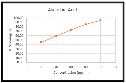
Figure no. 02: The scavenging ability of the standard (Red) on H2O2
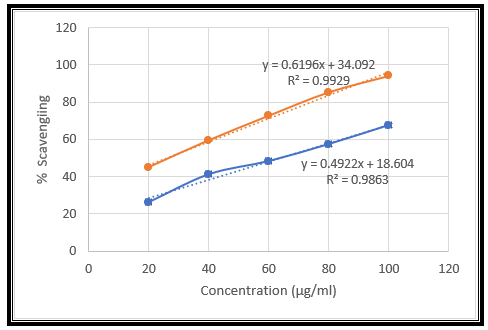
Figure no. 03: The scavenging ability of the methanolic extract (blue) and standard (Red) on H2O2
Table No.04 : Zone of inhibition of isolated compound by spread plate technique on Pseudomonas aeruginosa
|
Fraction no. |
|
Pseudomonas aeruginosa |
||
|
Code |
Zone of inhibition (mm) |
|||
|
|
|
1 |
2 |
Mean |
|
A |
1 |
11 |
10 |
10.5 |
|
B |
2 |
13 |
11 |
12.0 |
|
C |
3 |
16 |
14 |
15.0 |
|
D |
4 |
4 |
3 |
3.5 |
|
E |
5 |
17 |
15 |
16.0 |
|
F |
6 |
12 |
12 |
12.0 |
|
Ofloxacin |
7 |
12 |
11 |
11.5 |
Fraction F has equivalent activity as compared to standard Ofloxacin.
Figure no.04: Zone of inhibition of different fractions against Pseudomonas aeruginosa
Gram + ve Streptococcus agalactiae microorganism as zone of inhibition is more.
Table No. 05. Zone of inhibition of isolated compound by spread plate technique on Streptococcus agalactiae
|
Fraction no. |
|
Streptococcus agalactiae |
||
|
|
Code |
Zone of inhibition (mm) |
||
|
|
|
1 |
2 |
Mean |
|
A |
8 |
13 |
14 |
13.5 |
|
B |
9 |
12 |
11 |
11.5 |
|
C |
10 |
9 |
8 |
8.5 |
|
D |
11 |
10 |
12 |
11 |
|
E |
12 |
10 |
11 |
10.5 |
|
Ofloxacin |
14 |
22 |
24 |
23 |
Fraction B and Fraction C have equivalent or more antibacterial activity against
Gram – ve pseudomonas aeruginosa.
Figure no. 05. Zone of inhibition of different fractions against Streptococcus agalactiae
CONCLUSION
Preliminary phytochemical screening was carried out on the plant extract to identify the presence of major constituents. Standard qualitative tests were conducted to detect alkaloids, flavonoids, tannins, saponins, terpenoids, glycosides, phenols, and steroids, among others. The results revealed that the extract contains a diverse range of bioactive compounds. Specifically, Flavonoids, Glycosides, Alkaloids were identified, suggesting potential medicinal properties. The presence of flavonoids and phenolic compounds indicates antioxidant activity, while alkaloids suggest possible antimicrobial or anti-inflammatory potential. Terpenoids and glycosides may contribute to analgesic or cardiotonic effects, respectively. The in-vitro antioxidant activity of rhizome of Curcuma caesia extracts involves evaluating their ability to neutralize free radicals and prevent oxidative stress, which is linked to various chronic diseases and aging processes. In this study, different herbal extracts were subjected to in-vitro antioxidant assays such as H2O2 radical scavenging activity assay. The results demonstrated that herbal extracts exhibit significant antioxidant activity, which is closely related to their phenolic and flavonoid contents. The antimicrobial activity of the selected methanolic extract of rhizomes of Curcuma caesia was evaluated against a range of pathogenic microorganisms, including both Gram-positive and Gram-negative bacteria. The extract was prepared using standard methods such as solvent extraction (e.g., methanol). Antimicrobial testing methods like the agar well diffusion assay. The results demonstrated that the herbal extract exhibited notable inhibitory effects against tested microorganisms. The zone of inhibition varied depending on the fraction of the extract and the specific microbe tested. Generally, Gram-positive bacteria were more susceptible than Gram-negative strains, likely due to structural differences in their cell walls.
ACKNOWLEDGEMENT:
The author is very appreciative of Sahyadri College of Pharmacy for allowing him to complete the task. Author expresses sincere gratitude to respected advisor Dr. Ashpak M. Tamboli sir and Associate Professor Mr. Sagar S. Kale sir for her support, insightful counsel and careful attention.
REFERENCES


Amruta Kshirsagar*, Ashpak Tamboli, Sagar Kale, Manojkumar Patil, Vaishnavi Chavan, Prerana Panchwagh, Phytochemical and in-vitro Pharmacological Evaluation of Rhizomes of Curcuma caesia, Int. J. of Pharm. Sci., 2025, Vol 3, Issue 7, 320-333. https://doi.org/10.5281/zenodo.15790500
 10.5281/zenodo.15790500
