View Article

Abstract

Exosomes and extracellular vesicles (EVs) have emerged as next-generation drug delivery platforms owing to their natural origin, high biocompatibility, and ability to cross biological barriers. Derived from various cell types, these nanosized vesicles play vital roles in intercellular communication and can be engineered to carry therapeutic molecules such as small drugs, proteins, and nucleic acids with high specificity and stability. Recent advances in isolation, characterization, and surface modification techniques have enhanced their potential for targeted therapy in cancer, neurological, and inflammatory diseases. Compared to conventional nanocarriers, exosome-based systems offer superior biodistribution, cellular uptake, and reduced immunogenicity. Despite their potential, there are still significant obstacles to overcome, such as largescale production, standardization, and regulatory barriers. Exosomes and EVs may soon be recognized as a revolutionary and clinically feasible class of biological nanocarriers for precise drug delivery thanks to ongoing research into scalable manufacturing, functionalization, and clinical validation.

Keywords

Exosomes, Extracellular Vesicles, Nanocarriers, Drug Delivery, RNA Therapeutics, Targeted Therapy

Introduction

Cells mainly communicate with one another through chemical messengers, most notably extracellular vesicles (EVs).  In recent years, extensive research has focused on the potential of EVs in therapeutic medicine development.  Their distinct structural characteristics enable them to be modified to transport particular biomolecules like lipids, proteins, and nucleic acids—including messenger RNA (mRNA), microRNA (miRNA), other small non-coding  Genomic DNA (gDNA) and RNAs are derived from the parent cell.  Depending on their cellular  EVs are typically divided into three primary categories based on their size and origin: (a) exosomes (30–150  nm in diameter), (b) microvesicles or ectosomes (50 nm–1 µm), and (c) apoptotic bodies (50  nm–5 µm).[1] Extracellular vesicles (EVs) are membrane-bound lipid particles that are released by cells into the surrounding extracellular environment[2,3] The origin, release mechanisms, size range, molecular makeup, and biological functions of the three main subtypes of extracellular vesicles (EVs)—microvesicles (MVs), exosomes, and apoptotic bodies—are categorized.   [2,4]

 1. CLASSIFICATION OF EXTRACELLULAR VESICLES 

1.1EXOSOMES:


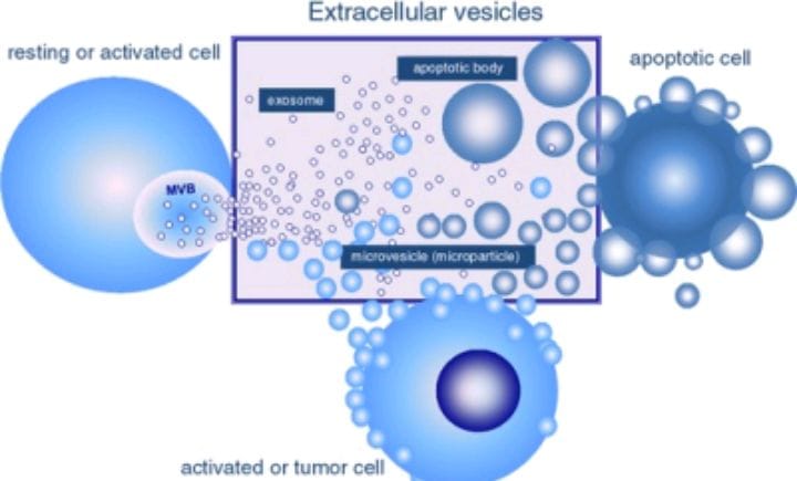
Figure 1. classification of extracellular vesicles


Intraluminal vesicles (ILVs), another name for exosomes, are membrane-bound structures with a single lipid bilayer surrounding them.  They have been found in a variety of bodily fluids, including plasma, urine, saliva, semen, breast milk, cerebrospinal fluid (CSF), bronchial secretions, serum, amniotic fluid, synovial fluid, lymph, bile, and gastric juice. They are released by almost all cell types. [5–19] Early endosomes, which are made when the plasma membrane buds inward, and multivesicular bodies (MVBs) are very important for cellular endocytosis and moving materials around.  They are in charge of sorting, recycling, storing, moving, and releasing proteins inside the cell. [20]  Exosomes are present in nearly all body fluids and originate from various cell types.  Their molecular contents often reflect disease-specific characteristics, including those linked to viral infections, neurodegenerative disorders (such as prion diseases, Alzheimer’s, and Huntington’s), and cancers.  Consequently, exosomes are being extensively studied as potential sources of novel biomarkers.  Many research efforts have aimed to elucidate their roles in intercellular communication, immune regulation, cellular development and differentiation, neuronal activity, signaling pathways, tissue regeneration, and different stages of viral replication. [21] The formation of exosomes starts in the endosomal compartment of the cell. [Fig. 1] It begins with early endosomes, which gradually mature into late endosomes or multivesicular bodies (MVBs). During this maturation, the endosomal membrane folds inward, creating intraluminal vesicles (ILVs) inside these organelles.[22] The multivesicular bodies (MVBs) then merge with the cell’s plasma membrane, releasing the intraluminal vesicles (ILVs), now called exosomes, into the extracellular space through exocytosis. Various cell types can secrete these exosomes under both normal and abnormal conditions.[23] The merging of MVBs with the plasma membrane and the movement of vesicles in time and space are managed by Rab GTPases. The endosomal sorting complex required for transport, or ESCRT, is the main molecular mechanism that plays a role in the formation of exosomes within endosomes.[24]

1.2 MICROVESICLES: 

Microvesicles (MVs) are a type of extracellular vesicle (EV) that come from the direct outward budding or shedding of the plasma membrane. Their diameter typically ranges from 100 nanometers to 1 micrometer.[2-4] The exact mechanism of MV formation is still unknown. It likely involves cytoskeletal elements like actin and microtubules. Molecular motors, such as kinesins and myosins, also play a role, along with fusion-related proteins like SNAREs and tethering factors.[25] The number of MVs released depends on the donor cell's condition and the surrounding environment.[2] Although the protein makeup of MVs varies based on the isolation method used, there is a set of proteins called “marker proteins.” These proteins are found in MVs regardless of where they come from, due to the common processes involved in their creation.[26] The presence of cytosolic and plasma membrane proteins in MVs is due to how they form. In comparison, proteins connected to organelles like the mitochondria, Golgi apparatus, nucleus, and endoplasmic reticulum are usually less common in MVs than in the whole-cell lysate. This is because these organelles do not take part in MV creation. [27,28]

1.3 APOPTOTIC BODIES:

Apoptotic bodies are membrane-bound vesicles that cells release when they go through programmed cell death. Their size usually ranges from about 50 nanometers to as large as 5 micrometers in diameter. Most apoptotic bodies tend to be on the larger side of this range.[4] These vesicles form when the cell contracts. This leads to higher internal pressure, which makes the plasma membrane separate from the cytoskeleton.[29] The molecular structure of apoptotic bodies is quite different from that of exosomes and microvesicles. Unlike these vesicles, apoptotic bodies usually contain intact cellular organelles, pieces of chromatin, and traces of glycosylated proteins.[4,30-32]

2. DRUG LOADING STRATEGIES –

In contrast, the post-loading technique means adding drugs to exosomes that have already been isolated. This method can be divided into two main strategies: passive loading and active loading.[33] Passive loading relies on the natural physical properties of the drug, which lets it move into exosomes on its own. In contrast, active loading uses specific methods, like electroporation, sonication, or extrusion, to help and improve the process of putting the drug inside the exosomes.[34,35] 

2.1 PASSIVE LOADING –

A simple way to put drugs into exosomes is through passive loading, especially with the incubation method. In this process, drugs enter exosomes because the outside drug concentration is higher. This method is easy to do and keeps the exosomes' structure intact. However, it mainly works with specific types of drugs, particularly hydrophobic compounds, which can interact well with the lipid membranes of exosomes. Thus, the loading efficiency relies heavily on the drug's hydrophobicity and how long the incubation lasts.[36] For instance, hydrophobic molecules like the polyphenolic compound curcumin and the anticancer drug paclitaxel can be effectively encapsulated in exosomes using a simple incubation process.[37,38] Compared to the pre-loading technique, the post-loading method offers better efficiency. This allows for more control over both the encapsulation efficiency and the drug-loading capacity of the final formulation.[39]


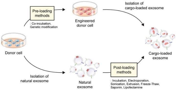
Figure 2. Drug- loading methods for exosome.


2.1.1 co- incubation (pre – passive loading)

Co-incubation involves putting the desired cargo into donor cells. These cells then release the cargo in exosomes. Later, researchers collect these exosomes from the coculture medium using ultracentrifugation. While this method is simple, it often leads to low drug-loading efficiency and limited control over encapsulation. This can change based on the physical and chemical properties of the drug and the type of donor cell used. To improve this efficiency, researchers have explored alternative strategies like mild electrical stimulation and ultraviolet exposure.[40, 41]

2.2 Active loading –

Physical Induction

To tackle the issues linked to passive loading, researchers have created various active loading methods. Physical induction uses external physical prompts to help therapeutic cargo enter exosomes. Techniques such as electroporation, sonication, freeze-thaw cycles, and extrusion are commonly used. These methods create temporary pores or apply mechanical stress to the exosomal membrane, allowing more efficient incorporation of drug molecules into the vesicles.[42]

2.2.1 Electroporation

Electroporation is a common and effective technique for getting large biomolecules, like miRNA and siRNA, into exosomes. In this method, an external electric field is applied to the exosomes. This generates a voltage strong enough to disrupt their phospholipid bilayer. As a result, transient pores form in the exosomal membrane. This temporarily increases permeability and allows the target molecules to enter. When exosomes and drug molecules experience a strong electric field, temporary pores form in the exosomal membrane. This allows the drug molecules to diffuse or pass through these openings and get encapsulated within the exosomes.[43]

3. Therapeutic applications –

3.1 Cardiovascular  disease

miRNA-21 is important for preventing cell death (apoptosis) and for promoting the growth of new blood vessels (angiogenesis) in heart-related issues.[44] miRNA-21 modulates apoptosis by targeting PDCD4 and AP-1 in cardiomyocytes. It also improves angiogenesis by activating the PTEN/Akt signaling pathway in endothelial cells. Recently, scientists encapsulated miR-21 within extracellular vesicles (EVs) from HEK293T cells. Almost 47.2% of these miR-21-loaded EVs measured between 30 and 150 nm. This size aligns with the known range for exosomes and is confirmed by markers like CD9, CD63, and CD81. These miR-21-enriched EVs effectively reduced PDCD4 protein expression, which is a known target of miR-21. They also decreased apoptosis in both cardiomyocytes and endothelial cells. This outcome was different from liposome-based systems, which did not show similar results. Additionally, delivering these miR-21 EVs directly into the heart tissue resulted in their presence in the damaged areas. This delivery helped improve cardiac repair and functional recovery.[45]

3.2 Cancer therapy:

Paclitaxel is a chemotherapy drug from the taxane family. It works against cancer by binding to and stabilizing microtubules. This stabilization stops microtubules from breaking down, which halts cell division and eventually leads to the death of cancer cells.[46-47] Paclitaxel is used to treat various cancers, including glioblastoma multiforme, breast cancer, ovarian cancer, lung cancer, and pancreatic cancer. However, its low solubility and dose-related toxicity limit its use in clinical settings. Early research on exosome-mediated delivery of paclitaxel involved mesenchymal stromal cells  (MSCs). When these cells were treated with paclitaxel, they released exosomes containing the drug. This method showed better anti-tumor activity than free paclitaxel.[48]  Curcumin is a natural compound that can help prevent the onset and spread of various cancers. However, its use in medicine is limited due to poor absorption and quick breakdown in the body. To overcome these issues and ensure it gets delivered effectively, scientists have created systems using exosomes to transport curcumin. Because curcumin is hydrophobic, it shows nearly a fivefold increase in solubility when encapsulated in exosomes that contain PBS, compared to PBS on its own. Additionally, exosome-loaded curcumin keeps over 80% of its stability after 150 minutes in PBS at pH 7.4, while free curcumin breaks down rapidly, retaining only about 25% of its initial concentration. When given either by injection into the body or orally, exosomal curcumin reaches 5 to 10 times higher levels in peripheral blood than curcumin given without exosomes.[37]

3.3 Neurodegenerative Disorders:

Alzheimer’s disease (AD) is a common neurodegenerative condition marked by the buildup of amyloid-β (Aβ) plaques, unusual phosphorylation of tau proteins, and the loss of neurons and synaptic connections. These harmful changes lead to a gradual and ongoing decline in cognitive abilities.[49] Alzheimer's disease (AD) has many aspects, so there are only a few drugs available for its treatment. This creates an urgent need for targeted and more effective treatment strategies. In AD, high levels of β-site amyloid precursor protein (APP)-cleaving enzyme 1 (BACE1) increase the breakdown of APP. This results in more amyloid plaques and the buildup of Aβ peptides, which are crucial in forming senile plaques.[50]  In one investigation, miR-29, which targets BACE1, was assessed as a potential treatment for Alzheimer’s disease. Researchers transfected HEK-293T cells and rat bone marrow-derived mesenchymal stem cells with recombinant expression vectors containing precursor sequences of miR-29a or miR-29b. They found that administering miR-29-enriched exosomes to an amyloid-β-induced Alzheimer’s model successfully prevented problems with spatial learning and memory. Supporting studies showed that miR-29b-loaded exosomes effectively reduced BACE1 expression in U87 glioblastoma cells. Moreover, another study found that exosomes loaded with curcumin had therapeutic effects by decreasing Tau hyperphosphorylation through modulation of the AKT/GSK-3β signaling pathway.[51]

 4.ADVANTAGES OF EXOSOMES/EVS:

    1. natural targeting ability-

Exosomes come from different types of cells and are found in almost all biological fluids. They have attracted a lot of interest because they can help track disease progression and may be useful in immunotherapy. Of the various sources, exosomes taken from human mesenchymal stem cells are relatively simple to isolate and have important therapeutic benefits. [52] Studies have shown that exosomes from bone marrow mesenchymal stem cells show good tolerance even after multiple doses and do not cause significant side effects. This suggests they could be a promising and safe treatment option for refractory graft-versus-host disease and other inflammation-related disorders.[53]

    1. Natural Origin & Biocompatibility:

EVs are made from cells. They have high compatibility and low toxicity compared to synthetic nanoparticles. [54]

    1. Enhanced Targeting & Uptake:

Surface proteins such as integrins and tetraspanins help with both homotypic and receptor-mediated uptake. This improves specificity. [55]

    1. Barrier Penetration:

Exosomes can cross biological barriers, including the blood-brain barrier. This ability allows for the treatment of neurological diseases. [56]

    1. Stability & Circulation:

Their natural membranes resist breaking down. This allows for longer circulation in the body.[55]

    1. Immune Evasion: 

EVs show self-markers like CD47. This reduces immune clearance and inflammation.[57] 

    1. Engineering Flexibility:

EVs can be modified to improve drug loading, targeting, and tracking. [57]

5. CHALLENGES AND LIMITATIONS –

Although significant progress has been made in new methods for isolating exosomes, some limitations still remain.

5.1 Cost and scalability:  Techniques like EXODUS and magnetic-based separation require significant initial investment and a complicated setup. This limits their ability to be used widely in regular clinical or industrial environments.[58]

5.2 Standardization and reproducibility:  A major concern in exosome research is the lack of consistency in experimental protocols, including isolation, purification, and characterization methods. This variation across laboratories results in poor reproducibility and restricts the ability to directly compare results from different studies. This, in turn, hampers the development of standardized analytical frameworks.[59]

5.3 Sample compatibility:  Many of the new isolation techniques show limited effectiveness when used with specific biological fluids, such as emulsified samples or cerebrospinal fluid. This limitation creates challenges in using these methods for different clinical or diagnostic applications, where the sample composition changes significantly.[58]

6. FUTURE PERSPECTIVES OF EXOSOME / EVS-

6.1 PRECISION MODIFICATION TECHNOLOGY:  

Advances in gene-editing tools are opening new possibilities for controlled engineering of exosomes. In the future, these technologies may allow precise genetic changes. This could help regulate the content and function of exosomes to improve their therapeutic and targeting abilities. A recent study pointed out how precision gene-editing technologies can modify exosomes for targeted therapeutic effects in Parkinson’s disease. By using genetic engineering, therapeutic molecules can be added to exosomal vectors and then delivered into target cells. The study also looked at the promising uses and challenges of using exosomes in neurological therapies.[60]

6.2 CELL ENGINEERING TECHNOLOGY:  

By manipulating the source cells using cellular engineering methods, we can control both the amount and quality of the exosomes they release. This control allows us to produce exosomes with desired traits for specific therapeutic or diagnostic uses. [61]

Nanotechnology:

Nanotechnology offers many chances to improve our understanding of cell functions and diseases. It also allows us to affect these processes at the molecular and cellular levels for better diagnosis and treatment. Recent research shows that we can use nanotechnology to change the size, shape, and surface features of exosomes, which in turn affects their biological roles and interactions in both healthy and unhealthy systems. The study emphasizes how nanotechnology-based methods can customize exosomal properties for targeted cancer treatment.[62]

CONCLUSION

Because they come from a natural source, are compatible with living tissues, and can carry different biomolecules, like proteins, nucleic acids, and small drugs, across biological barriers, exosomes and extracellular vesicles (EVs) have gained significant interest as new drug delivery systems [54–56]. They are better than many synthetic nanocarriers because of their natural targeting ability, stable circulation, and ability to avoid the immune system [55,57]. Despite these benefits, there are several drawbacks. These include high production costs, inconsistent isolation and characterization procedures, and limited compatibility with certain biological samples [58,59].  Recent developments in nanotechnology, cell engineering, and precision gene editing provide new ways to improve the therapeutic potential of exosomes. Cell engineering allows us to control exosome yield and composition [61], While gene-editing technologies allow precise manipulation of exosomal contents to improve targeting and therapeutic efficacy[60]. Additionally, nanotechnology enables us to change the surface and physical features of exosomes for better delivery in diseases such as cancer [62].  Future research should focus on developing production techniques that are affordable, repeatable, and scalable. It should also aim to establish widely recognized qualitycontrol standards. Exosome- and EV-based technologies are expected to play a crucial role in next-generation tailored and targeted treatments with further development [54– 62].

REFERENCES

  1. Ramos D, Albuquerque PR, Carmona V, Perfeito R, Nobre RJ, Parera de Almeida L.               Extracellular vesicles: novel promising delivery systems for therapy of brain diseases. J Control Release. 2017;262:247–58.
  2. Zaborowski, M.P.; Balaj, L.; Breakefield, X.O.; Lai, C.P.-K. Extracellular Vesicles:

Composition, Biological Relevance, and Methods of Study. Bioscience 2015, 65, 783– 797. [PubMed]

  1. Yáñez-Mó, M.; Siljander, P.R.-M.; Andreu, Z.; Zavec, A.B.; Borràs, F.E.; Buzas, E.I.; Buzas, K.; Casal, E.; Cappello, F.; Carvalho, J.; et al. Biological properties of extracellular vesicles and their physiological functions. J. Extracell. Vesicles 2015, 4, 27066. [PubMed]
  2. Borges, F.; Reis, L.; Schor, N. Extracellular vesicles: Structure, function, and potential clinical uses in renal diseases. Braz. J. Med. Biol. Res. 2013, 46, 824–830.
  3. Hornick, N.I.; Huan, J.; Doron, B.; Goloviznina, N.A.; Lapidus, J.; Chang, B.H.; Kurre, P. Serum Exosome MicroRNA as a Minimally-Invasive Early Biomarker of AML. Sci. Rep. 2015, 5, 11295.
  4. Akers, J.C.; Ramakrishnan, V.; Kim, R.; Skog, J.; Nakano, I.; Pingle, S.; Kalinina, J.; Hua, W.; Kesari, S.; Mao, Y.; et al. miR-21 in the extracellular vesicles (EVs) of Cerebrospinal Fluid (CSF): A Platform for Glioblastoma Biomarker Development. PLoS ONE 2013, 8, e78115. [CrossRef] [PubMed]
  5. Shi, R.; Wang, P.-Y.; Li, X.-Y.; Chen, J.-X.; Li, Y.; Zhang, X.-Z.; Zhang, C.-G.; Jiang, T.; Li, W.-B.; Ding, W.; et al. Exosomal levels of miRNA-21 from cerebrospinal uids associated with poor prognosis and tumor recurrence of glioma patients. Oncotarget 2015, 6, 26971. [PubMed]
  6. Goto, T.; Fujiya, M.; Konishi, H.; Sasajima, J.; Fujibayashi, S.; Hayashi, A.; Utsumi, T.;

Sato, H.; Iwama, T.; Ijiri, M.; et al. An elevated expression of serum exosomal microRNA191, −21, −451a of pancreatic neoplasm is considered to be efficient diagnostic marker. BMC Cancer 2018, 18, 116.

  1. Pisitkun, T.; Shen, R.-F.; Knepper, M.A. Identification and proteomic profiling of exosomes in human urine. Proc. Natl. Acad. Sci. USA 2004, 101, 13368–13373. 
  2. Caby, M.-P.; Lankar, D.; Vincendeau-Scherrer, C.; Raposo, G.; Bonnerot, C. Exosomallike vesicles are present in human blood plasma. Int. Immunol. 2005, 17, 879–887.
  3. Vojtech, L.; Woo, S.; Hughes, S.; Levy, C.; Ballweber, L.; Sauteraud, R.P.; Strobl, J.; Westerberg, K.; Gottardo, R.; Tewari, M.; et al. Exosomes in human semen carry a distinctive repertoire of small non-coding RNAs with potential regulatory functions. Nucleic Acids Res. 2014, 42, 7290–7304. [PubMed]
  4. Zlotogorski-Hurvitz, A.; Dayan, D.; Chaushu, G.; Korvala, J.; Salo, T.; Sormunen, R.; Vered, M. Human saliva-derived exosomes: Comparing methods of isolation. J. Histochem. Cytochem. 2015, 63, 181–189. [PubMed]
  5. Yuan, Z.;           Bedi,    B.;       Sadikot, R.T.   Bronchoalveolar         Lavage Exosomes in Lipopolysaccharide-induced Septic Lung Injury. J. Vis. Exp. 2018, 135, e57737. [PubMed]
  6. Dixon, C.L.; Sheller-Miller, S.; Saade, G.R.; Fortunato, S.J.; Lai, A.; Palma, C.; Guanzon, D.; Salomon, C.; Menon, R. Amniotic Fluid Exosome Proteomic Profile Exhibits Unique Pathways of Term and Preterm Labor. Endocrinology 2018, 159, 2229–2240. [PubMed]
  7. Li, Z.; Wang, Y.; Xiao, K.; Xiang, S.; Li, Z.; Weng, X. Emerging Role of Exosomes in the Joint Diseases.Cell. Physiol. Biochem. 2018, 47, 2008–2017. [PubMed]
  8. Grigor’Eva, A.E.; Tamkovich, S.N.; Eremina, A.V.; Tupikin, A.E.; Kabilov, M.R.; Chernykh, V.V.; Vlassov, V.V.; Laktionov, P.P.; Ryabchikova, E.I.; Kabilov, M. Exosomes in tears of healthy individuals: Isolation, identification, and characterization. Biochem. Suppl. Ser. B Biomed. Chem. 2016, 10, 165–172. 
  9. Milasan, A.; Tessandier, N.; Tan, S.; Brisson, A.; Boilard, E.; Martel, C. Extracellular vesicles are present in mouse lymph and their level differs in atherosclerosis. J. Extracell. Vesicles 2016, 5, 76. [PubMed]
  10. Yoon, S.B.; Chang, J.H. Extracellular vesicles in bile: A game changer in the diagnosis of indeterminate biliary stenoses? HepatoBiliary Surg. Nutr. 2017, 6, 408–410. [PubMed]
  11. Yoshida, Y.; Yamamoto, H.; Morita, R.; Oikawa, R.; Matsuo, Y.; Maehata, T.; Nosho, K.; Watanabe, Y.; Yasuda, H.; Itoh, F. Detection of DNA methylation of gastric juice-derived exosomes in gastric cancer. Integr. Mol. Med. 2014, 1, 17–21
  12. Borges, F.; Reis, L.; Schor, N. Extracellular vesicles: Structure, function, and potential clinical uses in renal diseases. Braz. J. Med. Biol. Res. 2013, 46, 824–830.
  13. Alenquer M, Amorim MJ. Exosome biogenesis, regulation, and function in viral infection. Viruses. 2015;7:5066–83.
  14. Hessvik NP, Llorente A. Current knowledge on exosome biogenesis and release. Cell Mol Life Sci. 2018;75:193–208.
  15. Beach A, Zhang HG, Ratajczak MZ, Kakar SS. Exosomes: an overview of biogenesis, composition and role in ovarian cancer. J Ovarian Res. 2014;7:14. https://doi.org/10.1186/1757-2215-7-14.
  16. Barile L, Vassalli G. Exosomes: therapy delivery tools and biomarkers of diseases. Pharmacol Ther. 2017;174:63–78.
  17. Cai, H.; Reinisch, K.; Ferro-Novick, S. Coats, Tethers, Rabs, and SNAREs Work Together to Mediate the Intracellular Destination of a Transport Vesicle. Dev. Cell 2007, 12, 671–682.
  18. Ostergaard, O.; Nielsen, C.T.; Iversen, L.V.; Jacobsen, S.; Tanassi, J.T.; Heegaard, N.H.H. Quantitative Proteome Profiling of Normal Human Circulating Microparticles. J. Proteome Res. 2012, 11, 2154–2163.
  19. Sinha, A.; Ignatchenko, V.; Ignatchenko, A.; Mejia-Guerrero, S.; Kislinger, T. In-depth proteomic analyses of ovarian cancer cell line exosomes reveals differential enrichment of functional categories compared to the NCI 60 proteome. Biochem. Biophys. Res. Commun. 2014, 445, 694–701.
  20. Christianson, H.C.; Svensson, K.J.; Van Kuppevelt, T.H.; Li, J.-P.; Belting, M. Cancer cell exosomes depend on cell-surface heparan sulfate proteoglycans for their internalization and functional activity. Proc. Natl. Acad. Sci. USA 2013, 110, 17380– 17385.
  21. Wickman, G.; Julian, L.; Olson, M.F.; Olson, M. How apoptotic cells aid in the removal of their own cold dead bodies. Cell Death Differ. 2012, 19, 735–742. 
  22. Théry, C.; Boussac, M.; Véron, P.; Ricciardi-Castagnoli, P.; Raposo, G.; Garin, J.; Amigorena, S. Proteomic Analysis of Dendritic Cell-Derived Exosomes: A Secreted Subcellular Compartment Distinct from Apoptotic Vesicles. J. Immunol. 2001, 166, 7309–7318.
  23. Escrevente, C.; Keller, S.; Altevogt, P.; Costa, J. Interaction and uptake of exosomes by ovarian cancer cells. BMC Cancer 2011, 11, 108.
  24. Kerr, J.F.R.; Wyllie, A.H.; Currie, A.R. Apoptosis: A Basic Biological Phenomenon with Wideranging Implications in Tissue Kinetics. Br. J. Cancer 1972, 26, 239–257. 
  25. Kimiz-Gebologlu, I.; Oncel, S.S. Exosomes: Large-scale production, isolation, drug loading efficiency, and biodistribution and uptake. J. Control. Release 2022, 347, 533– 543.
  26. Xi, X.M.; Xia, S.J.; Lu, R. Drug loading techniques for exosome-based drug delivery systems. Pharmazie 2021, 76, 61–67.
  27. Xu, M.; Yang, Q.; Sun, X.; Wang, Y. Recent advancements in the loading and modification of therapeutic exosomes. Front. Bioeng.Biotechnol. 2020, 8, 586130.
  28. Bellavia, D.; Raimondo, S.; Calabrese, G.; Forte, S.; Cristaldi, M.; Patinella, A.; Memeo, L.; Manno, M.; Raccosta, S.; Diana, P.; et al. Interleukin 3- receptor targeted exosomes inhibit in vitro and in vivo chronic myelogenous leukemia cell growth. Theranostics 2017, 7, 1333–1345.
  29. Sun, D.; Zhuang, X.; Xiang, X.; Liu, Y.; Zhang, S.; Liu, C.; Barnes, S.; Grizzle, W.; Miller, D.; Zhang, H.G. A novel nanoparticle drug delivery system: The anti-inflammatory activity of curcumin is enhanced when encapsulated in exosomes. Mol. Ther. 2010, 18, 1606– 1614. [PubMed]
  30. Salarpour, S.; Forootanfar, H.; Pournamdari, M.; Ahmadi-Zeidabadi, M.; Esmaeeli, M.; Pardakhty, A. Paclitaxel incorporated exosomes derived from glioblastoma cells: Comparative study of two loading techniques. Daru 2019, 27, 533–539.
  31. Burgio, S.; Noori, L.; Marino Gammazza, A.; Campanella, C.; Logozzi, M.; Fais, S.; Bucchieri, F.; Cappello, F.; Caruso Bavisotto, C. Extracellular vesicles-based drug delivery systems: A new challenge and the exemplum of malignant pleural mesothelioma. Int. J. Mol. Sci. 2020, 21, 5432.
  32. Fukuta, T.; Nishikawa, A.; Kogure, K. Low level electricity increases the secretion of extracellular vesicles from cultured cells. Biochem. Biophys. Rep. 2020, 21, 100713. 
  33. Ye, Z.; Zhang, T.; He, W.; Jin, H.; Liu, C.; Yang, Z.; Ren, J. Methotrexate-loaded extracellular vesicles functionalized with therapeutic and targeted peptides for the treatment of glioblastoma multiforme. ACS Appl. Mater. Interfaces 2018, 10, 12341– 12350.
  34. Zeng, H.; Guo, S.; Ren, X.; Wu, Z.; Liu, S.; Yao, X. Current strategies for exosome cargo loading and targeting delivery. Cells 2023, 12, 1416.
  35. Fu, S.; Wang, Y.; Xia, X.; Zheng, J.C. Exosome engineering: Current progress in cargo loading and targeted delivery. NanoImpact 2020, 20, 100261.
  36. Kura, B.; Kalocayova, B.; Devaux, Y.; Bartekova, M. Potential clinical implications of mir-1 and mir-21 in heart disease and cardioprotection. Int. J. Mol. Sci. 2020, 21, 700.
  37. Song, Y.; Zhang, C.; Zhang, J.; Jiao, Z.; Dong, N.; Wang, G.; Wang, Z.; Wang, L. Localized injection of mirna-21-enriched extracellular vesicles effectively restores cardiac function after myocardial infarction. Theranostics 2019, 9, 2346–2360.
  38. Yvon, A.M.; Wadsworth, P.; Jordan, M.A. Taxol suppresses dynamics of individual microtubules in living human tumor cells. Mol. Biol. Cell 1999, 10, 947–959. 
  39. Barbuti, A.M.; Chen, Z.S. Paclitaxel through the ages of anticancer therapy: Exploring its role in chemoresistance and radiation therapy. Cancers 2015, 7, 2360–2371.
  40. Pascucci, L.; Cocce, V.; Bonomi, A.; Ami, D.; Ceccarelli, P.; Ciusani, E.; Vigano, L.; Locatelli, A.; Sisto, F.; Doglia, S.M.; et al. Paclitaxel is incorporated by mesenchymalstromal cells and released in exosomes that inhibit in vitro tumor growth: A new approach for drug delivery. J. Control. Release 2014, 192, 262–270.
  41. Andrade-Guerrero, J.; Santiago-Balmaseda, A.; Jeronimo-Aguilar, P.; VargasRodriguez, I.; Cadena-Suarez, A.R.; Sanchez-Garibay, C.; Pozo-Molina, G.; MendezCatala, C.F.; Cardenas-Aguayo, M.D.; Diaz-Cintra, S.; et al. Alzheimer’s disease: An updated overview of its genetics. Int. J. Mol. Sci. 2023, 24, 3754. 
  42. Zong, Y.; Wang, H.; Dong, W.; Quan, X.; Zhu, H.; Xu, Y.; Huang, L.; Ma, C.; Qin, C. Mir29c regulates bace1 protein expression. Brain Res. 2011, 1395, 108–115.
  43. Wang, H.; Sui, H.; Zheng, Y.; Jiang, Y.; Shi, Y.; Liang, J.; Zhao, L. Curcumin-primed exosomes potently ameliorate cognitive function in ad mice by inhibiting hyperphosphorylation of the tau protein through the akt/gsk-3beta pathway. Nanoscale 2019, 11, 7481–7496.
  44. Yeo R, Lai R, Zhang B, et al. Mesenchymal stem cell: an efcient mass producer of exosomes for drug delivery. Adv Drug Delivery Rev. 2013;65:336–41.
  45. Kordelas L, Rebmann V, Ludwig A, et al. MSC-derived exosomes: a novel tool to treat therapy-refractory graft-versus- host disease. Leukemia. 2014;28:970–3.
  46. Kalluri R, LeBleu VS. The biology, function, and biomedical applications of exosomes. Science. 2020;367(6478):eaau6977.
  47. Vader P, Mol EA, Pasterkamp G, Schiffelers RM. Extracellular vesicles for drug delivery. Adv Drug Deliv Rev. 2016;106:148–156.
  48. Johnsen KB et al. A comprehensive overview of exosomes as drug delivery vehicles. J Control Release. 2019;298:217–230.
  49. Armstrong JP et al. Engineering extracellular vesicles to overcome biological barriers. ACS Nano. 2017;11(1):69–83.
  50. Chen Y, Zhu Q, Cheng L, et al. Exosome detection via the ultrafast-isolation system: EXODUS. Nat Methods. 2021;18(2):212–218. https://doi.org/10.1038/s41592-02001034-x.
  51. Zhang J, Gao Z, Xiao W, et al. A simplified and efficient extracellular vesiclebasedproteomics strategy for early diagnosis of colorectal cancer. Chem Sci. Published online October 15, 2024. doi:10.1039/d4sc05518g.
  52. Kojima R, Bojar D, Rizzi G, Hamri GC, El-Baba MD, Saxena P, et al. Designer exosomes Produced by implanted cells intracerebrally deliver therapeutic cargo for Parkinson’s Disease treatment. Nat Commun. 2018;9(1):1305.
  53. Zhang B, Wu X, Zhang X, Sun Y, Yan Y, Shi H, et al. Human umbilical cord Mesenchymal stem cell exosomes enhance angiogenesis through the Wnt4/beta-Catenin pathway. Stem Cells Transl Med. 2015;4(5):513–22.
  54. Li S, Xu J, Qian J, Gao X. Engineering extracellular vesicles for cancer therapy: recent Advances and challenges in clinical translation. Bio?mater Sci. 2020;8(24):6978–91.

Reference

  1. Ramos D, Albuquerque PR, Carmona V, Perfeito R, Nobre RJ, Parera de Almeida L.               Extracellular vesicles: novel promising delivery systems for therapy of brain diseases. J Control Release. 2017;262:247–58.
  2. Zaborowski, M.P.; Balaj, L.; Breakefield, X.O.; Lai, C.P.-K. Extracellular Vesicles:

Composition, Biological Relevance, and Methods of Study. Bioscience 2015, 65, 783– 797. [PubMed]

  1. Yáñez-Mó, M.; Siljander, P.R.-M.; Andreu, Z.; Zavec, A.B.; Borràs, F.E.; Buzas, E.I.; Buzas, K.; Casal, E.; Cappello, F.; Carvalho, J.; et al. Biological properties of extracellular vesicles and their physiological functions. J. Extracell. Vesicles 2015, 4, 27066. [PubMed]
  2. Borges, F.; Reis, L.; Schor, N. Extracellular vesicles: Structure, function, and potential clinical uses in renal diseases. Braz. J. Med. Biol. Res. 2013, 46, 824–830.
  3. Hornick, N.I.; Huan, J.; Doron, B.; Goloviznina, N.A.; Lapidus, J.; Chang, B.H.; Kurre, P. Serum Exosome MicroRNA as a Minimally-Invasive Early Biomarker of AML. Sci. Rep. 2015, 5, 11295.
  4. Akers, J.C.; Ramakrishnan, V.; Kim, R.; Skog, J.; Nakano, I.; Pingle, S.; Kalinina, J.; Hua, W.; Kesari, S.; Mao, Y.; et al. miR-21 in the extracellular vesicles (EVs) of Cerebrospinal Fluid (CSF): A Platform for Glioblastoma Biomarker Development. PLoS ONE 2013, 8, e78115. [CrossRef] [PubMed]
  5. Shi, R.; Wang, P.-Y.; Li, X.-Y.; Chen, J.-X.; Li, Y.; Zhang, X.-Z.; Zhang, C.-G.; Jiang, T.; Li, W.-B.; Ding, W.; et al. Exosomal levels of miRNA-21 from cerebrospinal uids associated with poor prognosis and tumor recurrence of glioma patients. Oncotarget 2015, 6, 26971. [PubMed]
  6. Goto, T.; Fujiya, M.; Konishi, H.; Sasajima, J.; Fujibayashi, S.; Hayashi, A.; Utsumi, T.;

Sato, H.; Iwama, T.; Ijiri, M.; et al. An elevated expression of serum exosomal microRNA191, −21, −451a of pancreatic neoplasm is considered to be efficient diagnostic marker. BMC Cancer 2018, 18, 116.

  1. Pisitkun, T.; Shen, R.-F.; Knepper, M.A. Identification and proteomic profiling of exosomes in human urine. Proc. Natl. Acad. Sci. USA 2004, 101, 13368–13373. 
  2. Caby, M.-P.; Lankar, D.; Vincendeau-Scherrer, C.; Raposo, G.; Bonnerot, C. Exosomallike vesicles are present in human blood plasma. Int. Immunol. 2005, 17, 879–887.
  3. Vojtech, L.; Woo, S.; Hughes, S.; Levy, C.; Ballweber, L.; Sauteraud, R.P.; Strobl, J.; Westerberg, K.; Gottardo, R.; Tewari, M.; et al. Exosomes in human semen carry a distinctive repertoire of small non-coding RNAs with potential regulatory functions. Nucleic Acids Res. 2014, 42, 7290–7304. [PubMed]
  4. Zlotogorski-Hurvitz, A.; Dayan, D.; Chaushu, G.; Korvala, J.; Salo, T.; Sormunen, R.; Vered, M. Human saliva-derived exosomes: Comparing methods of isolation. J. Histochem. Cytochem. 2015, 63, 181–189. [PubMed]
  5. Yuan, Z.;           Bedi,    B.;       Sadikot, R.T.   Bronchoalveolar         Lavage Exosomes in Lipopolysaccharide-induced Septic Lung Injury. J. Vis. Exp. 2018, 135, e57737. [PubMed]
  6. Dixon, C.L.; Sheller-Miller, S.; Saade, G.R.; Fortunato, S.J.; Lai, A.; Palma, C.; Guanzon, D.; Salomon, C.; Menon, R. Amniotic Fluid Exosome Proteomic Profile Exhibits Unique Pathways of Term and Preterm Labor. Endocrinology 2018, 159, 2229–2240. [PubMed]
  7. Li, Z.; Wang, Y.; Xiao, K.; Xiang, S.; Li, Z.; Weng, X. Emerging Role of Exosomes in the Joint Diseases.Cell. Physiol. Biochem. 2018, 47, 2008–2017. [PubMed]
  8. Grigor’Eva, A.E.; Tamkovich, S.N.; Eremina, A.V.; Tupikin, A.E.; Kabilov, M.R.; Chernykh, V.V.; Vlassov, V.V.; Laktionov, P.P.; Ryabchikova, E.I.; Kabilov, M. Exosomes in tears of healthy individuals: Isolation, identification, and characterization. Biochem. Suppl. Ser. B Biomed. Chem. 2016, 10, 165–172. 
  9. Milasan, A.; Tessandier, N.; Tan, S.; Brisson, A.; Boilard, E.; Martel, C. Extracellular vesicles are present in mouse lymph and their level differs in atherosclerosis. J. Extracell. Vesicles 2016, 5, 76. [PubMed]
  10. Yoon, S.B.; Chang, J.H. Extracellular vesicles in bile: A game changer in the diagnosis of indeterminate biliary stenoses? HepatoBiliary Surg. Nutr. 2017, 6, 408–410. [PubMed]
  11. Yoshida, Y.; Yamamoto, H.; Morita, R.; Oikawa, R.; Matsuo, Y.; Maehata, T.; Nosho, K.; Watanabe, Y.; Yasuda, H.; Itoh, F. Detection of DNA methylation of gastric juice-derived exosomes in gastric cancer. Integr. Mol. Med. 2014, 1, 17–21
  12. Borges, F.; Reis, L.; Schor, N. Extracellular vesicles: Structure, function, and potential clinical uses in renal diseases. Braz. J. Med. Biol. Res. 2013, 46, 824–830.
  13. Alenquer M, Amorim MJ. Exosome biogenesis, regulation, and function in viral infection. Viruses. 2015;7:5066–83.
  14. Hessvik NP, Llorente A. Current knowledge on exosome biogenesis and release. Cell Mol Life Sci. 2018;75:193–208.
  15. Beach A, Zhang HG, Ratajczak MZ, Kakar SS. Exosomes: an overview of biogenesis, composition and role in ovarian cancer. J Ovarian Res. 2014;7:14. https://doi.org/10.1186/1757-2215-7-14.
  16. Barile L, Vassalli G. Exosomes: therapy delivery tools and biomarkers of diseases. Pharmacol Ther. 2017;174:63–78.
  17. Cai, H.; Reinisch, K.; Ferro-Novick, S. Coats, Tethers, Rabs, and SNAREs Work Together to Mediate the Intracellular Destination of a Transport Vesicle. Dev. Cell 2007, 12, 671–682.
  18. Ostergaard, O.; Nielsen, C.T.; Iversen, L.V.; Jacobsen, S.; Tanassi, J.T.; Heegaard, N.H.H. Quantitative Proteome Profiling of Normal Human Circulating Microparticles. J. Proteome Res. 2012, 11, 2154–2163.
  19. Sinha, A.; Ignatchenko, V.; Ignatchenko, A.; Mejia-Guerrero, S.; Kislinger, T. In-depth proteomic analyses of ovarian cancer cell line exosomes reveals differential enrichment of functional categories compared to the NCI 60 proteome. Biochem. Biophys. Res. Commun. 2014, 445, 694–701.
  20. Christianson, H.C.; Svensson, K.J.; Van Kuppevelt, T.H.; Li, J.-P.; Belting, M. Cancer cell exosomes depend on cell-surface heparan sulfate proteoglycans for their internalization and functional activity. Proc. Natl. Acad. Sci. USA 2013, 110, 17380– 17385.
  21. Wickman, G.; Julian, L.; Olson, M.F.; Olson, M. How apoptotic cells aid in the removal of their own cold dead bodies. Cell Death Differ. 2012, 19, 735–742. 
  22. Théry, C.; Boussac, M.; Véron, P.; Ricciardi-Castagnoli, P.; Raposo, G.; Garin, J.; Amigorena, S. Proteomic Analysis of Dendritic Cell-Derived Exosomes: A Secreted Subcellular Compartment Distinct from Apoptotic Vesicles. J. Immunol. 2001, 166, 7309–7318.
  23. Escrevente, C.; Keller, S.; Altevogt, P.; Costa, J. Interaction and uptake of exosomes by ovarian cancer cells. BMC Cancer 2011, 11, 108.
  24. Kerr, J.F.R.; Wyllie, A.H.; Currie, A.R. Apoptosis: A Basic Biological Phenomenon with Wideranging Implications in Tissue Kinetics. Br. J. Cancer 1972, 26, 239–257. 
  25. Kimiz-Gebologlu, I.; Oncel, S.S. Exosomes: Large-scale production, isolation, drug loading efficiency, and biodistribution and uptake. J. Control. Release 2022, 347, 533– 543.
  26. Xi, X.M.; Xia, S.J.; Lu, R. Drug loading techniques for exosome-based drug delivery systems. Pharmazie 2021, 76, 61–67.
  27. Xu, M.; Yang, Q.; Sun, X.; Wang, Y. Recent advancements in the loading and modification of therapeutic exosomes. Front. Bioeng.Biotechnol. 2020, 8, 586130.
  28. Bellavia, D.; Raimondo, S.; Calabrese, G.; Forte, S.; Cristaldi, M.; Patinella, A.; Memeo, L.; Manno, M.; Raccosta, S.; Diana, P.; et al. Interleukin 3- receptor targeted exosomes inhibit in vitro and in vivo chronic myelogenous leukemia cell growth. Theranostics 2017, 7, 1333–1345.
  29. Sun, D.; Zhuang, X.; Xiang, X.; Liu, Y.; Zhang, S.; Liu, C.; Barnes, S.; Grizzle, W.; Miller, D.; Zhang, H.G. A novel nanoparticle drug delivery system: The anti-inflammatory activity of curcumin is enhanced when encapsulated in exosomes. Mol. Ther. 2010, 18, 1606– 1614. [PubMed]
  30. Salarpour, S.; Forootanfar, H.; Pournamdari, M.; Ahmadi-Zeidabadi, M.; Esmaeeli, M.; Pardakhty, A. Paclitaxel incorporated exosomes derived from glioblastoma cells: Comparative study of two loading techniques. Daru 2019, 27, 533–539.
  31. Burgio, S.; Noori, L.; Marino Gammazza, A.; Campanella, C.; Logozzi, M.; Fais, S.; Bucchieri, F.; Cappello, F.; Caruso Bavisotto, C. Extracellular vesicles-based drug delivery systems: A new challenge and the exemplum of malignant pleural mesothelioma. Int. J. Mol. Sci. 2020, 21, 5432.
  32. Fukuta, T.; Nishikawa, A.; Kogure, K. Low level electricity increases the secretion of extracellular vesicles from cultured cells. Biochem. Biophys. Rep. 2020, 21, 100713. 
  33. Ye, Z.; Zhang, T.; He, W.; Jin, H.; Liu, C.; Yang, Z.; Ren, J. Methotrexate-loaded extracellular vesicles functionalized with therapeutic and targeted peptides for the treatment of glioblastoma multiforme. ACS Appl. Mater. Interfaces 2018, 10, 12341– 12350.
  34. Zeng, H.; Guo, S.; Ren, X.; Wu, Z.; Liu, S.; Yao, X. Current strategies for exosome cargo loading and targeting delivery. Cells 2023, 12, 1416.
  35. Fu, S.; Wang, Y.; Xia, X.; Zheng, J.C. Exosome engineering: Current progress in cargo loading and targeted delivery. NanoImpact 2020, 20, 100261.
  36. Kura, B.; Kalocayova, B.; Devaux, Y.; Bartekova, M. Potential clinical implications of mir-1 and mir-21 in heart disease and cardioprotection. Int. J. Mol. Sci. 2020, 21, 700.
  37. Song, Y.; Zhang, C.; Zhang, J.; Jiao, Z.; Dong, N.; Wang, G.; Wang, Z.; Wang, L. Localized injection of mirna-21-enriched extracellular vesicles effectively restores cardiac function after myocardial infarction. Theranostics 2019, 9, 2346–2360.
  38. Yvon, A.M.; Wadsworth, P.; Jordan, M.A. Taxol suppresses dynamics of individual microtubules in living human tumor cells. Mol. Biol. Cell 1999, 10, 947–959. 
  39. Barbuti, A.M.; Chen, Z.S. Paclitaxel through the ages of anticancer therapy: Exploring its role in chemoresistance and radiation therapy. Cancers 2015, 7, 2360–2371.
  40. Pascucci, L.; Cocce, V.; Bonomi, A.; Ami, D.; Ceccarelli, P.; Ciusani, E.; Vigano, L.; Locatelli, A.; Sisto, F.; Doglia, S.M.; et al. Paclitaxel is incorporated by mesenchymalstromal cells and released in exosomes that inhibit in vitro tumor growth: A new approach for drug delivery. J. Control. Release 2014, 192, 262–270.
  41. Andrade-Guerrero, J.; Santiago-Balmaseda, A.; Jeronimo-Aguilar, P.; VargasRodriguez, I.; Cadena-Suarez, A.R.; Sanchez-Garibay, C.; Pozo-Molina, G.; MendezCatala, C.F.; Cardenas-Aguayo, M.D.; Diaz-Cintra, S.; et al. Alzheimer’s disease: An updated overview of its genetics. Int. J. Mol. Sci. 2023, 24, 3754. 
  42. Zong, Y.; Wang, H.; Dong, W.; Quan, X.; Zhu, H.; Xu, Y.; Huang, L.; Ma, C.; Qin, C. Mir29c regulates bace1 protein expression. Brain Res. 2011, 1395, 108–115.
  43. Wang, H.; Sui, H.; Zheng, Y.; Jiang, Y.; Shi, Y.; Liang, J.; Zhao, L. Curcumin-primed exosomes potently ameliorate cognitive function in ad mice by inhibiting hyperphosphorylation of the tau protein through the akt/gsk-3beta pathway. Nanoscale 2019, 11, 7481–7496.
  44. Yeo R, Lai R, Zhang B, et al. Mesenchymal stem cell: an efcient mass producer of exosomes for drug delivery. Adv Drug Delivery Rev. 2013;65:336–41.
  45. Kordelas L, Rebmann V, Ludwig A, et al. MSC-derived exosomes: a novel tool to treat therapy-refractory graft-versus- host disease. Leukemia. 2014;28:970–3.
  46. Kalluri R, LeBleu VS. The biology, function, and biomedical applications of exosomes. Science. 2020;367(6478):eaau6977.
  47. Vader P, Mol EA, Pasterkamp G, Schiffelers RM. Extracellular vesicles for drug delivery. Adv Drug Deliv Rev. 2016;106:148–156.
  48. Johnsen KB et al. A comprehensive overview of exosomes as drug delivery vehicles. J Control Release. 2019;298:217–230.
  49. Armstrong JP et al. Engineering extracellular vesicles to overcome biological barriers. ACS Nano. 2017;11(1):69–83.
  50. Chen Y, Zhu Q, Cheng L, et al. Exosome detection via the ultrafast-isolation system: EXODUS. Nat Methods. 2021;18(2):212–218. https://doi.org/10.1038/s41592-02001034-x.
  51. Zhang J, Gao Z, Xiao W, et al. A simplified and efficient extracellular vesiclebasedproteomics strategy for early diagnosis of colorectal cancer. Chem Sci. Published online October 15, 2024. doi:10.1039/d4sc05518g.
  52. Kojima R, Bojar D, Rizzi G, Hamri GC, El-Baba MD, Saxena P, et al. Designer exosomes Produced by implanted cells intracerebrally deliver therapeutic cargo for Parkinson’s Disease treatment. Nat Commun. 2018;9(1):1305.
  53. Zhang B, Wu X, Zhang X, Sun Y, Yan Y, Shi H, et al. Human umbilical cord Mesenchymal stem cell exosomes enhance angiogenesis through the Wnt4/beta-Catenin pathway. Stem Cells Transl Med. 2015;4(5):513–22.
  54. Li S, Xu J, Qian J, Gao X. Engineering extracellular vesicles for cancer therapy: recent Advances and challenges in clinical translation. Bio?mater Sci. 2020;8(24):6978–91.

Photo
Dr. Manisha S. Nangude
Corresponding author

Shivajirao S. Jondhle College of Pharmacy, Asangaon, Thane – 421 601, Maharashtra, India

Photo
T. A. Dalvi
Co-author

Shivajirao S. Jondhle College of Pharmacy, Asangaon, Thane – 421 601, Maharashtra, India

Photo
G. M. Choudhary
Co-author

Shivajirao S. Jondhle College of Pharmacy, Asangaon, Thane – 421 601, Maharashtra, India

Photo
A. S. Desai
Co-author

Shivajirao S. Jondhle College of Pharmacy, Asangaon, Thane – 421 601, Maharashtra, India

Photo
R. D. Damse
Co-author

Shivajirao S. Jondhle College of Pharmacy, Asangaon, Thane – 421 601, Maharashtra, India

Photo
G. N. Dhere
Co-author

Shivajirao S. Jondhle College of Pharmacy, Asangaon, Thane – 421 601, Maharashtra, India

Photo
G. S. Mahajan
Co-author

Shivajirao S. Jondhle College of Pharmacy, Asangaon, Thane – 421 601, Maharashtra, India

Dr. Manisha S. Nangude, T. A. Dalvi, G. M. Choudhary, A. S. Desai, R. D. Damse, G.N. Dhere, G. S. Mahajan, Exosomes And Extracellular Vesicles as Emerging Drug Delivery Platforms, Int. J. of Pharm. Sci., 2026, Vol 4, Issue 2, 2830-2841. https://doi.org/10.5281/zenodo.18682828

More related articles
Development and Evaluation of Polyherbal Anti-agin...
Avinash G.Mathpati, Dr.R.S Sakhare , Shivani V Kanole , Rutuja R ...
Overview Of The Use Of Wearable Artificial Intelli...
Ujwal Samadhan Chaudhari , Kishor V. Otari, Ajay Y. Kale, ...
Herbal Drug Formulation: Quality Aspects and Stand...
Ruturaj Bhosale, Seema Shinde, Dr. Nilesh Chougule, ...
Formulation, Development and Evaluation of Lip Serum...
A. S. Jadhav, P. B. Sutar, Dr. S. K. Mohite, ...
Advance Application of Artificial Intelligence (AI) In Pharmaceutical Sciences...
Vidya Karhale , Vedant Chimote , Mrudula Keche , Dr Nishan Bobade, ...
A Review On: Tinospora Cordfolia and Medicinal Uses...
Divya Navnath Arsule, Shinde S. B., ...
Related Articles
Nanorobots-The Future of Medicine ...
Dr. Paramita Dey, Susmita Ghosh, Shreya Das, ...
The Impact of Medicine Prices and Taxes on Access to Healthcare...
Dr. Sapna Khemnar, Gokul Cholke, Rushikesh Ghuge, Sunil Jadhav, ...
Development and Evaluation of Polyherbal Anti-aging Cream...
Avinash G.Mathpati, Dr.R.S Sakhare , Shivani V Kanole , Rutuja R Waghose , ...
More related articles
Development and Evaluation of Polyherbal Anti-aging Cream...
Avinash G.Mathpati, Dr.R.S Sakhare , Shivani V Kanole , Rutuja R Waghose , ...
Overview Of The Use Of Wearable Artificial Intelligence And Smart Devices In The...
Ujwal Samadhan Chaudhari , Kishor V. Otari, Ajay Y. Kale, ...
Herbal Drug Formulation: Quality Aspects and Standards...
Ruturaj Bhosale, Seema Shinde, Dr. Nilesh Chougule, ...