Bharat Institute of Technology, Mangalpally, Ibrahimpatnam, Rangareddy, Telangana, India.
An analytical technique known as gas chromatography-mass spectrometry(GC-MS) combines the separation powers of gas chromatography with the detection capabilities of mass spectrometry to enhance the efficiency of sample studies. This technique is well-recognised for its ability in unknown compound analysis. It enables the analysis of a broad range of compounds including organic acids, amino acids, sugars, fatty acids, sterols, and various xenobiotics. This paper provides information on how Gas Chromatography-Mass Spectrometry (GC-MS) plays a pivotal role across various scientific fields. Particularly, GC-MS is crucial in metabolomics, environmental analysis, pharmacology, and clinical diagnostics.
Gas chromatography–mass spectrometry (GC-MS) is an analytical technique that synergizes the attributes of gas chromatography and mass spectrometry, enabling the identification of diverse substances within a given sample. The groundwork for GC was established in 1950 through the gas-liquid chromatography system devised by James and Martin. The convergence of mass spectrometry (MS) as a detector alongside GC was conceptualized during the 1950s by Gohlke and McLafferty. In the years 1955-56, scientists from Dow Chemical, in a pioneering effort, successfully amalgamated gas chromatography (GC) and mass spectrometry (MS) to discriminate individual components within a mixture. This innovation marked the earliest instance of merging a separation technology with spectrometric methods, offering swift characterizations of chemical constituents. GC-MS endures as a preeminent, adaptable, and extensively employed tool for scrutinizing chemical compositions in contexts such as drug screening, forensics, environmental analysis, trace studies, and other potential applications. In 1957, Holmes and Morrell [1] were the first to showcase GC-MS coupling by employing an oscilloscope and a mass spectrometer to monitor a GC column's effluent behavior. In the laboratory setting, the sample solution is introduced into the GC inlet port, where it undergoes volatilization and is transported into a capillary column via an inert carrier gas. Constructed typically from silica, the capillary column features an inner coating serving as a stationary phase. As the sample traverses the column, compounds in the organic mixture segregate based on their interactions with the inner coating and the carrier gas. Distinct chemical properties and affinities with the stationary phase lead to enhanced separation as the sample progresses along the column. Gas Chromatography–Mass Spectrometry (GC-MS) is a powerful analytical technique used to identify and quantify compounds in a mixture. It combines two key technologies: gas chromatography (GC) and mass spectrometry (MS). This combination provides both the separation of compounds (GC) and their precise identification and quantification (MS)? of compounds based on their volatilities and interactions with a stationary phase inside a column. The sample is vaporized and carried by an inert gas (mobile phase) through a column coated with a liquid or polymer (stationary phase). Different compounds travel through the column at different rates, leading to their separation.
PRINCIPLE:
The mass spectrometer is a universal detector for gas chromatographs since any compound that can pass through a gas chromatograph is converted into ions in mass spectrometer. At the same time, the highly specific nature of mass spectrum makes the mass spectrometer a very specific gas chromatographic detector. Gas chromatography is an ideal separator, whereas mass spectrometry is excellent for identification. GC can well separate complex mixtures, and MS can detect these compounds. The combination of the two has a more favorite place, for example, both GC and MS can run in the gaseous state; thus, they can be connected directly, and the interface is very simple. Simply speaking, the performance of GC-MS is stable, and the reproducibility is good.[3] The aim of an interfacing arrangement is to operate both a gas chromatograph and a mass spectrometer without degrading the performance of either instrument. The problem is compatibility. One incompatibility problem is the difference in pressure required for the operation of a gas chromatograph and the mass spectrometer. Whereas the former operates at high pressures, the latter is designed to run under high vacuum. An associated problem is the presence of much carrier gas and little sample in the effluent from the gas chromatograph. If the gas chromatograph is using packed column the flow of carrier gas may be in excess of 30ml/min, which would collapse the vacuum of the mass spectrometer. Therefore carrier gas must be substantially removed and various designs have to be developed.
Gas chromatography is a common type of chromatography used in analytical chemistry for separating and analyzing compounds that can be vaporized without decomposition.[9,10] It is the most powerful and applicable separation technique for complex mixtures of volatile chemicals. Gas chromatography uses a gaseous mobile phase, or eluent, to carry the analyte being analyzed through a column packed or coated with a stationary phase. Some GC columns are up to 100 meters long.[11] The stationary phase in gas chromatography is commonly a packing of inert, small diameter particles (such as diatomaceous earth) with a nonpolar liquid coating them, or just a liquid coating on the inner surface of the column. This liquid is very thin layer (0.1 to 5 µm). The mobile phase is an inert gas such as Argon, Helium or Nitrogen that only carries the analyte molecules through the column. The carrier gas does not interact with the analyte and column packing material. Helium remains the most commonly used carrier gas in about 90% of instruments although hydrogen is preferred for improved separations. [9,12] The sample is injected into a long tubular column, the chromatography column. Drugs in a sample are separated from each other because some take longer tome to pass through the column than others. The retention time(time it takes to pass through the column) for an analyte is based on the time spent in the stationary phase vs. the mobile phase, with longer retention times for analytes with polarities closer to that of the stationary phase.
2. Mass spectrometry:
Mass spectrometry (MS) is an analytical technique that ionizes chemical species and sorts the ions based on their mass-to-charge ratio. A mass spectrum measures the masses within a sample. Mass spectrometry is used in many different fields and is applied to pure samples as well as complex mixtures. A mass spectrometer consists of three components: an ion source, a mass analyzer, and a detector. The ionizer converts a portions of the sample into ions. There is a wide variety of ionization techniques, depending on the phase (solid, liquid, gas) of the sample and the efficiency of various ionization mechanisms for the unknown species.[13] The detector records the charge induced or the current produced when an ion passes by or hits a surface. Typically, some type of electron multiplier is used, though other detectors including Faraday cups and ion-to-photon detectors are also used. Micro channel plate detectors are commonly used in modern commercial instruments.[14] The mass spectrum indicates the mass to charge ratio of the ions, not the molecular weight of the neutral species Where, e is the charge on an electron, Da is Daltons (1 Da = 1 amu) and z is the number of positive charges).
The essence of a mass spectrometric method revolves around the process of ionization of the molecule, with or without subsequent cleavage or fragmentation. The ionization of a molecule is an energy consuming process, which can be supplied by accelerated or thermal electrons (electron impact or electron capture), by photons (photoionization, corona discharge, laser beam), by atoms or ions accelerated by a high electrostatic field gradient or thermal impact, among other mechanisms. A rather large number of methods have been developed to transfer energy for the ionization process, to thermolabile, high- or lowmolecular weight, polar or non-polar molecules, in the gas phase (electron impact, EI, chemical ionization, CI, photoionization, PI, field ionization, FI) or in the condensed phase (field desorption, FD, laser desorption, LD, fast atom bombardment, FAB, plasma desorption, PD, secondary ion mass spectrometry, SIMS, matrix-assisted laser desorption ionization, MALDI). The ionization of neutral molecules is followed by fragmentation, or dissociative ionization, whose product ions can be separated with varying degrees of accuracy, depending on the "spectroscopic balance", i.e. analyzer used.
The mass selective detectors are divided into two groups. The first group corresponds to scanning analyzers. These include sector analyzers, e.g., magnetic deflection of single or double focus (when an electrostatic analyzer is added). The mass selective detectors are distinguished by their properties or specifications and analytical scope. The most important parameters include:
The classic configuration of a tandem mass spectrometer is the connection in series of MS1, collision- activated chamber and MS2, followed by a system for the detection and measurement of ionic currents. Tandem mass systems are divided into two large groups depending on the types of mass analyzers involved.[18] The first group is made up of tandemin-time mass spectrometers. These include linear and quadrupolar ion traps, orbital traps (orbitrap) and FTICR-MS. The second group of tandem mass (MS/MS) instruments is made up of the so-called tandem-in-space mass spectrometers. In these, at least 2 analyzers are separated in space[15]. A tandem mass spectrometer is one capable of multiple rounds of mass spectrometry, usually separated by some form of molecule fragmentation. Tandem MS can also be done in a single mass analyzer over time, as in a quadrupole ion trap. There are various methods for fragmenting molecules for tandem MS, including collision-induced dissociation (CID), electron capture dissociation (ECD), electron transfer dissociation (ETD), infrared multiphoton dissociation (IRMPD), blackbody infrared radioactive dissociation (BIRD), electron-detachment dissociation (EDD) and surface-induced dissociation (SID). The main tandem MS/MS scan modes are product ion, precursor ion, neutral loss, selected reaction monitoring, multiple reaction monitoring, and MS scans.[19] important
Application using tandem spectrometry is in protein identification.
INSTRUMENTATION AND WORKING OF GC-MS:
The gas chromatograph utilizes a capillary column which depends on the column’s dimensions as well as the phase properties (e.g. 5% phenyl polysiloxane) The difference in the chemical properties between different molecules in a mixture and their relative affinity for the stationary phase of the column will promote separation of the molecules as the sample travels the length of the column.
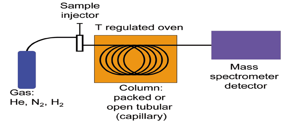
Gas supply:
Carrier gas is fed from the cylinders through the regulators and tubing to the instrument. It is usual to purify the gases to ensure high gas purity and gas supply pressure. Typically, Helium is used as carrier gas for hydrocarbon applications; however, hydrogen, argon and nitrogen are also used, depending on the application. A carrier gas must be dry, free of oxygen and chemically inert.
Injector :
Here the sample is volatilized and the resulting gas entrained into the carrier stream entering the GC column. To inject the sample into the analytical flow path, the sample injection valve is actuated and the carrier gas is switched so as to push the sample out of the sample loop and into the first column.
Column :
Gas Chromatography uses a gaseous mobile phase to transport sample components through columns either packed with coated silica particles or hollow capillary columns containing, the stationary phase coated onto the inner wall. The columns separate the gas mixture into its individual components using some physical characteristic. As the gas sample moves through the column, components with lower boiling points move more slowly than the components with higher boiling points. The speed at which this separation occurs is dependent on the temperature of the column. The length of the column determines the amount of separation of the components. Capillary GC columns are usually several meters long (10-120 m is typical) with an internal diameter of 0.10-0.50 mm, while packed GC columns tend to be 1-5 meters in length with either 2 or 4mminternal diameter. The diameter of the capillary column and the thickness of the stationary phase determine the β value (the distribution ratio of substance between the gas phase and the stationary phase), that is, the amount of substances distributed in the gas phase and the stationary phase. Column with thick-film stationary phase (low β value) is typically used for the analysis of volatile compounds, and thin-film column is beneficial for the analysis of less volatile compounds with high boiling point.
Detectors:
After the components have been separated by the chromatograph columns, they then pass over the detector. The detector is the device located at the end of the column which provides a quantitative measurement of the components of the mixture as they elute in combination with the carrier gas. Several types of detectors are available for gas chromatographs, including flame ionization detectors (for ppm-level hydrocarbons) and flame photometric detectors (for ppb- to ppm-level sulphur detection), but the most common detector used for most hydrocarbon gas measurements is the thermal conductivity detector (TCD). The other detectors are Electron- capture detector, Atomic Emission detector, GC Chemiluminescence detector.
Oven:
Gas chromatography have ovens that are temperature programmable, the temperature of the gas chromatographic ovens typically range from 50C to 4000C but can go as low as -250C with cryogenic cooling. The oven is designed to insulate the components from the effects of ambient temperature changes and maintain a very stable temperature internally. The temperature at which the oven is controlled is dependent on the application: the heavier the expected hydrocarbon mixture, the hotter the oven temperature. Natural gas applications have a typical oven temperature setting of around 800C.
ANALYTICAL METHOD DEVELOPMENT AND VALIDATION:
ANALYTICAL METHOD DEVELOPMENT:
The number of drugs introduced into the market is increasing every year. These drugs may be either new entities or partial structural modification of the existing one, very often there is a time lag from the date of introduction of a drug into the market to the date of its inclusion in pharmacopoeias. Basic criteria for new method development ofà drug analysis:
ANALYTICAL METHOD VALIDATION:
Analytical Method Validation is the collection and evaluation of data, from the process design stage throughout production, which establishes scientific evidence that a process is capable of consistently delivering quality products. Validation is an act of proving that any procedure, process, equipment, material, activity, or system performs as expected under given set of conditions and give the required accuracy, precision, sensitivity, ruggedness, etc. The biggest advantage of method validation is that it builds a degree of confidence, not only for the developer but also to the user.
VALIDATION PARAMETERS:
The various parameters according to the ICH Guidelines as follows.
ADVANTAGES OF GC-MS:
DISADVANTAGES:
CONCLUSION:
GC-MS is used to separate gas liquid different substances. It is widely used in industry and there are many applications like environmental monitoring, medicine, criminal forensics, sports anti-doping analysis. It contains different instrument contents and also some approaches. Its succinct, effective, automated system generates quick, repeatable, and efficient outcomes that play a critical role in the growth of science and technology. For greater future prospects, this adaptable analytical technique could be investigated.
REFERENCES


Dr. Namratha Sunkara, Dhandothkar Mounika, Daggula Lokesh, Domala Spandhana, Dudipala Gayathri, A Review on Gas Chromatography-Mass Spectrometry (GC-MS), Int. J. of Pharm. Sci., 2025, Vol 3, Issue 7, 1615-1622. https://doi.org/10.5281/zenodo.15864125
 10.5281/zenodo.15864125
