View Article

Abstract

This article offers a critical review of the evidence surrounding current therapeutic strategies for treating hyperuricemia. A variety of studies have been analyzed to assess the effectiveness of these treatments. Xanthine oxidase inhibitors, such as allopurinol and febuxostat, stand out as the most reliable and safe options for long-term management, making them the cornerstone of uric acid-lowering therapy. In contrast, the efficacy of uricosuric agents, which enhance renal excretion of uric acid, is significantly impacted by pharmacogenetic factors, meaning their effectiveness can vary between individuals based on genetic differences. Emerging therapies, including lesinurad and pegloticase, have demonstrated potential in the treatment of refractory hyperuricemia, particularly in acute cases. However, the current evidence supporting their use is limited to a few clinical trials, highlighting the need for more extensive, well-designed studies to establish clearer conclusions regarding their efficacy and safety in broader patient populations. These findings suggest a promising future for hyperuricemia treatment but emphasize the necessity for further research.

Keywords

Crystal deposition disease, Hyperuricemia, Gout, Uric acid.

Introduction

Hyperuricemia, the key factor in gout development, occurs when urate levels rise above saturation, leading to monosodium urate (MSU) crystal formation in joints. Uric acid, produced from purine metabolism, is balanced by daily excretion through feces and urine. Gout, a painful inflammatory arthritis, commonly affects the first metatarsophalangeal joint and results from excess urate deposition in tissues and joints. While hyperuricemia is the main cause of gout, only 5% of individuals with levels above 9 mg/dl develop the condition. Maintaining serum urate below 7 mg/dl is crucial for preventing gout and related conditions like diabetes, kidney damage, and cardiovascular disease velop luliconazole containing nanoemulgel and evaluate for topical drug delivery system.

 PATHOPHYSIOLOGY

Uric acid, the final product of purine metabolism in humans, exists mostly as monosodium urate (MSU) due to the lack of uricase. The primary cause of hyperuricemia is underexcretion of MSU, accounting for 90% of cases, though overproduction of urate can also contribute. Elevated uric acid levels lead to MSU crystal formation, which can occur in asymptomatic patients. The exact trigger for acute gouty arthritis is unclear but likely involves the shedding of MSU crystals into the synovial fluid, leading to complement activation and inflammation. Acute gout attacks are influenced by the solubility of uric acid, which decreases in cold temperatures and lower pH. Estrogen offers protection by promoting uric acid clearance, while conditions like hypertension and diabetes predispose individuals to gout by impairing kidney function. Uric acid's poor solubility in acidic urine can also lead to stone formation.

GENES RESPONSIBLE FOR URIC ACID PRODUCTION

The SLC22A12 gene encodes the URAT1 transporter on renal tubule membranes, while SLC2A9 encodes another transporter regulating uric acid (UA) excretion. Polymorphisms in these genes reduce UA excretion, raising serum uric acid (SUA) levels. ABCG2 is a UA transporter in kidney tubular cells and the gastrointestinal tract. SLC17A1 and SLC17A3 are key genes influencing SUA levels by acting as membrane transporters in the kidneys. Other genes affecting SUA levels include SLC22A11, GCKR (glucokinase regulatory protein), LRRC16A (Carmil), and PDZK1.

PRODUCTION OF URIC ACID

Urate crystals form when uric acid levels in the blood become too high, leading to supersaturation. This involves several steps:

  1. Elevated Uric Acid Levels:

Hyperuricemia occurs when uric acid levels exceed the solubility threshold (around 6.8 mg/dL), leading to crystal formation due to overproduction or underexcretion.

  1. Supersaturation:

Uric acid becomes less soluble in cooler temperatures and lower pH, causing it to shift from a dissolved to a solid state when its solubility limit is surpassed.

  1. Nucleation:

Small clusters of uric acid molecules form, influenced by factors like pH and temperature, serving as the foundation for larger crystals.

  1. Crystal Growth:

Crystals grow as more uric acid molecules aggregate, depending on the concentration of uric acid and other substances in joint fluid.

DIAGNOSIS

Hyperuricemia, or elevated uric acid levels, is diagnosed through a blood test. A serum uric acid level of 8 mg/dL or higher indicates hyperuricemia, with normal levels usually below 6.8 mg/dL. A 24-hour urine collection may also be done to assess uric acid levels, and a purine-restricted diet test helps determine if high levels are due to diet, overproduction, or underexcretion. A spot urine sample can measure the uric acid-to-creatinine ratio, with a ratio above 0.8 indicating overproduction of uric acid. Hyperuricemia itself doesn't cause symptoms but can be linked to conditions like gout, nephrolithiasis, rheumatoid arthritis, hypothyroidism, and others.

Steps involved in diagnosis process

1  Clinical Evaluation:

  • Patient History:

A comprehensive medical history is taken, focusing on past joint pain episodes, family history of gout, and lifestyle factors such as diet and alcohol use.

  • Physical Examination:

The affected joints are examined for signs of inflammation, particularly the big toe, which is commonly affected during gout attacks.

2 Laboratory Tests:

  • Serum Uric Acid Level:

Blood tests measure uric acid levels, with levels above 6.8 mg/dL indicating hyperuricemia. However, not all individuals with hyperuricemia develop gout.

  • Synovial Fluid Analysis:

In cases of joint inflammation, synovial fluid is aspirated and examined for needle-shaped monosodium urate crystals, which confirm gout.

  • Complete Blood Count (CBC):

This test checks for signs of infection or other inflammatory conditions.

3  Imaging Studies:

  • X-rays:

Used to rule out other causes of joint pain and may show joint damage or tophi in chronic cases.

  • Ultrasound:

Detects urate crystals in joints and identifies tophi.

  • Dual-Energy CT Scan:

This specialized imaging technique visualizes urate crystal deposits in joints and surrounding tissues.

Causes of hyperuricemia ang gout

Hyperuricemia is the condition of having elevated levels of uric acid in the blood, which can lead to gout. Gout is a form of inflammatory arthritis that occurs when urate crystals accumulate in joints, leading to intense pain and swelling. Below are the common causes of hyperuricemia and gout.

Increased Production of Uric Acid:

  • Dietary Factors:

Consuming purine-rich foods like red meat, organ meats, shellfish, and alcohol (especially beer) can lead to increased production of uric acid.

  • Genetic Predisposition:

Some people have genetic mutations that make their bodies produce more uric acid or eliminate it less efficiently.

  • Medical Conditions:

Conditions such as psoriasis, hemolytic anemia, and certain cancers can increase cell turnover, releasing more purines and increasing uric acid levels.                                                                                                                

Decreased Elimination of Uric Acid:

  • Kidney Dysfunction:

The kidneys may not filter uric acid efficiently, leading to its accumulation. Chronic kidney disease or other renal disorders can impair the kidneys' ability to eliminate uric acid.

  • Medications:

Drugs like diuretics (thiazides and loop diuretics), low-dose aspirin, and cyclosporine can reduce uric acid excretion.

  • Dehydration:

When the body is dehydrated, less uric acid is excreted through the urine, leading to its buildup.

Underlying Medical Conditions:

  • Metabolic Syndrome:

A cluster of conditions like hypertension, insulin resistance, and hyperlipidemia is associated with higher uric acid levels.

  • Hypertension:

High blood pressure is commonly associated with poor kidney function, leading to decreased uric acid elimination.

  • Diabetes and Insulin Resistance:

These conditions can impair kidney function, reducing uric acid excretion.
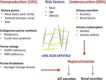


            Picture1.jpg


Mechanism of Hyperuricemia and Gout Disease

  1. Uric Acid Production and Eliminate:
  • Uric acid is produced from the breakdown of purines found in foods and tissues.
  • The liver metabolizes purines into uric acid, which is excreted by the kidneys.

Normal Uric Acid Levels:

  1. Men: 3.4-7.0 mg/dL
  2. Women: 2.4-6.0 mg/dL

2. Hyperuricemia:

  • Occurs when uric acid levels exceed normal ranges, leading to accumulation.

Causes:

  1. Increased Production:

High purine intake (from foods or alcohol), cell turnover (e.g., cancer treatments), genetic factors.

  1. Decreased Excretion:

Kidney dysfunction, medications (e.g., diuretics), genetic factors.

3. Gout Pathophysiology:

  • Gout is caused by monosodium urate (MSU) crystal deposition in joints and tissues due to prolonged hyperuricemia.

Stages of Gout Development:

  1. Asymptomatic Hyperuricemia:

Elevated uric acid without symptoms.

  1. Acute Gout Attack:
  • Crystallization of uric acid in joints leads to:
  • Crystal formation (MSU crystals in cooler joints, like the big toe).
  • Inflammatory response (immune activation, release of cytokines).
  • Symptoms: Sudden, severe pain, swelling, redness in affected joint.

4. Factors Contributing to Gout:

  • Diet:

High purine foods (red meat, shellfish), sugary drinks, alcohol (especially beer).

  • Obesity:

Increases uric acid production and decreases elimination.

  • Kidney Disease:

Impaired uric acid excretion.

  • Genetic Predisposition:

Genetic mutations affecting urate metabolism.

  • Medications:

Diuretics, aspirin, certain immunosuppressants reduce uric acid elimination.

  • Other Conditions:

Hypertension, metabolic syndrome, type 2 diabetes.

5. Treatment Strategies:

  • Acute Attack Management:
  1. NSAIDs for pain and inflammation.
  2. Colchicine to reduce immune response.
  • Long-term Management:
  1. Xanthine oxidase inhibitors (e.g., allopurinol, febuxostat) to reduce uric acid production.
  2. Lifestyle modifications:

Low-purine diet, weight loss, reduced alcohol intake, and adequate hydration.

CONCLUSION:

Gout is an inflammatory arthritis causing severe pain and swelling in joints. It happens due to high uric acid levels, forming crystals in joints. Treatment combines lifestyle changes and medications. Key changes include weight management, reducing alcohol, staying hydrated, and avoiding purine-rich foods. Medications like NSAIDs, corticosteroids, and urate-lowering drugs help control attacks. Early diagnosis and proper care prevent complications like chronic gout. With good management, people can reduce flare-ups and live healthy lives.

REFERENCES:

  1. Wortmann RL. Gout and hyperuricemia. In: Firestein GS, Budd RC, Harris ED Jr, et al, eds. Kelley’s Textbook of Rheumatology. 8th ed. Volume I. Philadelphia, PA: W. B. Saunders Company; 2008:1481–1506.
  2. Doghramji PP. Managing your patient with gout: a review of treatment options. Postgrad Med. 2011;123(3):56–71.
  3. Zgaga L, Theodoratou E, Kyle J, et al. The association of dietary intake of purine-rich vegetables, sugar-sweetened beverages and dairy with plasma urate, in a cross-sectional study. PLoS One. 2012;7(6):e38123.
  4. Halpern R, Fuldeore MJ, Mody RR, Patel PA, Mikuls TR. The effect of serum urate on gout flares and their associated costs: an administrative claims analysis. J Clin Rheumatol. 2009;15(1):3–7.
  5. Lieberman JA 3rd. Treatment and prophylaxis of gout flare in the clinic: an office-based approach to gout management. Postgrad Med. 2011;123(6):151–165.
  6. Pittman JR, Bross MH. Diagnosis and management of gout. Am Fam Physician. 1999;59(7):1799–1806, 1810.
  7. Terkeltaub R, Bushinsky DA, Becker MA. Recent developments in our understanding of the renal basis of hyperuricemia and the development of novel antihyperuricemic therapeutics. Arthritis Res Ther. 2006;8(suppl 1):S4.    
  8. Singh JA, Sarkin A, Shieh M, et al. Health care utilization in patients with gout. Semin Arthritis Rheum. 2011;40(6):501–511 
  9. .Neogi T, George J, Rekhraj S, Struthers AD, Choi H, Terkeltaub RA. Are either or both hyperuricemia and xanthine oxidase directly toxic to the vasculature? A critical appraisal. Arthritis Rheum. 2012;64(2): 327–338.
  10. Mazzali M, Kanellis J, Han L, et al. Hyperuricemia induces a primary renal arteriolopathy in rats by a blood pressure-independent mechanism. Am J Physiol Renal Physiol. 2002;282(6):F991–F997.
  11. Hsu CY, Iribarren C, McCulloch CE, Darbinian J, Go AS. Risk factors for end-stage renal disease: 25-year follow-up. Arch Intern Med. 2009;169(4):342–350.
  12. Feig DI, Kang DH, Johnson RJ. Uric acid and cardiovascular risk. N Engl J Med. 2008;359(17):1811–1821.

Reference

  1. Wortmann RL. Gout and hyperuricemia. In: Firestein GS, Budd RC, Harris ED Jr, et al, eds. Kelley’s Textbook of Rheumatology. 8th ed. Volume I. Philadelphia, PA: W. B. Saunders Company; 2008:1481–1506.
  2. Doghramji PP. Managing your patient with gout: a review of treatment options. Postgrad Med. 2011;123(3):56–71.
  3. Zgaga L, Theodoratou E, Kyle J, et al. The association of dietary intake of purine-rich vegetables, sugar-sweetened beverages and dairy with plasma urate, in a cross-sectional study. PLoS One. 2012;7(6):e38123.
  4. Halpern R, Fuldeore MJ, Mody RR, Patel PA, Mikuls TR. The effect of serum urate on gout flares and their associated costs: an administrative claims analysis. J Clin Rheumatol. 2009;15(1):3–7.
  5. Lieberman JA 3rd. Treatment and prophylaxis of gout flare in the clinic: an office-based approach to gout management. Postgrad Med. 2011;123(6):151–165.
  6. Pittman JR, Bross MH. Diagnosis and management of gout. Am Fam Physician. 1999;59(7):1799–1806, 1810.
  7. Terkeltaub R, Bushinsky DA, Becker MA. Recent developments in our understanding of the renal basis of hyperuricemia and the development of novel antihyperuricemic therapeutics. Arthritis Res Ther. 2006;8(suppl 1):S4.    
  8. Singh JA, Sarkin A, Shieh M, et al. Health care utilization in patients with gout. Semin Arthritis Rheum. 2011;40(6):501–511 
  9. .Neogi T, George J, Rekhraj S, Struthers AD, Choi H, Terkeltaub RA. Are either or both hyperuricemia and xanthine oxidase directly toxic to the vasculature? A critical appraisal. Arthritis Rheum. 2012;64(2): 327–338.
  10. Mazzali M, Kanellis J, Han L, et al. Hyperuricemia induces a primary renal arteriolopathy in rats by a blood pressure-independent mechanism. Am J Physiol Renal Physiol. 2002;282(6):F991–F997.
  11. Hsu CY, Iribarren C, McCulloch CE, Darbinian J, Go AS. Risk factors for end-stage renal disease: 25-year follow-up. Arch Intern Med. 2009;169(4):342–350.
  12. Feig DI, Kang DH, Johnson RJ. Uric acid and cardiovascular risk. N Engl J Med. 2008;359(17):1811–1821.

Photo
Dnyaneshwari Kure
Corresponding author

Gajanan maharaj college of pharmacy,nipani -bhalgaon, chh. Sambhaji nagar

Photo
Komal Chavan
Co-author

Gajanan maharaj college of pharmacy

Photo
Kavita Kulkarni
Co-author

Gajanan maharaj college of pharmacy

Photo
Ashish Shalik Faltankar
Co-author

Gajanan maharaj college of pharmacy

Komal Chavan, Kavita Kulkarni, Dnyaneshwari Kure, Ashish Shalik Faltankar, A Review Article On Hyperuricemia And Gout Disease, Int. J. of Pharm. Sci., 2024, Vol 2, Issue 10, 1178-1182. https://doi.org/10.5281/zenodo.13958184

More related articles
An Overview on Commonly Used Natural & Semisynthet...
Akshata More, Sahil Gupta, Akhilesh Singh, Vaishnavi Ranjhane, Ve...
Drug-Device Combinations And Materiovigilance In T...
Abhirami Jose, Jeeva Santhiya, eepika Kumar, Anjali M. A., Manjul...
Insights into immune thrombocytopenic purpura: a r...
Dhanya Dharman , Ahalya U, Praveena R Prasad, Shaiju S Dharan, ...
To Study Relationship Between Body Mass Index And Hypercholesterolemia...
Janvi kachhia , Amee Upadhyay, Janvi Kachhia, Amee Upadhyay, Jignasa Mansuriya, ...
Development and Characterization of a Cream Formulation Incorporating Vran Ropan...
Pallavi Nagorao Somthane, Vedantika Ramesh Salunkhe , Anand Anil Jadhav, Manoj Marotrao Shinde , Sa...
Formulation And Optimization Of Microemulsions For Improved Amlodipine Besylate ...
Mahadeo khose, Thakursing Dinesh Pawar , Dr. Nilesh Jadhav, Sugriv Ghodake, Gajanan Patingrao, Kiran...
Related Articles
Unravelling Endometriosis: Current Perspectives on Diagnosis and Treatment Innov...
Sakshi Kumari , Sonu Sharma, Shubhankar Kumar, Subham Nandi, Neeraj Kumar Mandal, Chandan Pal, Gaura...
Unraveling The Neurobiological Mechanisms And Therapeutic Challenges In Anxiety ...
Arnab Roy, Prof. (Dr) K. Rajeswar Dutt, Maheswar Prasad Deep, Abrarul Haque, Archana Rani, Nahida Kh...
History, Evidences of Therapeutic Application Of An Ancient Unani Procedure Fasd...
Bhoraniya Abdullah Ismail*, Rashid Qazi, Baig Ruqaiyya Khatoon Hushamuddin, Ansari Mushir, ...
Development And Validation of UV Spectroscopic Method for Estimation of Calcium ...
Kalyani Peluri, Ambika S., Pallavi K. C., Ruchitha M., Tejaswini V., ...
An Overview on Commonly Used Natural & Semisynthetic Polymers Used in The Floati...
Akshata More, Sahil Gupta, Akhilesh Singh, Vaishnavi Ranjhane, Vedika Nalawde, Sahil Singh, ...
More related articles
An Overview on Commonly Used Natural & Semisynthetic Polymers Used in The Floati...
Akshata More, Sahil Gupta, Akhilesh Singh, Vaishnavi Ranjhane, Vedika Nalawde, Sahil Singh, ...
Drug-Device Combinations And Materiovigilance In The Cardiology Department Of A ...
Abhirami Jose, Jeeva Santhiya, eepika Kumar, Anjali M. A., Manjula Devi A. S, R. Vickram Vignesh, ...
Insights into immune thrombocytopenic purpura: a rare hematological disorder ...
Dhanya Dharman , Ahalya U, Praveena R Prasad, Shaiju S Dharan, ...