Department of Pharmaceutics, Channabasweshwar Pharmacy College (Degree) Latur, Maharashtra- 413512.
Solid lipid nanoparticles (SLNs) have emerged as an advanced strategy in the development of antifungal drug delivery systems. The optimized composition of SLNs was characterized by evaluating various parameters, including particle size, drug entrapment efficiency, scanning electron microscopy (SEM), zeta potential, particle size distribution, and stability studies. Composed of biocompatible solid lipids, these nanoparticles offer notable advantages in improving the solubility, stability, and bioavailability of poorly water-soluble antifungal agents. The encapsulation of antifungal drugs within SLNs enhances their therapeutic efficacy by facilitating controlled release, minimizing side effects, and potentially addressing issues of drug resistance. Additionally, SLNs provide a platform for targeted drug delivery, enabling more precise treatment of fungal infections.
Solid lipid nanoparticles (SLNs) have emerged as an innovative, modern pharmaceutical approach in drug delivery systems (NDDS). Discovered in 1991, SLNs are a type of colloidal carrier, similar to polymeric nanoparticles, liposome emulsions, and other nanoparticle systems. The SLN approach is associated with enhancing drug permeation, ensuring a robust release profile, enabling targeted drug delivery, and offering excellent physical stability with low degradability. SLNs, typically ranging from 10 to 1000 nm in size, show promising effects in improving drug bioavailability. The formulation of SLNs is a key consideration in the development of colloidal drug delivery systems, presenting an alternative to traditional particulate carriers in NDDS.SLNs have been particularly useful in topical drug delivery, as they facilitate drug penetration into the skin, provide sustained drug release, and minimize systemic absorption. This system also reduces skin irritation due to the use of biocompatible excipients, many of which are approved or already used in commercially available pharmaceutical or cosmetic products.Fungal infections, caused by various fungal species, pose significant health risks, especially in immune compromised individuals, leading to high morbidity and mortality. These infections are particularly problematic in patients with hematologic disorders, those receiving allogeneic or autologous grafts, or those experiencing prolonged leukopenia. Fungal infections can affect the entire body and lead to severe, potentially lethal consequences for cellular systems.
Classification of solid lipid nanoparticle(2)
One kind of nanocarrier used in medicine delivery systems, cosmetics, and other biomedical applications is solid lipid nanoparticles. SLNs are categorised based on several criteria, such as their structure, composition, and method of manufacture.
Based on structure:
Based on composition:
Based on preparation method:
Ringworm(3)
Tinea corporis, also known as "ringworm," is a superficial fungal infection of the skin,other than affect the hands (tinea manuum), feet (tinea pedis), scalp (tinea capitis), bearded areas (tinea barbae), face (tinea faciei), groin (tinea cruris), or nails (onychomycosis or tinea unguium). It is most commonly caused by dermatophytes from one of three genera: Trichophyton (which causes infections on the skin, hair, and nails), Microsporum (which affects the skin and hair), and Epidermophyton (which affects the skin and nails).
Common Types of Ringworm:
Jock Itch:
Jock itch, medically referred to as tinea cruris, is a common fungal infection similar to ringworm. It typically causes an itchy, stinging, and burning rash on the skin in the groin, inner thighs, and buttocks. The infection may result in scaly, cracked skin or the development of bumps and blisters. "Tinea" is the medical term for ringworm, and "cruris" refers to the groin area.
Athletes Foot:
Athlete's foot, medically known as tinea pedis, is a cutaneous fungal infection caused by dermatophytes. It is characterized by itching, flaking, and fissuring of the skin. The infection may present in three distinct forms: maceration (white, soggy skin) between the toes, dryness and scaling on the soles of the feet, and redness with the appearance of vesicular eruptions on the foot. In dermatology, fungal skin infections are commonly referred to as "superficial" to differentiate them from systemic fungal infections.
Candidiasis:
Candidiasis is an opportunistic infection caused by Candida, a genus of fungi. Fungi are eukaryotic organisms that can exist in the form of yeasts, molds, or dimorphic fungi. Candida is a species of yeast. Candidiasis primarily occurs as a secondary infection in individuals with compromised immune systems. Other terms used interchangeably with candidiasis include candidosis, moniliasis, and thrush. Candida species are naturally present in various parts of the body, including the oral cavity, gastrointestinal tract, vagina, and penis. These fungi typically remain harmless but can become pathogenic under certain conditions. Candidiasis can affect multiple body sites, including the mouth, vagina, and penis. Oral candidiasis, commonly referred to as thrush, is characterized by the presence of white patches on the tongue, throat, and other areas of the mouth, often accompanied by soreness and difficulty swallowing. When Candida affects the vagina, the condition is commonly known as a yeast infection.Oral candidiasis can be pseudomembranous, erythematous, and chronic hyperplastic candidiasis. Pseudomembranous candidiasis is common in chronically ill patients and infants. It is presented as white, soft, slightly elevated plaques most commonly on the tongue and buccal mucosa. Plaques resemble curd and consist of tangled masses of fungal hyphae with intermingled desquamated epithelium, necrotic debris, keratin, leukocyte, fibrin, and bacteria. This white plaque, when wiped away, leaves an erythematous area.
Fungal Nail Infection:(4)
Fungal nail infection, also known as onychomycosis, is the invasion of the nail by a fungus. It is one of the most prevalent nail disorders in adults, typically affecting the toenails, although it can also involve the fingernails.The symptoms of fungal nail infection include nail thickening, crumbling, and discoloration. Sometimes the skin around the nail appears thickened or scaly Fungal infection may affect different parts of the nail. In most cases, the sides and tip of the nail are involved first. Sometimes, the top layer of the nail is covered with white markings. In rare cases, the base of the nail is primarily affected Fungal nail infections are sometimes painful, and, with toenail involvement, they can cause difficulty walking.
Skin:(5)
Skin is human body’s most broad organ. It treats various biological and chemical agents as Barrier to permeability opposite transdermal (TD) absorption. Skin is major factor in Regulating different drug delivery such as permeation and drug absorption through dermis.
Role of Skin
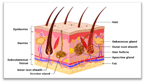
Fig no.01 : Structure of Human Skin:(6)
Skin is divided by three layers:
? Epidermis
? Dermis
? Hypodermis
Epidermis
The epidermis is skin’s outer layer, which is comparatively thin and hard. Epidermis at Upper layer is keratinocytes.
Five layers of epidermis:
Dermis
Dermis is usually elastic, but is abundant in blood channels, lymphatic vessels, and nerve Endings. A large network of dermal pours bind the essential circulation, with extensive Horizontal branching in the dermis from arterioles and venules to form plexuses and flow From capillaries to hair and sweat glands. Dermal capillaries help to extract antigens Outside of clear, fluid materials.
Hypodermis
The hypodermis tissue supports upper layer and inner layer like as dermis and epidermis. It should be used as storage of fat. Hypodermis layer supports temperature balancing,
Advantages of SLNs(7)
Disadvantages of SLNs
Application of SLNs:(8)
Controlled Release: SLNs can encapsulate both lipophilic and hydrophilic drugs, offering controlled release and improving the bioavailability of poorly soluble drugs.
Targeted Delivery: SLNs can be engineered to deliver drugs to specific sites in the body, which can enhance the therapeutic effect while minimizing side effects.
Enhanced Stability: SLNs help protect sensitive drugs from degradation, providing a stable formulation that can improve shelf life.
Table no.01:Excipients Of SLNs:(9)
|
Sr No |
Excipients |
Activity |
|
1 |
API |
Antifungal |
|
2 |
Glyceryl Monosterate, stearic acid |
Solid lipid |
|
3 |
Polaxomer 407 |
Surfactant |
|
4 |
Ethanol |
Solvent |
|
5 |
Water |
Solvent |
|
6 |
Methyl paraben |
preservative |
Preparation Techniques of SLN:(10)
High pressure Homogenization techniques:
High Pressure Homogenization (HPH) is a process in which a coarse oil-in-water (o/w) emulsion, produced after hot homogenization, is further homogenized under high pressure above the melting point (M.P.) of the lipid. This process converts the coarse emulsion of drug-loaded lipids into a nanoemulsion. During HPH, the sample temperature increases by approximately 10°C at a pressure of 5 × 10? Pa. Typically, 3-5 homogenization cycles at pressures ranging from 5 × 10? Pa to 5 × 10? Pa are sufficient to reduce the solid lipids to the nanometer scale.
Hot Homogenization techniques:
Hot homogenization is a technique in which homogenization of sample takes place at a Temperature higher than the melting point of lipid used. In this method, first liquefy the lipid over Its melting point and facilitate the solubilization of a lipophilic drug in liquefied lipid. Another Phase containing purified water mixed with surfactant was prepared and warmed closed to that of Drug loaded lipid phase. The melted phase was dispersed in hot aqueous surfactant mixture drop Wise and homogenized at high speed to make primary o/w type emulsion. This type of primary Emulsion has coarse size of particles. This emulsion was again homogenized at high pressure Above the liquefying temperature of lipid to convert the coarse emulsion of drug loaded lipid in Nanoemulsion form. This hot nanoemulsion was kept a side for some time to cool at Temperature where lipid solidify at room temperature and resultant mixture was filtered through membrane filter to gate Solid lipid nanoparticle.
Cold Homogenization techniques:
The cold homogenization technique is utilized in the formulation of solid lipid nanoparticles to address the challenges associated with the hot homogenization method. The hot homogenization process can lead to the degradation of thermolabile drugs, undesirable lipid transitions during recrystallization, and loss of drug in the aqueous phase. The initial step in cold homogenization is similar to that in hot homogenization, where the lipid is first liquefied above its melting temperature, and the drug is dispersed in the liquefied lipid. The lipid-drug phase is then cooled using dry ice or liquid nitrogen to solidify the lipid, resulting in coarse drug-loaded solid particles. These solidified particles are then comminuted using a grinding mill to a particle size range of 50 to 100 microns. The surfactant is dispersed in the aqueous phase, and the powdered solid particles are added to the surfactant mixture. The mixture is subsequently homogenized at high pressure at room temperature or lower to produce solid lipid nanoparticles.
High shear Homogenization:
Solid lipid nanoparticles can also be prepared using the ultrasonication method. This technique relies on the formation of cavities within the lipid during high-speed ultrasonication, which facilitates the encapsulation of the drug within the lipid core.
Solvent emulsification/evaporation techniques:
This method is widely used in the preparation of solid lipid nanoparticles. It enables the creation of an oily phase in an organic solvent by dispersing the drug in a liquefied lipid. The emulsification of the lipid is assisted by a surfactant in the aqueous phase. The emulsification of both phases is carried out using a mechanical stirrer at a speed of 1000 rpm. The organic phase is then evaporated through high-speed stirring at room temperature under reduced pressure. As a result, lipid nanoparticles are formed and separated due to precipitation.
Solvent in water diffusion techniques:
The key distinguishing characteristic of the solvent diffusion technique, compared to solvent evaporation, is the selection of the solvent used in the process. In solvent evaporation, volatile organic solvents are typically chosen, whereas in the solvent diffusion method, water-miscible solvents are preferred. This technique is applicable to both aqueous and organic phases.
Microemulsion Techniques:
The concept of microemulsion formation marked a significant advancement in the research of novel drug delivery systems (NDDS). In this method, the drug is partitioned between the aqueous and oily phases based on its partition coefficient. This approach results in a lower lipid content in the final formulation compared to the homogenization technique.
Double emulsion techniques:
This technique is based on the formation of an oil-in-water (o/w) type emulsion. In the process, a primary emulsion is first prepared by dissolving the hydrophilic drug in purified water. This aqueous phase is then dispersed into the melted lipid phase. The immiscible phases are emulsified with the assistance of a surfactant, resulting in the formation of the primary emulsion. The primary emulsion is then further dispersed into an aqueous phase containing a surfactant to form a double emulsion. The process typically involves solvent evaporation, during which the drug is entrapped in the lipid matrix after the solvent is removed.
Solvent injection method:(11)
This is a distinct technique in which solid lipid nanoparticles are precipitated from an aqueous organic phase. In this method, the lipid is melted and converted into a dispersion using a water-miscible organic solvent. A hydrophilic surfactant is dissolved in purified water to form the aqueous phase. The organic lipid phase is then rapidly injected into the aqueous phase containing water, with continuous agitation. The organic solvent mixes with the water, causing the lipid matrix, loaded with the drug, to separate into lipid nanoparticles. The resulting nanoparticles are separated by filtration. The surfactant present in the formulation provides stabilization to the developed nanoparticles.
Precipitation Method:
In this method, an organic solvent facilitates the dispersion of the lipid, which is then emulsified with the aqueous phase in the presence of an emulsifying agent, with continuous stirring. The continuous stirring increases the temperature of the dispersion, and the volatile nature of the solvent makes it conducive to evaporation. As the organic solvent evaporates, the lipid dissolved in it precipitates out, forming solid lipid nanoparticles. The nanoparticles are then washed with water.
Complex coacervation technique
A stable polymer solution is prepared by dissolving the polymer in water through heating, followed by cooling the mixture to room temperature. Lipid is then dispersed into the polymer solution, and the mixture is heated to a temperature at which the surfactant begins to form micelles, resulting in a transparent solution. A hot aqueous solution containing the drug is mixed with the polymer-lipid mixture. To achieve the desired pH, a pH inducer for the acidic range is added dropwise to the lipid dispersion of the drug. The prepared suspension is then cooled to room temperature, and the solid lipid nanoparticles (SLNs) are separated.
Membrane emulsification method
This novel technique is employed to produce solid lipid nanoparticles using microreactors, also known as the gas displacement method. In this process, the melted lipid in the dispersion medium is passed through a membrane to form smaller droplets. The lipid, dispersed in a solvent miscible with water, is injected as the inner core, while the aqueous phase serves as the outer coating material. As both phases come into close contact, water diffuses from the outer surface, resulting in the lipid becoming supersaturated. This supersaturation causes the lipid to precipitate in the form of lipid nanoparticles.
Preparation of SLNs:(12)
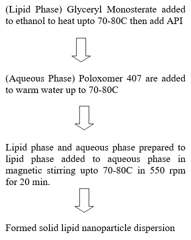
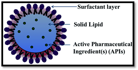
Fig no.02: structure of SLNs(13)
Fig no.03: Formulated SLN (10ml)

Fig No.04: Formulated Gel Base
Evaluation of Solid Lipid Nanoparticles(14)
Drug content analysis:
The drug content in the formulation was determined by using a 50% solvent solution, which was shaken well to ensure the complete dispersion of vesicles. The solution was then appropriately diluted with phosphate-buffered saline (PBS) at pH 7.4. The absorbance of the resulting solution was measured at 283 nm using UV-Vis spectroscopy.
The drug content was calculated based on the standard curve using the following formula:
Drug content = sample absorbance ×100
standard absorbance
Entrapment Efficiency (%):
The entrapment efficiency (EE) of the solid lipid nanoparticles (SLNs) loaded with the active pharmaceutical ingredient (API) was estimated using a modified version of the described method. Briefly, the prepared SLNs were dried at room temperature. A 5 mg sample of the dried SLNs was then dissolved in 10 ml of HPLC-grade ethanol. The solution was filtered using a 0.22 µm syringe filter to remove any particulate matter. The concentration of the API was determined spectrophotometrically at 283 nm. Measurements were performed in triplicate, and the formulation with the highest entrapment efficiency was selected for further evaluation. The entrapment efficiency was calculated using the following equation:
EE% = W (free drug) / W(Added drug)×100
In-vitro drug diffusion
The in vitro release of the active pharmaceutical ingredient (API) from different SLN dispersions was determined using the dialysis bag diffusion technique. An accurately weighed amount of API-loaded SLN dispersion, containing the drug equivalent to 2.5 mg, was transferred into a dialysis bag, which was then sealed. The sealed dialysis bag was suspended in a beaker containing 250 ml of phosphate-buffered saline (PBS) at pH 7.4. The system was stirred continuously at a constant speed of 50 rpm and maintained at 37°C ± 0.5°C to simulate physiological conditions.
Zeta potential:
The zeta potential of the formulation was measured using a Zetasizer (Horiba SZ, Japan). Zeta potential is a critical parameter that indicates the stability of the formulation, as it reflects the electrostatic charge on the surface of the particles. An optimal zeta potential value is typically considered to be ±30 mV, as this range suggests good stability due to sufficient repulsive forces preventing particle aggregation.
Size distribution studies:
The particle size of solid lipid nanoparticles (SLNs) can be determined using dynamic light scattering (DLS). This technique measures the fluctuations in the intensity of light scattered by the particles in the sample. Small particles undergo random thermal motion, known as Brownian motion, which causes variations in the scattering pattern. A laser light source illuminates the sample contained in a cuvette, and the scattered light is detected by photodetectors positioned at specific angles, typically 90° (right angle) or 173° (back angle). The collected optical signals exhibit random intensity fluctuations corresponding to the movement of the particles, which are analyzed to determine the particle size distribution.
SEM studies:
Solid lipid nanoparticles (SLNs) can be easily visualized using scanning electron microscopy (SEM). SEM is a powerful imaging technique that uses a focused beam of electrons to produce high-resolution images of sample surfaces. It enables imaging at magnifications that are not achievable with traditional optical microscopes. A typical SEM can achieve magnifications of over 30,000×, whereas conventional light microscopes typically reach magnifications of up to 1,000×. The images produced by SEM are displayed in black and white, as the technique relies on electron scattering rather than light to create images.
Stability Study:(15)
According to ICH guidelines [ICH Q1A(R2), ICH Q1B], stability studies of the API-loaded solid lipid nanoparticles (SLNs) were conducted for the optimized formulation. The studies were performed under different storage conditions: room temperature (25 ± 2°C / 60 ± 5% RH) and accelerated conditions (40 ± 2°C / 75 ± 5% RH) for a duration of 30 days. The physical appearance, pH, percentage drug entrapment efficiency, and in vitro release profile were evaluated as a function of the storage conditions. SLNs stored at 4°C were found to be stable for 28 days compared to those stored at room temperature and under accelerated conditions.
CONCLUSION:
Solid lipid nanoparticles for topical antifungal activity represent a promising approach for improving the efficacy and stability of antifungal treatment.
REFERENCES


Joshi Maharudra R., Shaikh Nasheer S.*, Kazi Wasim B., Anantwal Akshat B., Limaye Akhilesh S., Rajput Amrapali V., Solid Lipid Nanoparticles: A Novel Approach for Fungal Therapy, Int. J. of Pharm. Sci., 2025, Vol 3, Issue 4, 1902-1912 https://doi.org/10.5281/zenodo.15223034
 10.5281/zenodo.15223034
