Department of Pharmacy, I. E. T., M. J. P. Rohilkhand University, Bareilly.
An innovative drug delivery method called polymer-lipid hybrid nanoparticles (PLNs) blends the biomimetic qualities of lipid-based carriers with the structural advantages of polymeric nanoparticles. These hybrid nanoparticles are perfect for targeted therapeutics, such as gene delivery and cancer treatment, since they provide improved drug stability, bioavailability, and controlled release. PLNs are made up of an exterior PEGylated lipid layer to extend circulation, a lipid monolayer for stability, and a polymer core that encapsulates the medication. For particular therapeutic purposes, several varieties have been created, including erythrocyte membrane-camouflaged nanoparticles, monolithic PLNs, and polymer core lipid shells. High-pressure homogenization, emulsification-solvent evaporation, and nanoprecipitation are preparation techniques. Despite the fact that PLNs have several benefits, issues including batch-to-batch uniformity, large-scale production, and possible cytotoxicity still exist. Research is also being done to incorporate stimuli-responsive drug release mechanisms, optimize their design, and increase drug loading efficiency. PLNs offer better medication delivery with fewer adverse effects, making them a revolutionary technique in nanomedicine. These nanoparticles have enormous promise for next-generation pharmaceutical applications and personalized medicine with further development, guaranteeing safer and more efficient therapies.
About 90% of the drugs used in the field of pharmaceutics are solid particles. Drug nanoparticles that can be used in a variety of innovative ways are now possible thanks to the development of nanotechnology. Novel drug delivery techniques that increase treatment efficacy while reducing negative effects are now feasible. Drug distribution aims to change the biochemistry of the body. All biological processes are based on the cell and the metabolic activity that occurs within it. Targeting specific biological functions at their native sizes is now possible because to the creation of nanoparticles [1]. Nanotechnology is a promising medical technology that can significantly impact the distribution of a range of therapeutics, including peptides, genomes, small molecular RNA therapeutics, and medical imaging agents, while also potentially improving the systemic pharmacokinetics and therapeutic index of many drugs [2]. The matrix composition, pH of the microenvironment, and ambient temperature all affect how released the payloads are [3], [4]. These payloads are covalently affixed to the surface of the nano carriers or encapsulated within them once they have been systemically assimilated. Some potential benefits of nano carriers include the ability to overcome a number of inherent obstacles in vivo, the improvement of a drug's pharmacodynamic and pharmacokinetic properties holistically without changing its molecular structure, more effective and efficient molecular targeting of cells and tissues, targeted and non-targeted drug delivery to the target, such as the nucleus, cytosol, etc., and improved drug therapy [5, 6]. The formulation problems that drugs categorised as belonging to classes II and IV in the Biopharmaceutical Classification System face are now addressed by a variety of preparation procedures. When modifying drug release patterns and the bioavailability of sparingly soluble medications, expanding lipid excipient production with unique properties necessitates a more adaptable and wide-ranging use [7]. Figure 1 illustrates lipid-based preparations that can be used in a variety of ways to affect how well active components are absorbed. They can alter the intestinal environment, initiate the lymphatic transport of the active components, and interact with enterocyte-based transport [8]. Drug processing is significantly improved when a drug is immersed in the lipid-formed matrix of the drug delivery device [9–13].
One or more bilayers of natural or manufactured amphipathic lipids are dispersed in water to form liposomes, which are vesicles. Since their creation, they have primarily been used for targeted distribution due to their enhanced biocompatibility, efficacy, and safety features. Polyethylene glycol (PEG) can be applied on their surface to lengthen the half-life of circulation [14].
When injected into the body, biodegradable, amphiphilic copolymer blocks with different hydrophobicities self-assemble to create polymeric nanoparticles. Polymeric nanoparticles' core–shell structure prolongs the time that drugs are released into the bloodstream and makes it easier to encapsulate hydrophobic substances. To selectively distribute drugs, they might have ligands affixed to their surfaces. When injected into the body, biodegradable, amphiphilic copolymer blocks with different hydrophobicities self-assemble to create polymeric nanoparticles. Polymeric nanoparticles' core–shell structure prolongs the time that drugs are released into the bloodstream and makes it easier to encapsulate hydrophobic substances. To selectively distribute medications, they might have ligands affixed to their surfaces [15].
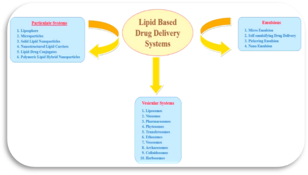
Fig. 1: Various approaches of Lipid Based Drug Delivery Systems
In order to overcome disadvantages such as structural disintegration, a shorter circulation period, and material leakage, a new generation of delivery systems known as polymer-lipid hybrid nanoparticles has been developed using the unique properties of polymeric nanoparticles and liposomes that contributed to their early therapeutic effectiveness [16]. As seen in figure 2, this hybrid system has the potential to be a dependable drug delivery technique due to its appropriate tissue and molecular targets, high encapsulation efficiency, improved drug release kinetics, and unique drug absorption [17].
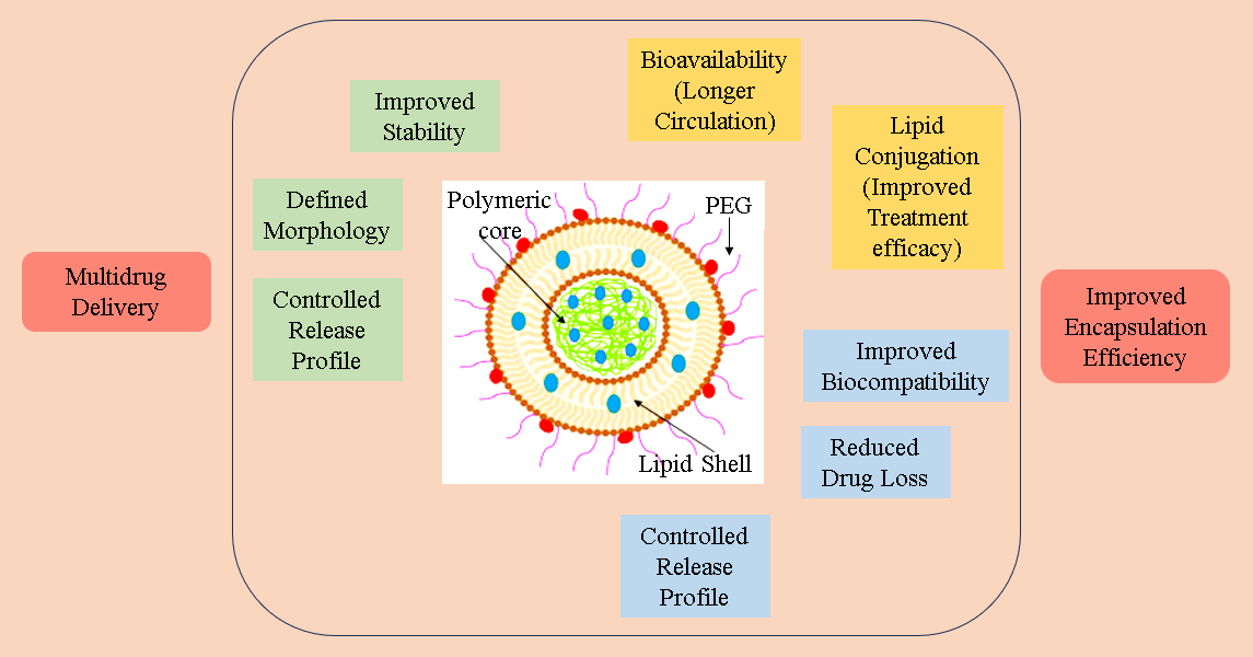
Fig. 2: Main advantages associated with the individual components (polymeric core, lipid shell, external pegylated lipid layer) and the overall structure of the Polymer-lipid hybrid nanoparticles.
Polymer-lipid hybrid nanoparticles (PLNs)
Polymer-lipid hybrid nanoparticles, which are conceptually derived from both liposomes and polymeric nanoparticles, are core-shell nanostructures of the second generation in which a polymer core is encased in a lipid layer [18]. Each of the two most common types of nanoparticles lipid-based (liposome) and polymeric has specific drawbacks when it comes to drug delivery. In contrast, polymeric materials are susceptible to polycytotoxicity, disintegration, and the addition of harmful solvents during processing. It shares characteristics with liposomes, including an unstable physical and chemical state, inadequate drug loading, and rapid drug release. They have therefore been combined to produce polymer-lipid hybrid nanoparticles in an effort to maximise their benefits and minimise their drawbacks. In order to provide better targeting and a desired drug release profile, the Polymeric Lipid Hybrid Nanoparticle system combines the structural advantages of biodegradable polymeric carriers (polymeric nanoparticles) with the benefits of lipid-based nanocarriers (liposomes), such as enhanced drug loading capability and biomimetic properties [19][20]. In order to address the numerous oral delivery problems, integrated hybrid carrier systems are required. Since 1995, parenteral formulations of polymer-lipid hybrid nanoparticles have been marketed for use in medicines. The main objective of these formulations' development was to combine the advantageous features of lipid-based and polymeric systems [21]. Recent developments in the molecular understanding of multifunctional polymers and lipid-based excipients in drug delivery applications have led to the emergence of polymer-lipid hybrid nanoparticles for the oral administration of various therapeutic agents [22].
Structural Classification of Polymer-lipid hybrid nanoparticles
Various classes of Polymer-lipid hybrid nanoparticles have been identified shown in
Polymer core lipid shells
The simplest type of polymer-lipid hybrid nanoparticles to create is this one. One or more lipids (lipid PEG and a lipoidal shell) envelop a polymer core [23]. A unique, dependable delivery system that can be used for a variety of topical and systemic conditions is created by combining the structural advantage of the biodegradable polymer core with the lipids' simulated consistency. The hybrid structure is created by the polymer and lipid covering. For targeted cancer treatment, a drug carrier with enhanced therapeutic efficacy and continuous distribution that consists of a lipid monolayer shell and a biodegradable polymer core holds great promise. Improved drug loading efficiency, serum stability, controlled drug release and particle size, and the capacity to load multiple drugs are among the advantages of polymer-lipid hybrid nanoparticles [24].
Monolithic Polymer-lipid hybrid nanoparticles
PEG molecules in this type of lipid or lipid nanoparticles are distributed within a polymeric core matrix composed of drug molecules [25], which is a development that can be compared to a colloidal of their design; however, they are not sufficiently versatile to allow high density PEGylation to change their structure [24]. This type of nanoparticle is also known as monolithic polymer-lipid hybrid nanoparticles.
Erythrocyte membrane-camouflaged polymeric nanoparticles
Biomimetic nanoparticles are erythrocyte vesicle nanoparticles. These sub-100 nm nanoparticles coated with erythrocyte cell membrane (RBC) polymers are used to make vesicles that reproduce or mimic the intricate surface chemistry of erythrocyte membranes. Because these nanoparticles can stay in the bloodstream for a long period, they may be able to carry drugs across the membrane barrier efficiently. These nanoparticles, such as cell membrane disguised nanoparticles as a medication carrier for cancer treatment, have been the subject of numerous recent investigations. As a possible candidate for a therapeutic platform called "Erythrocyte-Membrane-Camouflaged Nanocarriers," which provide variable Paclitaxel release kinetics via macromolecules, the potential application of biomimetic-cell-camouflaged polymer nanocarriers with properties identical to the physicochemical adaptability of synthetic polymers and the functional versatility of natural cell membranes is being investigated [26].
Polymer caged liposomes
In order to preserve cell stability, this method uses polymers coated on the liposome surface to self-possess. When it comes to processing proteins, nucleic acids, and chemotherapeutic drugs, nanoscale polymer caged liposomes (20–200 nm) have remarkable pharmacokinetic properties. The pH-responsive stable polymer-caged liposome could alter these pH-sensitive nanoparticles [27]. A pH-sensitive polymer-caged liposome delivery system with exceptional stability is still pending patent. Another study used polymer-caged liposomes to characterise photoactive medication release processes. PEG coating makes it possible to distribute different proteins, peptones, and peptides in a customised way for targeted intracellular movement [24].
Core-shell type lipid polymer-lipid hybrid nanoparticles
This kind of nanoparticle consists of an open inner core surrounded by a thick lipid coating, a polymeric coating, and an outer core composed of lipid PEG. Because core-shell lipid polymer hybrid nanoparticles combine the biomimetic advantages of biodegradable polymeric nanoparticles with the mechanical advantages of liposomes, they have emerged as a powerful and efficient delivery mechanism [27]. A membrane made of phospholipid membranes envelops the biodegradable polymeric nucleus of CS-Polymer-lipid hybrid nanoparticles. The polymer and lipid complexes are often made by combining liposomes with polymeric nanoparticles. The polymeric core surface is encased in a lipid or lipid multilayer bilayer [24].
Table 1: Types of Polymer-Lipid Hybrid Nanoparticles and Their Characteristics
|
Type of PLN |
Description |
Reference |
|
Polymer Core Lipid Shells |
Consists of a polymer core covered with one or more lipid layers (lipoidal shell and lipid PEG). This hybrid structure enhances drug loading efficiency, serum stability, and controlled drug release. It shows promise for targeted cancer therapy. |
[23], [24] |
|
Monolithic Polymer-Lipid Hybrid Nanoparticles |
Also known as monolithic PLNs, these have PEG molecules or lipid nanoparticles dispersed within a polymeric core matrix containing drug molecules. They are not adaptable enough for high-density PEGylation. |
[24], [25] |
|
Erythrocyte Membrane-Camouflaged Polymeric Nanoparticles |
Biomimetic nanoparticles coated with erythrocyte cell membranes. These nanoparticles can mimic the surface chemistry of erythrocytes, prolong circulation time, and effectively transport drugs for cancer treatment. They offer adaptable properties for therapeutic platforms like Paclitaxel release kinetics. |
[26] |
|
Polymer Caged Liposomes |
Involves coating liposomes with polymers to maintain cell stability. These pH-sensitive nanoparticles allow for improved pharmacokinetics, targeted intracellular delivery, and drug release modifications. A patent is pending for a pH-sensitive polymer-caged liposome delivery system. |
[24], [27] |
|
Core-Shell Type Lipid Polymer-Lipid Hybrid Nanoparticles |
Comprises an external lipid PEG core, a polymeric coating, and an open inner core surrounded by a lipid bilayer. This combination enhances stability and biomimetic properties, making them effective drug carriers. |
[24], [27] |
Elucidating the Structure and Formation Mechanism of Polymer-lipid hybrid nanoparticles
Polymer-lipid hybrid nanoparticles offer a distinctive structural amalgamation by fusing features from liposomes and polymeric SLNs. Three essential components make up their basic architecture, as seen in Figure 1: 1) a drug-containing polymer core, 2) a lipid monolayer around the polymer core, and 3) an exterior lipid–PEG layer that acts as a steric stabiliser to extend the systemic circulation of polymer-lipid hybrid nanoparticles by avoiding immune clearance. As a molecular barrier, the central lipid monolayer reduces drug loss during the formulation of polymer-lipid hybrid nanoparticles and protects the core from deterioration by preventing water from diffusing into the inner core. Investigations into the intricate molecular interactions that cause lipids and polymers to fuse are still ongoing. Interestingly, different production processes for polymer-lipid hybrid nanoparticles show unique mechanisms of creation. For instance, in single-step techniques, the polymer spontaneously self-assembles into a monolayer around the core after precipitating from the organic solvent when added to aqueous environments containing lipids. At the same time, PEGylated lipids self-assemble, with the PEG chain extending outward into the aqueous environment and a lipid moiety sticking to the surface of the polymer core. On the other hand, the two-step process entails the development of the bilayer structure and its attachment to the core, which is followed by the breakdown of the bilayer as a result of hydrophobic interactions between the lipid chains and the polymer. Taking into account hydrophobic, van der Waals, and electrostatic interactions, this hybrid formation is thermodynamically advantageous [28][29]. Because of their core–shell structure, polymer-lipid hybrid nanoparticles have strong structural integrity, storage stability, and controlled release capabilities. Furthermore, the lipid and outer PEGylated layers are responsible for their high biocompatibility and improved bioavailability [30][31].
Methods of Preparation of Polymer-lipid hybrid nanoparticles
Common methods for creating polymer-lipid hybrid nanoparticles include high-pressure homogenisation, spray drying, emulsification-solvent evaporation (ESE), and nanoprecipitation.
The two main techniques for creating polymer-lipid hybrid nanoparticles are single-step and double-step processes; both of these planning techniques are covered below:
There are many advantages to single-step PLN preparation versus two-step methodology, including superior scalability, cost-effectiveness, and compatibility with traditional preparation techniques. The two-step process requires all of your time and energy to remove polymeric nanoparticles from lipid vesicles. Most of the errors occur in the single PLN preparation step of the two-step process. A one-step network cloning mechanism was created to address these issues. For example, the two-step process requires lipid vesicles and polymeric nanoparticles, whereas the one-step one does not. As a result, the one-step approach just requires mixing the polymer and lipid solutions. Then, utilising emulsification-solvent-evaporation or nanoprecipitation—two techniques that have been systematically used to create non-hybrid polymeric nanoparticles—the materials can easily self-assemble to form a PLN structure [32].
The dual-step process is a commonly employed technology in the early stages of PLN synthesis to create monolayer, bilayer, or multilayer shells for Polymer-lipid hybrid nanoparticles. This technique combines cationic lipid vesicles with anionic polymeric nanoparticles through electrostatic interactions. The processes of melt emulsification, solvent injection, solvent evaporation, nanoprecipitation, and emulsification via high-pressure homogenisation are commonly used to create polymer-lipid hybrid nanoparticles. Through the processes of hydration and sonication, the polymeric core and lipid shell are combined to produce the structure of the lipid-core polymer. Usually, there are two steps in this process. First, lipid nanoparticles were produced using a variety of techniques, such as melt emulsification, solvent emulsification, solvent injection, ultrasonication, and high-pressure homogenisation (both hot and cold). High-pressure ultrasonic homogenisation was used to combine the lipid nanoparticles and the polymer solution to create lipid-polymer hybrid nanoparticles [33].
Two-step approaches fall into two categories: standard two-step procedures and non-traditional two-step methods. We talk about these below.
Traditional methods are used for most small-scale manufacturing of polymer-lipid hybrid nanoparticles. Polymeric nanoparticles were produced by emulsification solvent evaporation, high-pressure homogenisation, or nanoprecipitation. The traditional two-step procedure can be used to two types [44]. A dried thin lipid film made by hydrating the thin film or a performed lipid vesicle made by dissolving the lipid in an organic solvent (like chloroform) and evaporating it in a rotary evaporator can be combined with previously made polymeric nanoparticles to create the polymer-lipid hybrid nanoparticles. The purification stage uses differential centrifugation to extract free lipids from polymer-lipid hybrid nanoparticles. For example, PLGA was used to create cationic lipid vesicles under continuous stirring or bath sonication at 30ºC in a hybrid nanoparticle production process. The produced particle has a diameter of 200–400 microns and a potential surface charge of 20–30 microvolts [24].
The main use of the nonconventional method is the massive synthesis of polymer-lipid hybrid nanoparticles. In a novel method, polymer-lipid hybrid nanoparticles were produced by soft lithography particle moulding and spray drying. After spray drying to produce nanoparticles in the 400–500 nm size range, they were dispersed in an organic dichloromethane solvent containing different lipids. Spray drying (SD) and spray freeze drying (SFD) were applied to the lipoid polymeric suspension to produce powdered limestone nanoparticles. Polymer-lipid hybrid nanoparticles for the transport of genetic material were made using the soft lithography moulding technique known as particle replication in non-wetting templates (PRINT) [34]. The organic solvent polymer PLGA, or genetic material such as siRNA, is dissolved and deposited onto a polyethylene terephthalate sheet. It is then heated in conformal contact with a PRINT mould, which causes the polymer to pour into the mould and solidify when the temperature is dropped to room temperature. The nanoparticles were then extracted from the mould and separated from PVA-coated PET sheets using an aqueous lipid solution to produce polymer-lipid hybrid nanoparticles [34].
Large amounts of polymer-lipid hybrid nanoparticles smaller than 100 nm can be produced using this self-assembled nanoprecipitation process. Using the self-assembly nanoprecipitation technique, lipid, polymer, and drug are combined in a single phase to create a monolayer of nanoparticles. A self-assembled nanoprecipitation method is used to dissolve the polymer and the medication to be encapsulated in water or another organic solvent prior to the creation of the polymer-lipid hybrid nanoparticles. Then, to dissolve it and provide a homogeneous distribution, lipid or lipid-PEG water is heated to 65 to 70°C. The resultant solution was then continuously churned or sonicated. The lipid or lipid-PEG molecule self-assembled with hydrophobic contact across the polymer's core after the polymeric drug solvent was introduced dropwise to precipitate the polymer. The polymeric material was supplemented with a lipid tail that was insoluble in water. Lipid or lipid PEG was used to spherically fix the polymer-lipid hybrid nanoparticles, which were formed by attaching a water-miscible head to the surrounding layer. The solvent was then evaporated after centrifuging the excess lipid and polymer from the Polymer-lipid hybrid nanoparticles [24].
The emulsification solvent evaporation (ESE) method comes in two varieties: single emulsification and double emulsification. The lipid is dispersed in the water using a variety of techniques, such as homogeneous dispersion, bath sonication, and stirring. After that, it is heated to a certain temperature in order to dissolve the medication and organic solvent polymer. After the organic solution was applied dropwise in the aqueous procedure, a lipid coating was added and mixed for five minutes to decrease the polymeric particles to tiny particles [24].
This method involves extracting the encapsulating polymer or active medicinal component into the organic phase (oil phase). The combination was then placed in a lipid water dispersion medium, and an oil in water emulsion (o/w) was created by ultrasonic content stirring. The lipid or lipid PEG self-assembles around the polymer core after the organic solvent is evaporated using a rotary evaporator at lowered pressure. Because of the stable emulsion formation, this method is more frequently used than nanoprecipitation in the formulation of various polymer-lipid hybrid nanoparticles. Drug and gene delivery using this technique was previously announced. Researchers recently reported using lipid-polymer nanohybrid nanoparticles loaded with nucleic acids to cure cancer [35].
The best technique for creating polymeric nanoparticles is the double emulsification solvent evaporation process. This technique is important for creating core-shell type polymer-lipid hybrid nanoparticles as well as polymeric nanoparticles. It improves the capacity to encapsulate hydrophilic or hydrophobic drugs. Finding drugs that are readily soluble in water but insoluble in the organic phase is the main goal of this approach. They can't dissolve together because of a hydrophobic polymer core. A common use of this technique is the production of water-in-oil-in-water (w/o/w) emulsions. To create a water-in-oil (w/o) emulsion, a medication is first mixed with an aqueous polymer solution and an organic solvent that contains a lipid. Following that, the fresh water-in-oil-in-water (w/o/w) emulsion is moved to a different aqueous media. Polymer-lipid hybrid nanoparticles were created by evaporating the oil phase of the emulsion with a rotary evaporator [36]. The same process is used to make nucleic acid-loaded polymer-lipid hybrid nanoparticles, which are used as innovative nanotherapeutics in the treatment of cancer. With the best possible drug load capacity, this technique effectively produces core-shell polymer-lipid hybrid nanoparticles that can be employed to deliver one drug or a combination of pharmaceuticals [24].
Commonly Used Materials in Synthesis of Polymer-lipid hybrid nanoparticles
Polymer Lipid Hybrid Nanoparticles are synthesised by utilising various polymers and lipids (shown in table 2). Commonly used polymers and lipids in formulating Polymer-lipid hybrid nanoparticles are given below:
Table 2: Various commonly used Lipids and Polymers in the formulation of Polymer-lipid hybrid nanoparticles
|
S. No. |
Lipid Component |
Polymer Component |
Drug |
References |
|
1 |
DSPE-PEG (2000) |
PLGA |
Docetaxel |
[37] |
|
2 |
DSPE-PEG-3000, stearyl amine |
PLA |
Methotrexate & betacarotene |
[38] |
|
3 |
Lipoid S75 |
Chitosan |
Camptothecin & Curcumin |
[39] |
|
4 |
DSPE-PEG-Malemide, PSC and cholesterol |
PLGA |
Salinomycin |
[40] |
|
5 |
Lecithin, cholesterol |
mPEG-PLGA |
Curcumin |
[41] |
Characterization of Polymer-lipid hybrid nanoparticles
The physicochemical characteristics of nanoparticles, such as their size, shape, and zeta potential, affect the in vivo profile of polymeric hybrid nanoparticles. Atomic force microscopy (AFM), transmission electron microscopy (TEM), and scanning electron microscopy (SEM) can all be used to assess the surface shape and structure of nanoparticles. Polymer-lipid hybrid nanoparticles' actual physical size, size distribution, structure, and surface appearance are evaluated using transmission electron microscopy (TEM) and scanning electron microscopy (SEM). The physical size and size distribution may be shown by the dried nanoparticles attached to the silicon wafer substructure for imaging using electron microscopy (SEM). The inner core-shell structure of the polymer-lipid hybrid nanoparticles was examined using transmission electron microscopy (TEM) [42], [43].
At average sizes, vesicles have a high zeta potential. One of the most crucial elements affecting the lifespan of nanoparticles in the bloodstream and their ability to passively assemble in tumour tissues is their size. It is well accepted that the particle size range of 10 to 150 nm is advantageous and ideal for the systemic dispersion of medication. To determine the sizes of particles or molecules ranging from 1 nm to many microns, zeta sizer devices employ dynamic light dispersion. The dispersion of electrophoretic light and the molecular weight of static light diffusion determine the zeta potential and electrophoretic stability. At average sizes, vesicles have a high zeta potential. One of the most crucial elements affecting the lifespan of nanoparticles in the bloodstream and their ability to passively assemble in tumour tissues is their size. It is well accepted that the particle size range of 10 to 150 nm is advantageous and ideal for the systemic dispersion of medication. To determine the sizes of particles or molecules ranging from 1 nm to many microns, zeta sizer devices employ dynamic light dispersion. The dispersion of electrophoretic light and the molecular weight of static light diffusion determine the zeta potential and electrophoretic stability. The real mean values of zeta potential (mV), particle size (nm), polydispersity index (PDI), and particle size distribution are determined using the Malvern Zeta sizer (Nano ZS instrument) under optimal testing and temperature conditions. The zeta potential is one tool that may be used to determine the surface charge of nanoparticles (colloids) in the solution. The concentrations of polymers and lipids are two factors that can alter the hybrid polymer nanoparticles' particle sizes and polydispersity index. The formulation's particle size may have a significant impact [24].
The two most important properties for describing polymer-lipid hybrid nanoparticles that are suggested to the drug loading range for any polymer or excipient are entrapment efficiency (EE) and drug loading capacity (LC). It deals with the prepared PLN's actual drug entrapment capacity. The lipid or polymeric shell that envelops the medication increases its loading capacity. The hydrophobic polymer core contains most hydrophobic medications [44]. The ion interaction between pharmaceuticals is used by tiny molecules, and polymers may be a crucial tactic for raising drug charges. Drug loading ability (DL) and entrapment effectiveness (EE) can be impacted by phenolic density, aqueous phase pH, drug lipid load interactions, affinity, solubility, manufacturing techniques, and polymer and lipid phase miscibility. The importance of drug loading and trapping is also influenced by the preparation procedure (EE). The volume of polymer lipid hybrid nanoparticles encasing the medicine can be ascertained by means of dialysis, centrifugation, membrane filtering, and high-performance liquid chromatography (HPLC) methods [24]. The drug loading capacity (DL) and entrapment efficiency (EE) mathematical expressions are as follows:
%EE = amount of drug loaded in polymer lipid hybrid nanoparticles X 100
Total amount of drug added in formulation
%DL = Amount of drug loaded in polymer lipid hybrid nanoparticles X 100
Total weight of polymer lipid hybrid nanoparticle
When it came to hydrophilic medications, the hydrophobic product rapidly divided between the lipid surfaces, increasing the amount of medication encapsulated in close proximity to the hydrophilic drug.
Drug permeability, drug solubility, polymer degradation rate, and particle size interaction between polymer and drug potential particle charge can all affect drug release profiles [45]. By detecting the diffusion and release of chemically conjugated hydrolysed medications, polymer-lipid hybrid nanoparticles are utilised to quantify drug release from a mechanically confined material. Dialysis, high performance liquid chromatography (HPLC), a mass spectrometer, and a semi-permeable membrane are used in the majority of in-vitro drug release investigations. With a large volume of liquid, the dialysis procedure for assessing drug release from nanoparticles can be carried out at 37°C. At 37°C, nanoparticles are introduced to a massive volume of release media. The drug molecules are released from the nanoparticles and permeate the releasing environment through the diffusion process. Any released medication and any leftover medication in the form of nanoparticles are collected by centrifuging the solution for 30 minutes at 1200 RPM. High-performance liquid chromatography (HPLC), an analytical mass spectrometer technique, is used to quantify the supernatant [46].
Both physical and chemical stability are necessary for polymer-lipid hybrid nanoparticle stability studies for both polymer-lipid hybrid solution and free dry nanoparticles in order to evaluate drug loss and modifications in nanoparticle composition during storage. Numerous factors, including microscopic examination, entrapment efficiency, zeta potential, particle size and polydispersity index, physical and chemical evaluation, optimisation, and research of a generated nanocarrier system, must be considered when evaluating formulated polymer-lipid hybrid nanoparticles [24]. As a result, by using the self-modified properties of the nanoparticles on the lipid nanoparticles on the lipid monolayer surface to increase the solubility and stability of DSC and TGA nanoparticles, researchers can investigate how temperature and time affect the thermal properties of nanoparticles [47]. To examine drug losses from nanoparticles and structural alterations under storage settings, stability studies are conducted on lyophilised and suspended nanoparticles [24].
Drug-loaded polymer-lipid hybrid nanoparticles' cytotoxicity and cellular absorption are in vitro characteristics that are assessed in vivo for target cell evaluation. To evaluate the hybrid polymer-lipid Drug-loaded nanoparticles can be targeted using fluorescence microscopy, such as a confocal laser scanning microscope, and fluorescence microscopy, such as isothiocyanate fluorescence (FITC) with cells. The cell is visible once the extraneous particles have been removed. The absorption of generated nanoparticles by in vivo cells is usually controlled by the endocytosis mechanism. The surface of polymer lipid hybrid nanoparticles that target ligands is conjugated to the nanoparticles in order to facilitate receptor-controlled endocytosis. The size, shape, kind of drug load, and structure of nanoparticles all affect how they interact with the biological system. These elements may have an impact on cytotoxicity, cell uptake, and biological interactions [48].
Certain techniques can be used to evaluate in-vitro or in-vivo cytotoxicity, including:
The incubation cycle must be completed for a predetermined amount of time in order to assess cell cytotoxicity. After 72 hours of various cultures, appropriate assays are conducted to evaluate the cell's capacity to release medicines, such as the ATP and MTT assays. Drug side effects are directly reduced by nanoparticles [48].
CONCLUSION
Combining the benefits of lipid-based and polymeric nanoparticles, polymer-lipid hybrid nanoparticles (PLNs) have completely transformed the field of nanomedicine. Their distinct core-shell structure, which is made up of a polymer core, lipid monolayer, and PEGylated lipid outer layer, allows for targeted release, longer blood circulation time, and improved drug delivery effectiveness. These nanoparticles' hybrid nature makes them ideal for a variety of pharmaceutical applications because it enables better drug encapsulation, enhanced stability, and controlled drug release. Depending on the therapeutic use, the various PLN types—such as polymer core lipid shells, monolithic PLNs, erythrocyte membrane-camouflaged nanoparticles, polymer-caged liposomes, and core-shell lipid polymer hybrids—offer varied benefits. Each of these structural variations improves serum stability, bioavailability, and adjustable release kinetics to increase drug delivery performance. They are promising possibilities for the treatment of diseases like cancer, infectious diseases, and neurological disorders because of their capacity to incorporate functional ligands or biomimetic coatings, which further improves their targeting capabilities. Researchers have a variety of formulation possibilities thanks to the PLN preparation techniques, which include high-pressure homogenisation, emulsification-solvent evaporation, and nanoprecipitation. It is still difficult to optimise these procedures for large-scale manufacturing, nevertheless. Important factors that need for more study and technical developments include ensuring batch-to-batch consistency, preserving stability during storage, and enhancing reproducibility in synthesis. Despite their encouraging promise, a number of obstacles need to be overcome before they may be widely used in therapeutic settings. Their quick conversion into commercial medications is constrained by issues with cytotoxicity, interactions with the immune system, and the difficulty of large-scale manufacture. Furthermore, thorough preclinical and clinical testing is necessary to guarantee the safety and effectiveness of nanoparticle-based medication compositions before they can receive regulatory approval. By investigating new biocompatible polymers and lipids, enhancing drug loading techniques, and incorporating smart nanotechnology characteristics like stimuli-responsive drug release mechanisms, future research should concentrate on improving PLNS formulations. PLN design optimisation and in vivo behaviour prediction may also be greatly aided by developments in artificial intelligence (AI) and machine learning. To sum up, PLNs have the potential to revolutionise contemporary medication delivery strategies. PLNs can open the door for novel precision medicine therapies, targeted therapy, and next-generation pharmaceutical formulations with further development in formulation methods and a better understanding of their pharmacokinetics and biodistribution. They are positioned as a major actor in the future of nanomedicine due to their capacity to maximise therapeutic efficiency while overcoming conventional drug delivery obstacles.
REFRENCES
Lipid–polymer hybrid nanoparticles as a next-generation drug delivery platform: state of the art, emerging technologies, and perspectives
. International Journal of Nanomedicine, Volume 14, 1937–1952. https://doi.org/10.2147/ijn.s198353
Lipid–polymer hybrid nanoparticles as a next-generation drug delivery platform: state of the art, emerging technologies, and perspectives
. International Journal of Nanomedicine, Volume 14, 1937–1952. https://doi.org/10.2147/ijn.s198353

Ajay Pratap Singh*, Neeraj Kumar Rathour, Saurabh Mishra, A Comprehensive Review on Polymer-Lipid Hybrid Nanoparticles, Int. J. of Pharm. Sci., 2025, Vol 3, Issue 3, 1388-1403. https://doi.org/10.5281/zenodo.15031392
 10.5281/zenodo.15031392
