Malla Reddy Institute of Pharmaceutical Sciences, Secunderabad, India
Coumarin is an organic compound with chemical structure consisting of a benzene ring fused to an alpha-pyrone. Its chemical formula is C9H6O2, featuring two functional groups: an alkene and an ester. Coumarin derivatives are important source of heterocyclic compounds of pharmacological interest, as they show a wide spectrum of biological activity viz antibacterial, antifungal, antiviral, antioxidant, antimalarial, herbicidal, and antitumour activities are exhibited by coumarins. The vast majority of medications are available to treat a wide range of illnesses, but they are not without drawbacks, such as toxicity, resistance, and various adverse effects. The creation of novel chemical entities with greater activity and increased efficacy is required to address these problems. The present study involves in synthesizing four novel coumarin derivatives and characterization of the synthesized derivatives by using Mass, IR and NMR spectras and evaluating the anti-bacterial activity of the prepared compounds.
Organic chemists have increasingly focused on heterocyclic chemistry due to its significant applications in the synthesis of various biologically active derivatives. While nitrogen, oxygen, and sulphur are the predominant heteroatoms, heterocyclic rings incorporating other heteroatoms are also extensively recognized. The synthesis of heterocyclic compounds and their associated biological activities: Pharmacologically significant heterocyclic compounds are crucial in combating diseases that impact humans, animals, and plants, leading to the discovery of novel molecules with potential biological effects [1]. Coumarins belong to the benzopyrone class of heterocyclic compounds, characterized by a fused structure of a six-membered α-pyrone ring and a benzene ring. These compounds are commonly found in a variety of natural products as benzo derivatives. An effective approach for synthesizing coumarins involves the reaction between resorcinol and ethyl acetoacetate through Pechmann condensation, resulting in pyrano coumarin derivatives that exhibit high yield and atom economy [2]. Various techniques are available for the synthesis of coumarins, including Pechmann condensation [3], Knoevenagel reaction [4], Claisen rearrangement [5], Baylis-Hillman reaction [6], and Wittig reaction [7].
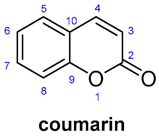
Fig.1 Structure Of Coumarin
Coumarins are heterocyclic compounds known for their diverse medicinal properties, including antifungal [8], antibacterial [9], antioxidant [10], and anti-inflammatory activities [11]. Additionally, coumarin and its derivatives exhibit anticancer effects against various cancer types, such as prostate, renal, breast, laryngeal, lung, colon, central nervous system (CNS), leukemia, and malignant melanoma [12]. Furthermore, coumarin-based derivatives have demonstrated potential as anti-HIV agents, capable of inhibiting multiple stages of the HIV replication cycle, including the attachment of the virus to host cells and the fusion of cell membranes [13]. Coumarins, classified as 2H-1-benzopyran-2-ones, represent significant oxygen-containing fused heterocycles utilized in pharmaceuticals and dyes [14]. The term 'coumarin' is derived from 'coumarou,' the common name for the Tonka bean (Dipteryx odorata Willd, Fabaceae), from which coumarin was first extracted in 1820[15]. These compounds belong to a family of lactones characterized by a benzopyrone structural framework, which can be obtained through both natural extraction from plants and total synthesis in laboratory settings [16]. The addition of various groups as fused components to the parent coumarin modifies its properties, resulting in the formation of more functional derivatives [17].
Table 1
The Physical Properties of Coumarin
|
Molecular Formula |
C9H6O2 |
|
Molecular Weight |
146.1427g/mol |
|
Density |
935Kg/m3 |
|
Melting point |
68-73C |
|
Solubility in Water |
0.17g/100ml at 20C |
|
Nature |
Colourless to white solid crystals, flakes, or powder |
|
UV absorbance |
275nm |
|
CAS Register Number |
91-64-5 |
MATERIALS AND METHODS
All the materials and reagents were obtained from commercial sources such as Pine chem industry and thermos chemicals Lmt. Precoated TLC silica gel plates purchased from S.D. Fine were used to confirm the components purity and the reactions progression. EZ Melt automatic melting point equipment and an open capillary tube were used to assess the melting points of all the compounds. All the compounds that are synthesized were characterized by spectral analysis like Mass spectrometry and IR spectrometry. The final compounds were screened for their anti-bacterial activity.
Procedures
Synthesis of Coumarin
75 ml of concentrated sulphuric acid is placed in a beaker which is immersed in ice bath until the temperature of the acid falls to below 100C. Required quantity of phenols [P. Cresol, Hydroquinone] is added to 22ml of ethyl acetoacetate and stirred continuously until complete solution is obtained in another beaker. This solution is added complete to concentrated sulphuric acid, so that the temperature of the reaction mixture doesn’t rise above 100C.The reaction mixture is kept at room temperature with occasional stirring and poured it with vigourous stirring into a mixture of crushed ice. The precipitate is collected by filtration through action pump.It is then washed with cold water. The precipitate is dissolved in 10% NAOH and reprecipitated by adding dilute sulphuric acid or dilute hydrochloric acid. The precipitate is collected by filtration at the pump. Re-Crystallisation is done from ethanol and the percentage yield is reported.
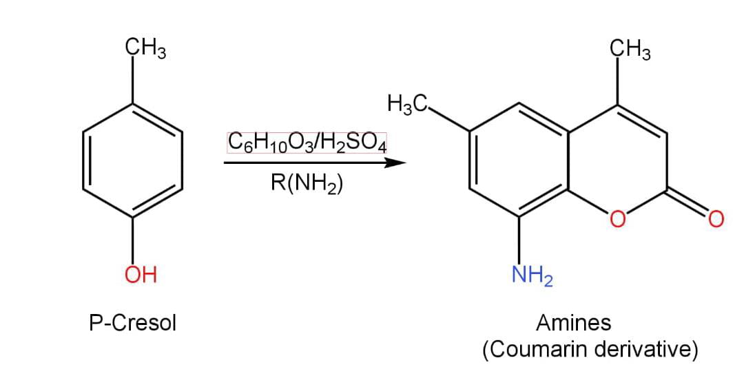
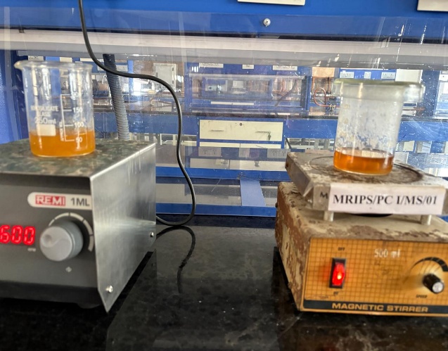
Fig 2. Scheme for The Synthesis of Coumarin
Synthesis of derivative A: P. cresol
A reflux condenser was set up with a 50 mL round bottom flask and a magnetic stir bar. 2 drops of concentrated HCl is added to 2.0 g of P. Cresol, 1.76 g of dimethylamine hydrochloride as well as 0.66 g of paraformaldehyde was added to the solution and refluxed for 2 hours. The solution was left to cool in a water bath to room temperature. While the solution was still somewhat warm, 11 mL of acetone was added and the solution cooled/was gently stirred. When crystallization occurred, it sat at room temperature for 15-20 minutes, then placed in an ice bath for 10 minutes. The crystals were filtered and washed with 2 mL of ether.
Synthesis of derivative B: Hydroquinone
A reflux condenser was set up with a 50 mL round bottom flask and a magnetic stir bar. 2 drops of concentrated HCl was added to 2.0 g of Hydroquinone, 1.76 g of dimethylamine hydrochloride as well as 0.66 g of paraformaldehyde was added to the solution and refluxed for 2 hours. The solution was left to cool in a water bath to room temperature. While the solution was still somewhat warm, 11 mL of acetone was added and the solution cooled/was gently stirred. When crystallization occurred, it sat at room temperature for 15-20 minutes, then placed in an ice bath for 10 minutes. The crystals were filtered and washed with 2 mL of ether.
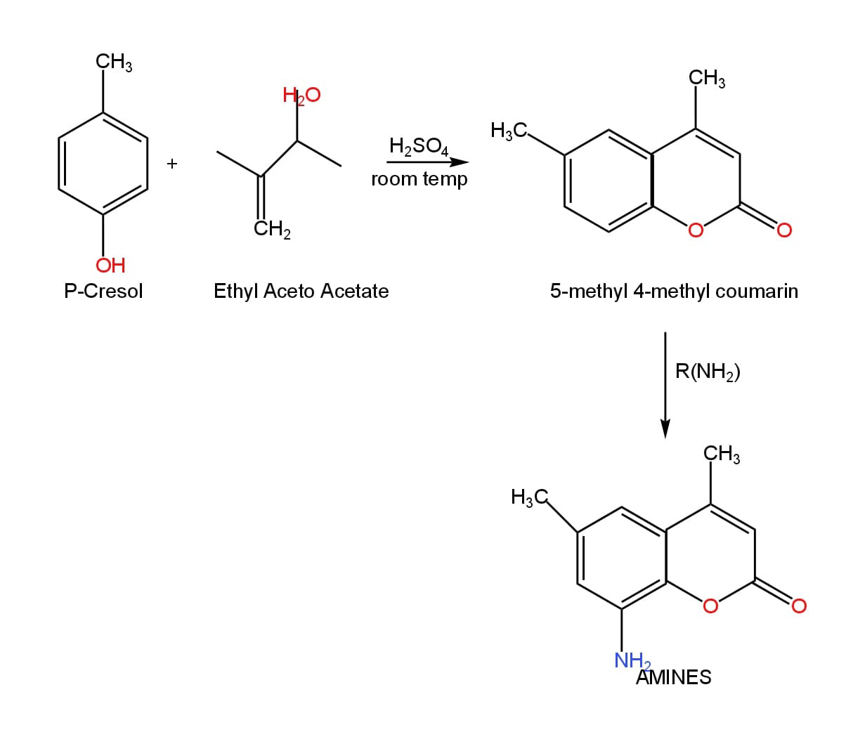
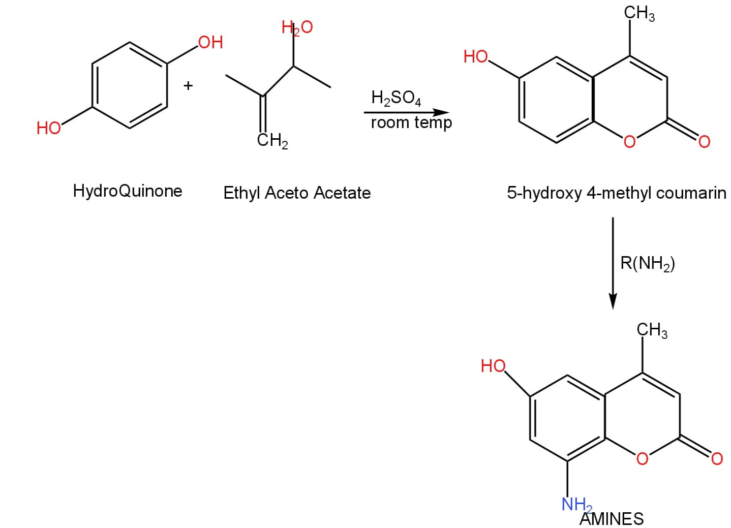
Fig.3 Scheme for Synthesis of Coumarin Derivatives
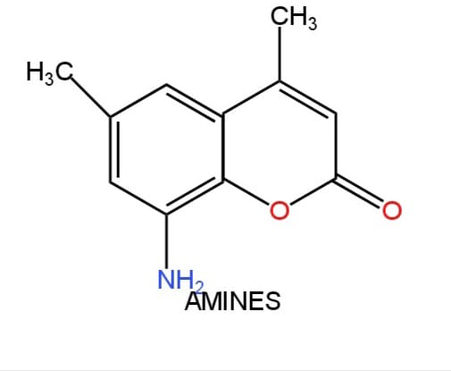
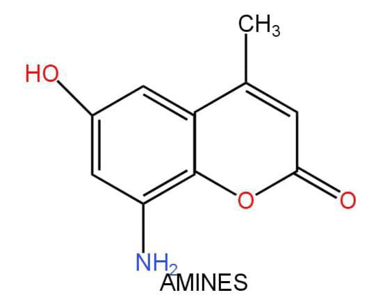
[B]
Fig.4 Structures of Coumarin Derivatives [A] And [B]
Antibacterial Activity
All the synthesized Coumarin derivatives were screened for their antibacterial activity. The invitro antibacterial activity of the synthesized compounds were assessed against Bacillus subtilus and Escherichia coli by cup plate method on nutrient agar medium. The nutrient agar plate was inoculated with test organism, with a depth of 4-5mm and then allow it to solidify. Ensure that the layers of medium are uniform in thickness. Divide the nutrient agar plate into four (4) equal parts. Then with the help of sterile borer make four cavities, one in each portion. Fill the cavity with the test solution and slowly incubated the plates at 37?c for 24 hours. Accurately measure the diameter or areas of circular inhibition zones. The antibacterial activity was determined by measuring the diameter of the inhibition zone in mm [18-25]. Bacteria: The different species of bacteria used to carry out the antibacterial activity tests are Bacillus subtilis, Escherichia coli. Antibiotic assay media, sterile discs are used
Culture media:
Table II
Antibiotic Assay Media
|
Composition: |
g/ lit |
|
Peptone |
6 |
|
Pancreatic digest of casein |
4 |
|
Yeast extract |
3 |
|
Beef extract |
1.5 |
|
Dextrose |
1 |
|
Agar |
15 |
Final PH (After sterilization) 7.9 + 0.1
Generally, the antibacterial activity of the compound is expressed in terms of its ability to inhibit the growth of bacteria in nutrient broth or agar. The bacterial growth inhibition can be measured by two methods.
They are:
Dilution method: This method is used to determine the minimal concentration of antibacterial to inhibit or kill the microorganism. This minimal concentration is called as Minimal inhibitory concentration (MIC). This method is measured by two methods, one broth dilution method and agar dilution method
Diffusion method: This is of two types; one is paper disc diffusion method and agar diffusion method (or) cup plate method. This method followed here for testing the anti-bacterial Cup plate method.
Cup plate method: Media are made up of the final concentrations used in the usual methods of microbiological assay, but with addition of 1-5 per cent of agar. Eleven-ml, portions of medium are placed in Pyrex bacteriological test tubes, autoclaved at 10th, for 10 minutes and stored in the refrigerator until required. Just before use the tubes are heated in the boiling water- bath to melt the medium and then held in a bath at 45?C until needed. The tubes are now inoculated with 1ml of a suitable suspension of organism, after thorough mixing by several inversions, the medium is poured into Petri dishes, which are allowed to cool on a flat bench. It is essential to use dishes of which the circular inner surface is plane and level Four holes are cut in the agar by means of a sharp 10-mm. Cork borer, the underside of the plates are marked to permit subsequent identification of the cups. Three drops of test or standard solutions are now added to the appropriate cups from a pipette of the type used in ampicillin estimations, in which the dropper has a platinum tube of 0.0365 inch external 0.0295-inch internal diameter and about 12mm, long, fused into the tip at an angle of about 130 degree to take up a position normal to the Petri dish during delivery. A pipette of this type has been found to give drops highly uniform in size. The plates are incubated at a temperature suitable for the organism used. The width of the zones of growth may be measured after 18 hours incubation, but it has been found that under the conditions so far investigated, the diameters are not altered by longer incubation. During preparation of the plates strict asepsis is unnecessary, normal cleanliness and the use of oven dried apparatus have so far avoided all trouble from contaminating organisms and it has also been found unnecessary to sterilise either test or standard solutions.
Cup plate method:
Preparation of antibiotic culture media: The culture media was prepared according to the composition and the final was adjusted to PH 7.40+ 0.01 after sterilization.
Preparation of Petri plates: The prepared antibiotic culture media was poured into the Petri plates to get a depth of 4mm. About 25-30ml of antibiotic culture media was poured in Petri plates with diameter of 100mm.
Preparation of antibiotic stock solution: Antibiotic may be received as powder or tablets. Powder was weighed accurately dissolved in the appropriate diluents to yield the required concentrations using sterile glass ware. The standard drug Ampicillin powder of 10 mg was dissolved the 10 ml. Solvent to get a concentration of 1mg/ml. This was used as stock solution. From this different dilution were prepared. The different concentrations prepared were 100μg/ml, 500 µg/ ml, 1000 µg/ ml. Cup plate method: The antibacterial activity was done by using the cup plate method.
Table III
Zone of Inhibition of The Derivatives
|
Zone of Inhibition ZOI (mm) |
||
|
Compound
|
Species |
Concentration (µg/ml) |
|
100 500 1000 |
||
|
Compound1(coumarin) Compound 1a (Derivative A) Compound 1b (Derivative B) Ampicillin |
Bacillus subtilis |
9 11 18 11 12 21 8 9 12 10 15 18 |
|
Compound 1 (coumarin) Compound 1a (Derivative A) Compound 1b (Derivative B) Ampicillin |
Escherichia coli |
6.0 7.0 8.5 7.2 8.5 9.0 5.1 7.5 8.5 7.5 8.0 10.0 |
RESULTS AND DISCUSSIONS
Results and discussion of synthesized compounds
P. Cresol [Compound 1]
The percentage yield: 72%, m. p 35.5 °C, the formation of compound was evident from the IR spectrum by the appearance of broad peak at the 3416.02 , due to the presence of stretching vibrations of C-H bonds in the aromatic ring and also due to the presence of hydroxyl group [-OH] [Fig….], Mass spectrum [ESI MS] by the appearance of molecular ion peak [M] at m/z [%] 120 [Fig….],NMR spectrum by the appearance of triplet at 7.5ppm protons on C5,C4 Carbons, triplet at 7.9 ppm protons on C5,C7 Carbons[Fig…..].
Hydroquinone [Compound2]
The percentage yield: 83%, m. p 172 °C, the formation of compound was evident from the IR spectrum by the appearance of broad peak at the 3074.67 cm-1 , due to the presence of stretching vibrations of C-H bonds in the aromatic ring and also due to the presence of hydroxyl group [-OH] [Fig….], Mass spectrum [ESI MS] by the appearance of molecular ion peak [M] at m/z [%] 110 [Fig….],NMR spectrum by the appearance of triplet at 7.8 ppm protons on C5,C4 Carbons, triplet at 8.0 ppm protons on Carbons[Fig….].
Results and discussion of Antibacterial Activity:
The compound 1a at 100μg/ml conc. Showed good activity compared to 1b, where the ZOI is (11mm) in B. SUBTILIS. The ZOI of 1b, was found to be (8mm). The compound 1a at 500μg/ml conc. Showed good activity compared to 1b, and where the ZOI is (12mm) in B. SUBTILIS. The ZOI of 1b, was found to be (9mm). The compound 1a at 1000μg/ml conc. Showed good activity compared to 1b, and where the ZOI is (21mm) in B. SUBTILIS. The ZOI of 1b, was found to be (12mm). The compound 1a at 100μg/ml conc. Showed good activity compared to 1b, and where the ZOI is (7.2mm) in E. COLI. The ZOI of 1b, was found to be (5.1mm). The compound 1a at 500μg/ml conc. Showed good activity compared to 1b, and where the ZOI is (8.5mm) in E. COLI. The ZOI of 1b, was found to be (7.5mm). The compound 1a at 1000μg/ml conc. Showed good activity compared to 1b, and where the ZOI is (9mm) in E. COLI. The ZOI of 1b, was found to be (8.5mm). The compound 1a was showing good anti -bacterial activity than compound 1b in B. SUBTILIS AND E. COLI species
CONCLUSION:
The proposed compound coumarin and its derivatives were synthesised and the derivatives are characterised by the spectral data [IR and MASS] and further evaluated for antibacterial activity. Even though all the synthesized coumarin derivatives showed remarkable ZOI [Zone Of Inhibition] with standards and have good antibacterial activities, the compound 1a was showing good antibacterial activity than 1b, in B. subtilis species and E. coli species.
ACKNOWLEDGEMENTS
Authors are very thankful to Department of Chemistry, Mallareddy Institute Of Pharmaceutical Sciences for providing laboratory facilities and support to furnish our work.
REFERENCES


B. Swapna*, B. Rajkamal, P. Rasagnya, T. Poojitha, Synthesis and Biological Evaluation of Novel Coumarin Derivatives as Antimicrobial Agents, Int. J. of Pharm. Sci., 2025, Vol 3, Issue 6, 5693-5702. https://doi.org/10.5281/zenodo.15768973
 10.5281/zenodo.15768973
