Aditya Pharmacy College, Beed
Standardization of herbal drugs is essential to ensure their quality, safety, efficacy, and reproducibility. Herbal medicines often exhibit significant variations in chemical composition due to differences in plant sources, environmental factors, harvesting times, and processing methods. Chromatographic techniques such as High-Performance Thin Layer Chromatography (HPTLC), High-Performance Liquid Chromatography (HPLC), and Gas Chromatography (GC) play a pivotal role in the standardization process by enabling the qualitative and quantitative analysis of bioactive constituents. These methods allow the identification of marker compounds, fingerprint profiling, and detection of adulterants, thereby ensuring batch-to-batch consistency. This paper reviews the application of various chromatographic techniques in the standardization of herbal drugs and highlights their importance in promoting the acceptance of herbal medicines in modern healthcare systems.
Introduction Herbal drugs have been used since ancient times as medicines for the treatment of a range of diseases in different ancient civilization of world. Medicinal plants have played a key role in world health and very idea of curative effect has been obtained from herbal treatment. In spite of the great advances observed in modem medicine in recent decades, plants still make an important contribution to health care. Medicinal uses of plants are exploited worldwide. Over the past decade, interest in drugs derived from plants, especially the Phyto-therapeutic ones, has increased expressively. It is estimated that about 25% of all modem medicines are directly or indirectly derived from plants (1-4). In some particular cases, such as antitumoral and antimicrobial drugs, about 60% of the medicines currently available on the market and most of those in the late stages of clinical trials are derived from natural products, mainly from plants. The new series of anticancer therapeutics like Vinblastine etc.
are developed from Vinca rosa plant. According to the World Health Organization (WHO) , because of poverty and lack of access to modem medicine, about 65-80% of the world's population which lives in developing countries depends essentially on plants for primary health care. Currently, the major pharmaceutical companies have demonstrated renewed interest in investigating plants as sources for new lead structures and also for the development of standardized Phyto-therapeutic agents with proved efficacy, safety and quality (2,6-9). Herbal medicinal preparations are normally very popular in developing countries with a long tradition in the use of medicinal plants and also in some developed countries such as Germany, France, Italy and the United States where appropriate guidelines for registration of such medicines exist.
1.2 Herbal pharmacopoeias The World Health Organization (WHO) has published guidelines in order to define basic criteria for evaluating the quality, safety, and efficacy of herbal medicines aimed at assisting national regulatory authorities, scientific organizations and manufacturers in this particular area (5).
Furthermore, the WHO has prepared Pharmacopoeia monographs on herbal medicines and the basis of guidelines for the assessment of herbal drugs. WHO Guidelines for Quality Standardized Herbal Formulations (20, 21) a. Quality control of crude drugs material, plant preparations and finished products.
b. Stability assessment and shelf life. c. Safety assessmentj documentation of safety based on experience or toxicological studies. d. Assessment of efficacy by ethno medical information and biological activity evaluations. The bioactive extract should be standardized on the basis of active principles or major compounds along with the chromatographic fingerprints (TLC, HPTLC, HPLC and GC).
Microbial contamination –
Total viable aerobic count, pathogenic bacteria like enterobacteria, E. coli, salmonella, Pseudomonasaeruginosa, Staphylococcus aureus, etc. and presence of afflatoxins etc.
2.6 Radioactive contamination. Several regulatory models for herbal medicines currently exist, including prescription drugs, over-the-counter drugs, traditional medicines and dietary supplements. Thus, the need to establish global and/or regional regulatory mechanisms for regulating herbal drugs seems obvious.

Isolated compounds from plants of Arecaceae family:
Compounds isolated from stem of the Pothos acandens include hemiterpene glucosides like Canthoside B (Kanchanpoom et al., 2002), Pothobanoside A, Pothobanoside B, Pothobanoside C (Muhit et al., 2015), Phenyl isobutanol compounds like Pothobanol (Muhit et al., 2015), Flavones like Vicenin-2 (Islam et al., 2014), Scoparin 2-O-xyloside, Vitexin 2-O-xyloside (Fiasson et al., 1989), Neoschaftoside (Xie et al., 2003). Isoschaftoside (Wada et al., 2000), Flavonols like Isorhamnetin 3-Ogentiobioside (Huang et al., 2010), Kaempferol 3-O-gentiobioside (Iwashina et al., 2000), Quercetin 3-O- gentiobioside (Liao et al., 2012), Glycosides like Canthoside A (Kanchanpoom et al., 2002). Zizybeoside I (Takashima et al., 2007), Hydroquinone glycoside like Markhamioside F (Kanchanpoom et al., 2002). Diketopiperazines Glycoside like Eleutherazine B (Li et al., 2010). Alkaloid like 1,2,3,4- tetrahydro 3-carboxy-2 carboline (Zhang et al., 2013). Whole plant contains diterpenoid compounds like (3β)-ent-kaurane-3,16,17-triol-3-β-D glucopyranoside (Ferreira and Oliveira, 2010), Methyl Pothoscandensate (Liu et al., 2012), (3β)- ent kaurane-3,16,17-triol (Xue-Xiang et al., 2015), Alkaloids like N-trans-p cumaroyltyramine (Sembiring et al., 2010), N-trans- cinnamoyltyramine (Torel et al.,1985), N-trans-feruloyltyramine (Kanada et al., 2012), Indole alkaloids like (–)-serotobenine (Sato et al., 1985) and (+)-syringaresinol (Hussain et al., 2014) which was a lignan. Whole aerial part of the plant contains 5- oxoundecyl-3 hydroxy pentanoate (Gupta et al., 2014). GC-MS analysis of the ethanolic leaf extract of the plant showed the presence of 19 compounds. They are dodecanoic acid, tetra decanoic acid, hexadecenoic acid, octadecanoic acid, 2 methyl-1-Hexadecanol, 2-hexa- decanol,1,1-diethoxy- Butane, d-Mannose, 2,3 dihydro-3,5-dihy droxy-6-methyl- 4H- Pyran-4-one, hexanoic acid, ethyl ester, 1,1,3-triethoxy- propane, (Z,Z)-9,12-octadecadienoic acid, phytol, 1,4 dimethyl-7-(1- methylethyl)azulene, (Z)-2-(9- octadecenyloxy)ethanol, 3,7,11,15- tetramethyl-hexadecan-1-ol, (Z,Z,Z)-9,12, 15- octadecatrienoic acid, 1,2- benzene dicarboxylic acid, diisooctyl ester, 1-monolino-leoylglycerol trimethylsilyl ether (Lalitharani et al., 2009).
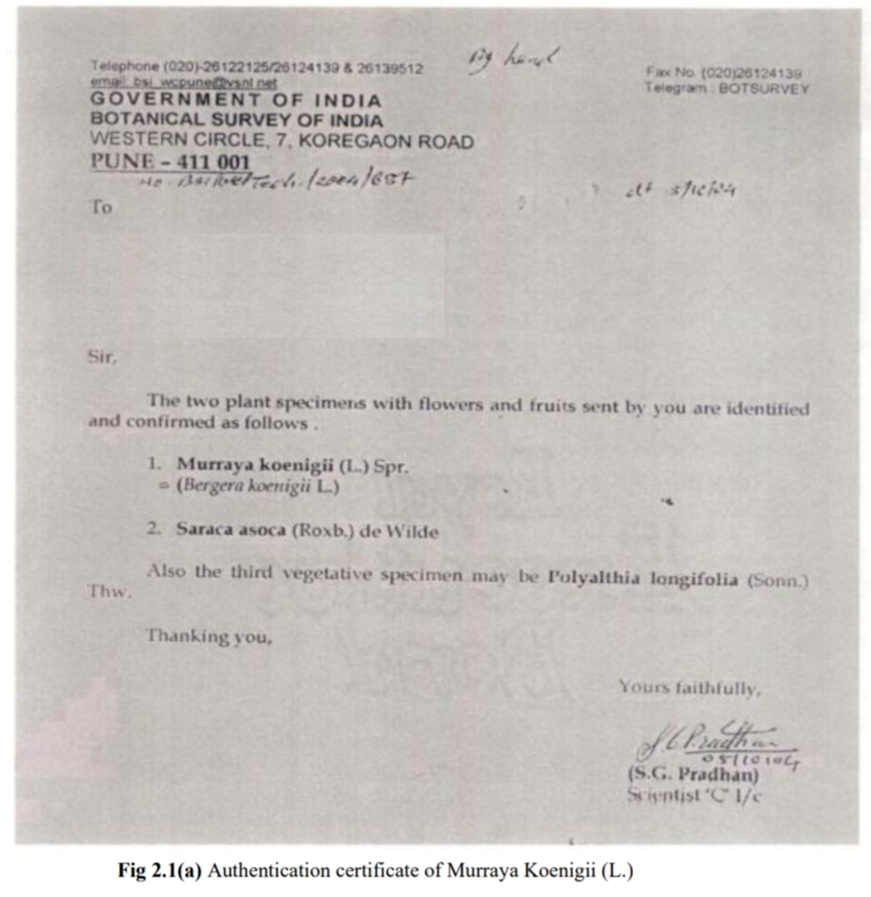
AUTHENTICATION:
Authentication certificate the plant used for present study is collected from Shahpur region of Maharashtra state and authenticated by Botanical survey of India, Government of India, Western circle, Koregaon Road, Pune-411001
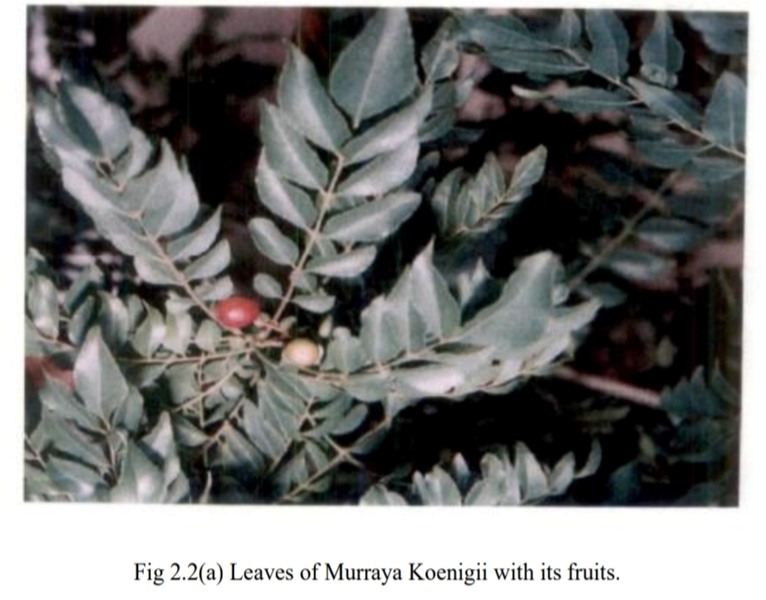
Murraya Koenigii (L.) Sprang.
Synonymy: Bergera Koenigii (L.) Roxb.
Family: Rutaceae
English name: Curry leaf-tree
Indian names: karepaku (Andhra Pradesh); narasingha, bishahari (Assam); barsanga, kartaphulH (Bengal); gorenimb, kadhihmbdo (Gujarat); mitha neem, gandhla, gandhela, gandhelu (Himachal Pradesh); kathnim, mitha neem, kurry patta gandhela, barsanga (Hindi); karibeva (Karnataka); kariveppilei (Kerala); gandhela, gandla, gani (Kumaon); bassan, basango, bhursanga (Orissa); surabhinimba, kalasaka, mahanimb (Sanskrit); karivempu, karuveppilei (Tamilnadu).
Botanical Description:
A small spreading shrub, about 2.5 meters high; the main stem, dark green to brownish, with numerous dots on it; its bark can be peeled off longitudinally, exposing the white wood underneath; the girth of the main stem is 16 cm. Leaves, exstipulate, bipinnately compound, 30 cm long, each bearing 24 leaflets, having reticulate venation; leaflets, lanceolate, 4.9 cm long, 1.8 cm broad, having 0.5-cm-long petiole. Flowers, bisexual, white, funnel-shaped, sweetly scented, stalked, complete, ebracteate, regular, actinomorphic, pentamerous, hypogynous, the average diameter of a fully opened flower being 1.12 cm; inflorescence, a terminal cyme, each bearing 60 to 90 flowers; calyx, 5-lobed, persistent, inferior, green; corolla, white, polypetalous, inferior, with 5 petals, lanceolate; length, 5 mm; androecium, polyandrous, inferior, with 10 stamens, dorsifixed, arranged into circles of five each; smaller stamens, 4 mm. long whereas the longer ones, 5 to 6 mm; gynoecium, 5 to 6 mm long; stigma, bright, sticky; style, short; ovary, superior. Fruits, round to oblong, 1.4 to 1.6 cm long, 1 to 1.2 cm in diameter; weight, 880 mg; volume, 895 microliters; fully ripe fruits, black with a very shining surface; pulp. Wistaria blue 640/2; the number of fruits per cluster varying from 32 to 80. Seed, one in each fruit, 11 mm long, 8 mm in diameter.
Chemical Constituents:
Leaves contain a volatile essential oil, resembling the oil of Aegle marmelos, a resin and a crystalline principalglucoside named Koeinigin; seeds yield an oil. The Strong odiferous oil occurs in the leaves and the seeds of Murraya koenigii (L.) Spreng. The chemical examination of this essential oil exhibited a strong antibacterial and antiftingal activity. The acetone extract of the fresh leaves of Murraya koenigii resulted in the isolation of three bioactive carbazole alkaloids, mahanimbine, murrayanol, and mahanine . Five carbazole alkaloids were isolated from the Dichloromethane extract and their structures were identified to be euchrestine B, bismurrayafoline E, mahanine, mahanimbicine, and mahanimbine.
Pharmacological Activities: Analgesic, astringent, antidysentery, antioxidant, febrifuge, hypolipidemic, hypoglycemic, for improvement of vision, to treat night-blindness, and for regulation of fertility. Formulations and preparations: Infusions and Decoctions.
ISOLATION AND PURIFICATION OF- PROTEINS:
Chemistry of proteins:
The term protein is derived from Greek word meaning “primary” or “holding first place”. Proteins are indeed extremely important constituents of living cells: quantitatively, they generally represent half the dry weight of cells, and from a qualitative point of view, besides the structural proteins, other proteins having a capital biological function, especially the enzymes. All proteins contain the four elements: C, H, O and N. Many contain Sulphur. Phosphorus is present insome. The nitrogen content of proteins being around 16%, its appropriate quantity can be estimated by a simple nitrogen titration after isolating the proteins. Proteins are macromolecules, formed by the condensation of a large number of units (50 to several thousands) called amino acids. As indicated by their name, amino acids contain one acidic group and one primary amino group in a position with respect to the carboxyl group. Excepting proline and hydroxyproline, their general formula is; Whatever their origin (viral, bacterial, plant or animal), we find in proteins about twenty amino acids which differ by their radical R and may be classified as follows
Aliphatic amino acids:
a. Hydrocarbon chain amino acids e.g. Glycine, Alanine, Valine.
b. Hydroxyl amino acids e.g. Serine, Threonine.
c. Sulphur containing amino acids e.g. Cysteine
d. Dicarboxylic amino acids and their amides e.g. Aspartic acid. Glutamic acid.
e. Basic amino acids e.g. Lysine, Arginine
3.1.2 Cyclic amino acids:
a. Aromafic amino acids e.g. Tyrosine, Phenylalanine.
b. Hetrocylic amino acids e.g. Proline.
All proteins are composed of a primary structure consisting of amino acids with the amino
acids joined together by a peptide linkage in general peptides are classified as: a. Oligonucleotides: dipeptides, tripeptides, which do not give the Biuret reaction, are called Oligonucleotides.
b. Polynucleotides: from tetrapeptides onwards, which gives the characteristic Biuret reactions are called polypeptides.
Classification of proteins:
Different types of classification for proteins have been proposed
Classification based on molecular shape
Classification based on solubility
Classification based on composition
Classification based on molecular shape:
This class includes Fibrous and Globular proteins. Fibrous proteins are also called Scleroproteins. They consist of fibers and are practically insoluble. Collagens and Keratins are some examples of this class. Globular Proteins are called spheroproteins on account of their spherical and ovoidal shape they are generally more easily soluble. This group generally includes albumins and globulins.
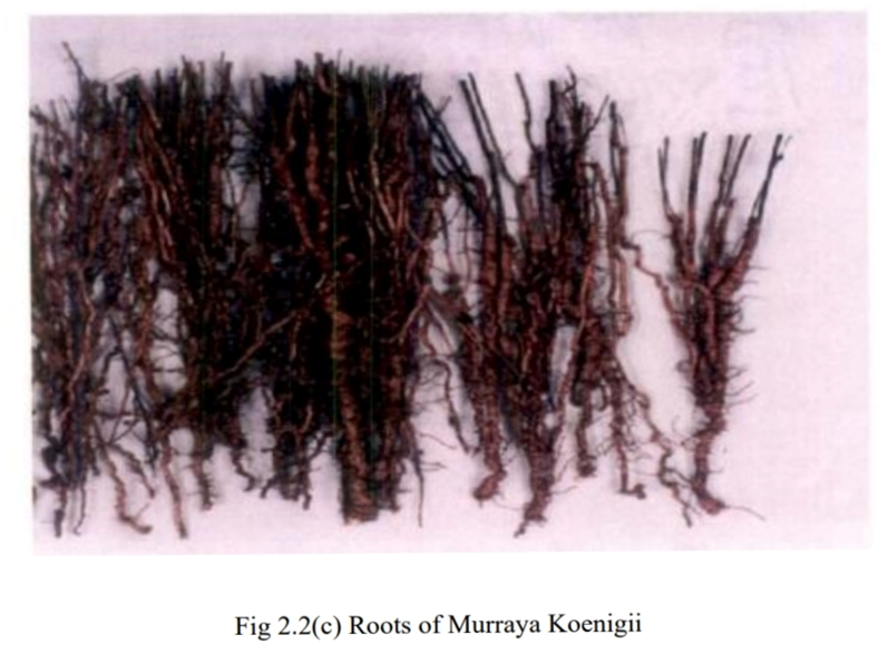
Isolation and Purification of proteins of Murraya koenigii :
Preparation of Extract
Equipments: 100-1000|xl Micropipette, Cooling centrifuge, Electronic balance, pH meter & etc.
Chemicals: Tris/Glycine buffer.
Protein Extraction Buffer
• Tris/Glycine pH ~_5.5 o 30.28 gms
Tris 144.2 gms Glycine
up to one liter with distilled water.
Protein extract:
Isolation and Purification of proteins of Murraya koenigii :
Preparation of Extract
Equipments: 100-1000|xl Micropipette, Cooling centrifuge, Electronic balance, pH meter & etc. Chemicals: Tris/Glycine buffer. Protein Extraction Buffer • Tris/Glycine pH ~_5.5 o 30.28 gms Tris 144.2 gms Glycineup to one liter with distilled water. UProtein extract: • Isolation of proteins was done from plant parts such as Leaves, Bark, and Roots individually.
• Homogenize the individual plant part into a tissue homogenizer. Weigh the homogenized
material. Extraction buffer is added exactly twice the weight of homogenized material &
shaken for 12 hours. (For extract to be liquid, but concentrated, the Extracting buffer is used
sparingly.)
• Extract was centrifuged in cooling centrifuge at -4*^C & 8000 rpm for 15 minutes to sediment
the plant debris.
• Transfer the liquid proteins extract to a fresh centrifuge tube. Discard the plant debris pellet.
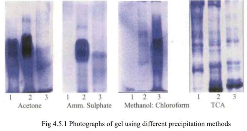
4.2 Purification of proteins
Trichloroacetic acid (TCA) Precipitation
• To 10ml of individual protein extract for each plant p a rt, 1ml of freshly prepared ice cold
Trichloroacetic acid (TCA) having concentration of 72g of TCA crystals in 100 ml of distilled
water was added.
• Mixed and kept in ice for 12 hours (cold room at -20°C).
• Centrifuged at 4°C for 15 minutes at 8000 rpm. • Supernatant was removed and 1.5ml of distilled water was added to wash the pellet and remove
the residual acid. Vortexed thoroughly & centrifuged at -4°C for 15 minutes. Supernatant was removed and discarded. Same step was repeated one more time.
• 1.5ml of ice-cold acetone was added to wash the pellet. Vortexed thoroughly & centrifuged at -4°C for 15 minutes. Supernatant was removed and discarded. Same step was repeated one
more time.
• Pellet was dried for 20 minutes in Speed Vac at 37°C.
• Individual pellets were collected from all other sample tubes of same sample and homogenized
to acquire protein powder and stored at ambient temperature.
• Using above method protein powder of individual plant parts such as Leaves, Bark and Roots
were prepared.
Quantitative determination of proteins:
Spectrophotometric determination of plant proteins:Lowry’s method of protein estimation [14] w as used to determine the protein percentage of above prepared batch of isolated proteins from individual parts of the same plant. This method was used because of its sensitiveness to interfering substances. The assay was carried out using Bangalore Genei Pvt.Ltd “Protein Estimation Kit by Lowrys Method” Catalogue No KT-18.
Principle:
Lowry’s assay is based on the reduction by protein of the phosphomolybdic-tungstic mixed acid chromogen in the Folin-Ciocalteu’s phenol reagent, resulting in an absorbance maximum at 660 nm. The Folin-Ciocalteu’s phenol reagent reacts primarily with tyrosine residues in the protein, which can lead to variation in the response of the assay to different proteins.
Standards and Reagents:
• 5000 ppm BSA standard was prepared by reconstituting one vial containing 5mg BSA with 1 ml distilled water.
• Complex forming reagent was prepared by mixing one volume of copper sulphate lsolution (solution I) to 100 volumes of alkaline tartarate solution (solution II) just hyy 66before use.
• Folin reagent (solution III) was used directly.
Procedure:
• Standard BSA, samples and distilled water were pipetted referring to the protocol
given in following table and adjusted the volume to 0.2ml.
• 2ml of complex forming reagent was added and mixed thoroughly using cyclomixer.
• 0.2ml of Folin Ciocalteu’s reagent was added and mixed thoroughly using cyclomixer and incubated it for 30 minutes at room temperature.
• Optical density was read on spectrophotometer at 660 nm
Observations Table 1:
Calibration curve readings for standard bovne serum albumin

Preparation of samples:
• Accurately weighed 1.00 mg of protein powder isolated from individual parts of the same plant was transferred to a 1.5 cm^ Eppendorf tubes and diluted with 100 ^1 of 4x running buffer. The Eppendorftubes were sonicated for 60 minutes and centrifuged at high speed of 15,000 rpm for 15 minutes. The supernatant was transferred to another Eppendorf and used for further analysis.30 |il of the above supernatant was mixed with 10) al of sample buffer before the sample loading. • Sample preparation for unhygienic impurities:
The unhygienic impurities studied here are insect debris and bird excreta. The protein extract for the standard insect debris (caterpillar) and bird excreta was prepared as given for the individual plant parts in the Chapter 03. The extract obtained was then precipitated and the protein powder was prepared as per the procedure given in the Chapter 03. A Murraya koenigii leaf that was originally contaminated with caterpillar debris and bird excreta separately was used to prepare the sample to detect the presence of unhygienic impurities. The protein extract of this contaminated raw material was prepared as per the protocol followed for the pure raw material. The sample was further processed using the Preparation of samples:
• Accurately weighed 1.00 mg of protein powder isolated from individual parts of the same plant was transferred to a 1.5 cm^ Eppendorf tubes and diluted with 100 ^1 of 4x running buffer. The Eppendorf tubes were sonicated for 60 minutes and centrifuged at high speed of 15,000 rpm for 15 minutes. The supernatant was transferred to another Eppendorf and used for further analysis.30 |il of the above
supernatant was mixed with 10) al of sample buffer before the sample loading.
• Sample preparation for unhygienic impurities:
The unhygienic impurities studied here are insect debris and bird excreta. The protein extract for the standard insect debris (caterpillar) and bird excreta was prepared as given for the individual plant parts in the Chapter 03. The extract obtained was then precipitated and the protein powder was prepared as per the procedure given in the Chapter 03. A Murraya koenigii leaf that was originally contaminated with caterpillar debris and bird excreta separately was used to prepare the sample to detect the presence of unhygienic impurities. The protein extract of this contaminated raw material was prepared as per the protocol followed for the pure raw material. The sample was further processed using the precipitation technique described in Chapter 03 to obtain the protein powder of the contaminated raw leaves of Murraya koenigii. Accurately weighed 1.00 mg of protein powder isolated from the possible contaminant of insect debris (caterpillar) and that of the sample of Murraya koenigii leaves contaminated with insect debris was transferred to different 1.5cm^ Eppendorf tubes and was diluted with 100|ul of 4x Extraction buffer separately. The Eppendorf tubes were sonicated to 60minutes and centrifuged at high speed of 15,000rpm for l0 minutes. The supernatant was transferred to another orEppendorf tube and used for further analysis. 30|l of the above supernatant was mixed with l0 jil of Sample buffer before the sample loading. Thus, the protein samples for the standard insect debris and the leaves in presence of the insect debris were prepared. The same procedure was followed to prepare the protein samples for bird excreta and the leaves contaminated with bird excretaprecipitation technique described in Chapter 03 to obtain the protein powder of the contaminated raw leaves of Murraya koenigii. Accurately weighed 1.00 mg of protein powder isolated from the possible contaminant of insect debris (caterpillar) and that of the sample of Murraya koenigii leaves contaminated with insect debris was transferred to different 1.5cm^ Eppendorf tubes and was diluted with 100|ul of 4x Extraction buffer separately. The Eppendorf tubes were sonicated for 60minutes and centrifuged at high speed of 15,000rpm for l0 minutes. The supernatant was transferred to another Eppendorf tube and used for further analysis. 30|l of the above supernatant was mixed with l0 jil of Sample buffer before the sample loading. Thus, the protein samples for the standard insect debris and the leaves in presence of the insect debris were prepared. The same procedure was followed to prepare the protein samples for bird excreta and the leaves contaminated with bird excreta.
According to USP
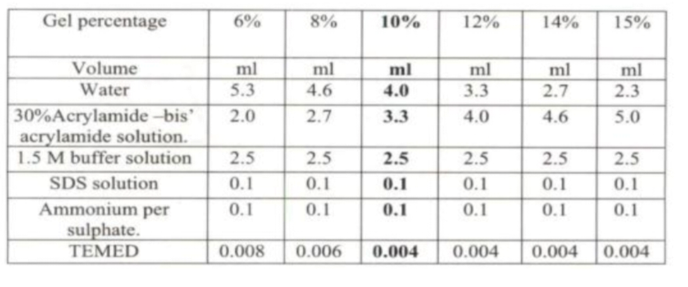
pH of Running Buffer Selection:
This has little effect on filly ionized compound such as inorganic salts, but for organic compounds pH determines The extent of ionization. The ionization of organic acids increases as pH increases whereas the reverse applies for Organic bases; therefore, their degree of migration will be pH dependent. Considering the above facts both the Acidic pH at 5.5 and basic pH at 8.0 were checked. The gel showing the profile at the different pH is given below:
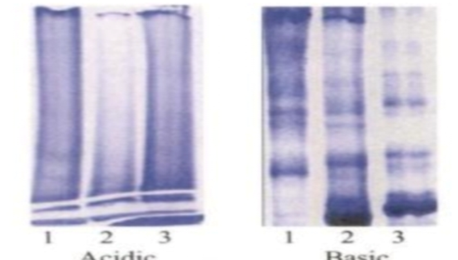
Fig 4.5.3 Photographs of gel using acidic and basic running buffers In the basic pH, sample proteins were quite stable and optimum results were obtained within minimum Amount of time. Hence pH of running buffer was selected as 8.3.
Temperature:
From the above-discussed effect of temperature both the condition of running samples in room temperature and cold temperature at 180C were tried out.
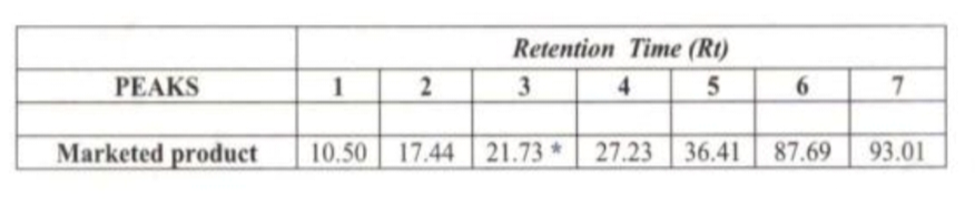
RESULT:
Retention times of the proteins of a marketed produc:-
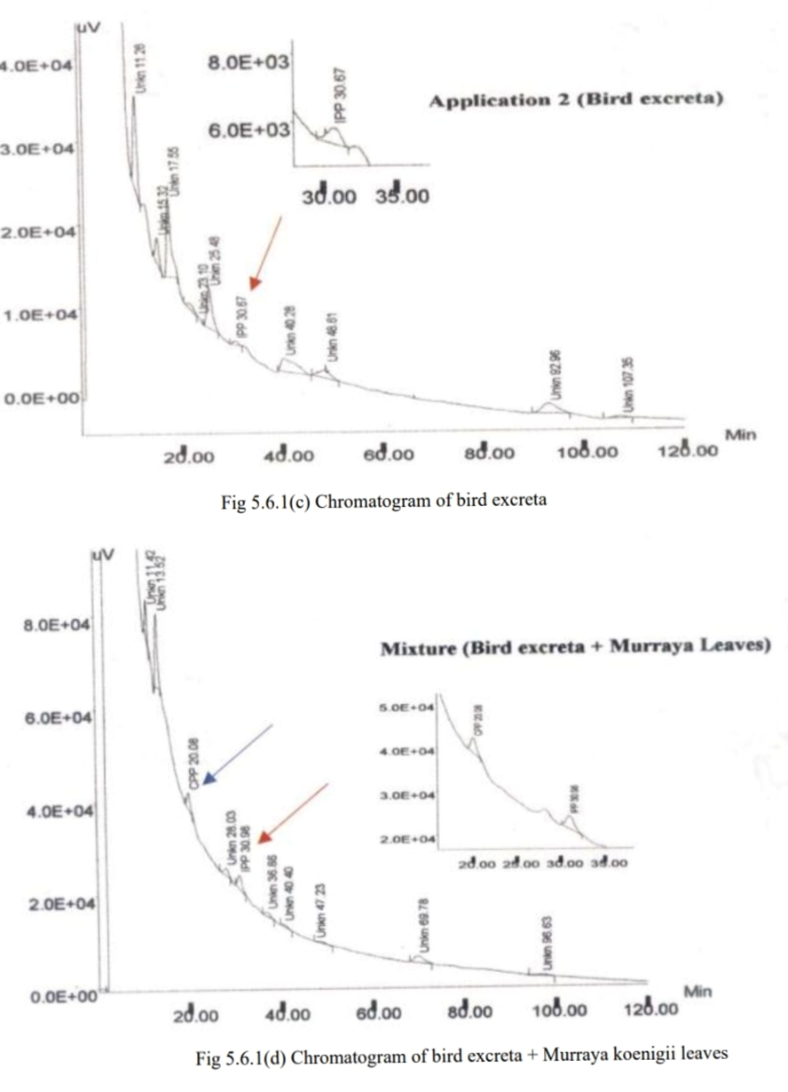
Unhygienic Impurities
Insect debris and Bird excretaSince, a distinct fingerprint pattern was obtained for the different parts of the same plant it was decided to check whether a distinct pattern could be obtained for possible contaminants like insect debris (catepillar) and bird excreta. If a characteristic profile would be obtained then this could serve as a quality control parameter in the first stage of production itself the sample preparation and the chromatographic conditions for the protein samples of insect debris and bird excreta were kept the same as that obtained for Murraya koenigii. Injections were given for the powders obtained from the possible contaminants first. These protein powders from each of the contaminants were then mixed with that of Murray koenigii leaves separately and the patterns obtained were checked. The patterns obtained with the protein powders of the possible contaminants as well as their mixtures with Murraya koenigii leaves are show.
REFERENCES


Kale Rajashri*, Puja Aher, Standardization Of Herbal Drug Using Chromatographic Techniques, Int. J. of Pharm. Sci., 2025, Vol 3, Issue 6, 2581-2594. https://doi.org/10.5281/zenodo.15652861
 10.5281/zenodo.15652861
