IIMT College of Pharmacy, Greater Noida, Uttar Pradesh-201306.
Breast cancer is one of the leading causes of death among women worldwide, making early detection and accurate diagnosis crucial for saving lives. Artificial intelligence (AI) is becoming an important tool in improving the way breast cancer is detected and diagnosed. This review looks at how AI, through technologies like machine learning and deep learning, is helping analyze mammograms, ultrasounds, and tissue samples with greater precision and efficiency. It also explains the steps involved in preparing data, such as cleaning and enhancing images, to make AI models perform better. Important metrics like sensitivity, specificity, and accuracy are used to measure the success of these systems. Although AI offers many benefits, challenges remain. Issues like the quality of available data, understanding how AI models work, ethical concerns, and the difficulty of bringing these systems into everyday clinical practice need to be addressed. The article also explores the current use of AI in healthcare and its potential to provide personalized care for patients. Future trends include combining imaging, genetic information, and other medical data to create more comprehensive AI tools. Making these technologies available in low-resource areas could reduce healthcare inequalities. This review highlights the need for collaboration between healthcare providers, AI researchers, and policymakers to unlock the full potential of AI in fighting breast cancer. Furthermore Mammogram-based automated breast cancer detection plays a critical role in precise cancer diagnosis and treatment planning, ultimately saving lives. Mammography is a simple yet effective diagnostic for detecting breast cancer. Few extensive surveys have been provided to quickly analyses approaches for identifying breast cancer using mammography
Introduction & Background
Breast cancer is one of the most significant public health concerns globally, contributing substantially to cancer-related deaths among women [1]. Early detection is a cornerstone in reducing mortality and improving survival rates. Traditional screening techniques, such as mammography, have served as the primary diagnostic methods for decades. However, these approaches face notable challenges, including variability in interpretation among radiologists, limited sensitivity and specificity, and dependence on resources, which can hinder their widespread effectiveness [2]. These shortcomings underscore the urgent need for innovative diagnostic tools capable of delivering consistent, accurate, and efficient results. In response to these challenges, artificial intelligence (AI) has emerged as a transformative technology in breast cancer detection [3]. By leveraging large datasets and sophisticated algorithms, AI technologies—particularly machine learning (ML) and deep learning (DL)—have shown remarkable potential in enhancing the accuracy and efficiency of diagnostic processes. AI-powered systems excel at analyzing complex medical imaging data, such as mammograms, ultrasounds, and histopathological slides, enabling faster and more reliable detection of abnormalities [4]. These advancements not only address the limitations of traditional methods but also pave the way for significant improvements in diagnostic precision and consistency. This review provides an in-depth exploration of the current progress and future directions in AI applications for breast cancer detection, emphasizing their transformative potential in overcoming existing barriers and improving patient outcomes.
Artificial Intelligence Technologies in Breast Cancer Detection
Artificial intelligence (AI) technologies, particularly machine learning (ML) and deep learning (DL), have revolutionized breast cancer detection by transforming medical imaging analysis. ML models, such as support vector machines (SVMs), random forests, decision trees, and k-nearest neighbors (k-NN), are commonly employed to analyze breast imaging data [5]. These algorithms excel in identifying patterns and classifying images as benign or malignant by leveraging large datasets and learning complex features. Their simplicity and interpretability make them valuable tools in diagnostic applications [6] [7]. Deep learning, a specialized branch of ML, has gained prominence for its advanced capabilities in automated feature extraction and hierarchical representation learning from raw data. Convolutional Neural Networks (CNNs) have been particularly successful in breast cancer detection, as they are adept at processing intricate image data with high precision [7]. Other deep learning frameworks, such as Recurrent Neural Networks (RNNs), are being explored for analyzing sequential data like time-series imaging, while Generative Adversarial Networks (GANs) show promise in generating synthetic data for training and improving model robustness [8]. Recent advancements include transformer-based models, which are being adapted for breast cancer imaging tasks, and federated learning approaches, enabling collaborative training on distributed datasets while preserving patient data privacy [9]. These AI-driven advancements not only enhance detection accuracy but also alleviate the workload of radiologists, ensuring faster and more reliable diagnostic outcomes. The ongoing evolution of AI technologies underscores their critical role in advancing breast cancer detection and improving patient care [10].

Figure 1:- Steps For Breast Cancer Detection
Artificial Intelligence Applications in Breast Cancer Detection
Artificial intelligence (AI) has made significant strides in breast cancer detection across various imaging techniques, including mammography, ultrasound, MRI, and histopathology. Mammography, commonly used for breast cancer screening, benefits from AI algorithms that detect early indicators of cancer, such as microcalcifications, architectural distortions, and masses, with remarkable precision [5]. In ultrasound imaging, AI enhances the ability to differentiate between benign and malignant lesions by analyzing characteristics such as texture, shape, and acoustic properties. This improves the diagnostic accuracy of ultrasound examinations [7]. For MRI scans, AI algorithms assist in identifying small, aggressive tumors and evaluating the vascularity of lesions, which is crucial for staging and treatment planning [8]. In histopathology, AI-powered image analysis helps pathologists examine breast tissue samples more efficiently by detecting malignant cells and automating the processes of grading and subtyping tumors [9].
Moreover, AI is increasingly applied to various facets of breast cancer detection. In mammography interpretation, AI helps radiologists identify suspicious lesions, thereby reducing false positives and improving the overall accuracy of mammogram readings [5]. AI systems are also used to analyze breast ultrasound images, which aids in lesion detection and characterization, boosting the specificity of ultrasound evaluations [7]. In MRI analysis, AI algorithms improve the interpretation of subtle abnormalities and provide a more detailed assessment of tumor extent [8]. For histopathology, AI assists pathologists by enhancing the accuracy and efficiency of analyzing breast tissue samples [9]. Additionally, AI-driven risk prediction models are being developed to integrate factors such as genetic information, family history, and lifestyle choices, enabling better stratification of patients based on their likelihood of developing breast cancer [10]. These advancements are not only making breast cancer detection more accurate but also significantly improving early diagnosis and personalized treatment plans for patients.
Data Preprocessing and Image Enhancement for AI
Effective data preprocessing and image enhancement techniques play a vital role in optimizing AI performance for breast cancer detection. These techniques are necessary to ensure that AI models receive high-quality input data, which is essential for accurate predictions and diagnoses. One of the primary preprocessing steps is noise reduction, where filters are applied to remove unwanted noise and artifacts from medical images, thus improving their quality for further analysis. Additionally, contrast enhancement techniques are used to adjust image contrast, making subtle features more visible and helping AI algorithms identify potential abnormalities more effectively. Segmentation is another crucial technique, where regions of interest within the breast images are isolated. This allows the AI model to focus its analysis on the most relevant areas, reducing the risk of false positives from non-relevant regions [11]. In the same vein, data augmentation is employed to increase the size and diversity of training datasets by generating synthetic data, which can significantly improve the robustness of AI models. This is especially important in cases where the availability of labeled data is limited. Standardization is also critical, as it ensures that image intensities are normalized and image formats standardized across different imaging devices and protocols, creating uniformity and consistency for AI models to analyze [12]. Moreover, high-quality data is essential for training effective AI models, and the preprocessing steps are designed to enhance data quality. Normalization ensures that intensity values are consistent, while noise reduction removes artifacts that could hinder model performance. Augmentation helps the model generalize better by simulating various imaging conditions, thus improving its performance across a range of scenarios. Image enhancement techniques like contrast adjustment, histogram equalization, and edge detection are widely used to highlight diagnostically relevant patterns in the images. Furthermore, advanced techniques such as generative adversarial networks (GANs) are applied for data augmentation and the generation of synthetic images, addressing the challenge of limited labeled datasets. Finally, segmentation techniques are used to help isolate regions of interest in images, improving the focus of AI analysis and enhancing its ability to detect cancerous lesions effectively [13].
Evaluation Metrics for AI in Breast Cancer Detection
To assess the performance of AI algorithms in breast cancer detection, several evaluation metrics are commonly utilized to measure their accuracy and clinical applicability [14]. Sensitivity (true positive rate) and specificity (true negative rate) are fundamental metrics used to gauge the AI system's ability to correctly identify positive and negative cases, respectively. Sensitivity reflects how well the model detects actual cancer cases, while specificity measures its ability to rule out non-cancerous cases. Additionally, the Area under the Receiver Operating Characteristic (ROC) curve (AUC) is a widely used metric that evaluates the overall performance of AI models across various classification thresholds, offering a comprehensive view of the model’s discrimination ability [15]. Other important metrics include Positive Predictive Value (PPV) and Negative Predictive Value (NPV), which assess the probability that a positive or negative result is truly accurate, respectively. These metrics are crucial for understanding the reliability of the AI predictions. The F1 score combines precision (the accuracy of positive predictions) and recall (the ability to capture all true positives), providing a balanced measure of the model's performance, particularly when there is an uneven class distribution. For tasks like lesion localization, the Dice coefficient is a valuable metric for evaluating the overlap between AI-generated segmentations and the ground truth annotations, helping ensure that the algorithm can accurately identify and delineate abnormal tissue [16]. In addition to these metrics, model evaluation is critical for ensuring the clinical applicability of AI systems. Comparative analysis with traditional methods helps validate the efficacy of AI in real-world scenarios. Calibration techniques are employed to align AI predictions with clinical expectations, while external validation on independent datasets is essential to test the model's robustness across different populations and imaging conditions [17]. By using these metrics and validation methods, researchers and clinicians can better understand the performance of AI algorithms, ensuring they are both effective and reliable for breast cancer detection.
|
Aspect |
Traditional Workflow |
AI-Enhanced Workflow |
|
Speed |
Relatively slower due to manual processes. |
Faster with automated analysis and decision support. |
|
Accuracy |
Dependent on clinician interpretation. |
Enhanced accuracy with AI-assisted image analysis. |
|
Personalization |
Treatment based on clinical experience. |
Personalized treatment plans based on AI predictions. |
|
Resource Efficiency |
May require more imaging and tests. |
Optimized use of resources through AI-driven prioritization. |
Figure 2:- comparative study between traditional and AI- enhanced workflow based on various aspects
Challenges and Limitations of AI in Breast Cancer Detection
Despite the promising advancements in AI-driven breast cancer detection, several challenges and limitations must be addressed to fully realize its potential in clinical practice. One significant hurdle is data quality and quantity. Access to large, diverse, and high-quality datasets is essential for training and validating AI models, ensuring that these systems are capable of detecting cancer across different populations and imaging conditions. Without such comprehensive data, the generalizability of AI models may be compromised. This issue is compounded by data limitations, where insufficient, imbalanced, or unrepresentative datasets limit the model's ability to perform well across various demographic groups, making it difficult to apply AI universally in breast cancer detection [17]. Another key challenge is interpretability. Many AI models, especially deep learning-based systems, are often referred to as "black-box" models because their decision-making processes are not easily understood by humans [18]. This lack of transparency can hinder clinician trust and acceptance of AI-assisted diagnoses. To address this, explainable AI (XAI) approaches are being developed to provide clearer insights into how AI systems arrive at their conclusions, which is crucial for clinical acceptance and regulatory approval. Moreover, generalizability remains a concern, as AI models must be able to perform consistently across various patient populations, imaging devices, and clinical settings. This is closely related to the challenge of integration into existing workflows. Seamlessly incorporating AI tools into clinical practice without disrupting established systems or increasing the workload for healthcare professionals is a critical factor for successful adoption [19]. AI systems must be user-friendly, with interfaces designed for easy use by clinicians, and clear guidelines for implementation. Additionally, ethical and legal considerations must be addressed, particularly around data privacy and algorithmic bias. As AI systems often rely on patient data, safeguarding privacy and ensuring compliance with regulations such as GDPR and HIPAA are essential [20]. Furthermore, the issue of liability in AI-assisted decision-making needs careful consideration to establish accountability frameworks for AI systems' actions and outcomes.
Clinical Integration of AI in Breast Cancer Diagnosis
The integration of AI into clinical practice for breast cancer detection requires careful attention to multiple factors to ensure its successful implementation and adoption. Validation studies are crucial, as large-scale, multi-center clinical trials help demonstrate the efficacy and safety of AI-based detection systems in real-world settings. These studies are necessary to build trust among clinicians and regulatory bodies. Regulatory approval is another significant hurdle, as AI-based medical devices and software must navigate complex approval processes to ensure they meet safety and performance standards. Once approved, training and education for healthcare professionals is essential to ensure they are equipped to effectively use and interpret AI-assisted detection tools. Additionally, adequate infrastructure development is necessary to support AI systems in healthcare facilities, including robust IT infrastructure, secure data handling, and integration with existing clinical workflows [21]. Incorporating AI into clinical practice also involves fostering a collaborative approach between clinicians, AI developers, and researchers to ensure that the tools are designed to meet clinical needs and are user-friendly. The integration of AI systems as decision-support tools has shown promise in enhancing radiologists' work by reducing their workload, minimizing diagnostic errors, and improving overall efficiency. Furthermore, seamless integration with electronic health records (EHRs) can facilitate real-time decision-making, allowing for personalized treatment plans based on AI-generated insights [22]. To assess the practical impact, pilot programs and clinical trials are being conducted to evaluate AI's role in improving patient outcomes and providing valuable insights for its wider adoption. These initiatives are essential for refining AI tools and ensuring they meet the needs of both healthcare professionals and patients in real-world clinical settings.
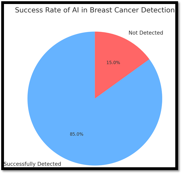
Figure 3:- Success Rate Of Artificial Intelligence In Breast Cancer Detection
Future Trends in AI for Breast Cancer Detection
Several emerging trends are shaping the future of AI in breast cancer detection, each contributing to the evolution of more precise, efficient, and accessible diagnostic tools. One of the most promising developments is multimodal AI, which integrates information from various imaging modalities (such as mammography, MRI, and ultrasound) alongside clinical data to provide a more comprehensive and accurate assessment of breast cancer. This approach has the potential to improve diagnostic accuracy by combining different data sources, allowing for a holistic view of the patient’s condition [23]. Another key trend is federated learning, a distributed learning approach that enables AI models to be trained on decentralized data across multiple locations. This method addresses privacy concerns by ensuring that sensitive patient data never leaves its local site, making it possible to collaborate on a larger scale while maintaining data security. In parallel, explainable AI (XAI) is advancing rapidly, focusing on enhancing transparency in AI decision-making processes. As AI tools become more integrated into clinical workflows, it is crucial for clinicians and patients to understand how AI arrives at its conclusions. This fosters trust and facilitates better collaboration between AI systems and healthcare professionals [24].
AI-powered personalized screening is another exciting area, with AI models being developed to tailor screening protocols to individual patients. By assessing specific risk factors such as genetics, family history, and lifestyle, these AI systems can customize screening frequencies and methods for each patient, improving early detection and potentially saving lives. Furthermore, edge computing is transforming the landscape by enabling AI algorithms to run directly on devices (such as smartphones or portable imaging equipment). This allows for real-time analysis at the point of care, reducing reliance on cloud-based infrastructure and making the technology more accessible in remote or underserved areas. In addition to these trends, the integration of AI with wearable devices and mobile health applications is poised to further extend the reach of AI-powered solutions. These technologies enable continuous monitoring of health parameters, facilitating early intervention and personalized care. The potential of AI to be deployed in low-resource settings could help bridge healthcare disparities by providing affordable, accurate diagnostic tools to underserved populations, improving access to care globally [25].
Current Perspective on Breast Cancer Detection by AI
The current perspective on AI in breast cancer detection is one of cautious optimism, with a growing body of research supporting its potential to enhance both the accuracy and efficiency of breast cancer screening and diagnosis. AI has already shown promising results in tasks like mammography interpretation, where its performance can rival or even surpass that of experienced radiologists in some instances [26]. Despite these advances, AI is still in a transitional phase, and for now, it primarily serves as an assistive tool rather than a fully autonomous diagnostic system. Radiologists and other healthcare professionals continue to play a central role in the decision-making process, with AI working to augment their capabilities rather than replace them [27]. There is an increasing awareness of the need for thorough validation of AI systems across diverse clinical settings and patient populations. As AI technology continues to develop rapidly, there are ongoing efforts to address challenges such as data quality, algorithmic bias, and seamless integration into clinical workflows. At the same time, regulatory bodies are adapting to this rapid pace, establishing frameworks for evaluating and approving AI-based medical devices, ensuring they meet necessary safety and efficacy standards [28]. Collaboration between clinicians, AI researchers, and developers is becoming ever more crucial to ensure that AI tools remain clinically relevant, user-friendly, and aligned with the real-world needs of healthcare. Equally important is the attention being given to the ethical implications of AI in healthcare, including concerns about transparency, accountability, and patient privacy [29]. AI's transformative potential is already evident, as it continues to enhance diagnostic accuracy and efficiency. The focus is now on improving model reliability through better training techniques, expanding data access via collaborative efforts, and addressing ethical concerns to ensure that AI is used responsibly. The increasing acceptance of AI by healthcare professionals and patients alike reflects it’s potential to significantly impact breast cancer detection and care [30].
CONCLUSION
Artificial intelligence (AI) has become a transformative force in the detection and diagnosis of breast cancer, offering remarkable potential to enhance the accuracy, efficiency, and consistency of screening processes. The current landscape of AI in breast cancer detection is marked by rapid technological advancements, promising research outcomes, and increasing clinical interest. Through the use of machine learning and deep learning algorithms, AI has shown promise in improving lesion detection, classification, and risk assessment across various imaging modalities, including mammography, ultrasound, MRI, and histopathology. These AI-driven tools have the potential to support healthcare professionals by reducing workload, minimizing interpretation variability, and improving patient outcomes.
However, while the applications of AI in breast cancer detection are vast, several challenges remain that must be addressed for its optimal implementation. Issues such as ensuring high-quality and diverse datasets for training AI models, improving model interpretability, overcoming generalizability concerns, and navigating complex regulatory frameworks all need attention. Ethical considerations, including data privacy and algorithmic bias, are also central to the adoption of AI in clinical practice, requiring careful thought as these systems become more integrated into healthcare. Looking ahead, several emerging trends are expected to shape the future of AI in breast cancer detection. Multimodal AI systems, federated learning, explainable AI, and personalized screening protocols are set to further refine the capabilities of AI, offering more comprehensive and individualized approaches to detection. The integration of edge computing and real-time analysis will also play a significant role in enhancing the speed and accessibility of AI-powered diagnostics. For AI to fully realize its potential, collaboration between healthcare professionals, AI developers, researchers, and regulatory bodies is essential. This includes continued research, large-scale clinical validation studies, and ongoing education and training for medical personnel. AI should be viewed as a complementary tool, not a replacement for human expertise. Ultimately, AI has the capacity to enhance early detection, reduce diagnostic errors, and improve patient outcomes in breast cancer care. As the field progresses, its continued evaluation, refinement, and ethical implementation will be key to maximizing its impact on global healthcare.
REFERENCES


Aanchal Dahiya*, Dr. Nakul Gupta, Dr. Md. Sarfaraz Alam, Kamal Singh Bani, Preeti Bhunia, Aditi Singh, Showing Artificial Intelligence in The Sensing of Breast Cancer, Int. J. of Pharm. Sci., 2025, Vol 3, Issue 5, 4398-4408. https://doi.org/10.5281/zenodo.15528154
 10.5281/zenodo.15528154
