Department of Pharmaceutics, Paavai College of Pharmacy and Research, Namakkal-637018, Tamil Nadu, India
Pulsatile drug delivery systems (PDDS) represent a paradigm shift in pharmaceutical dosage form design by enabling programmed drug release after a predetermined lag time followed by rapid and complete drug release. Unlike conventional immediate or sustained-release formulations, PDDS are specifically designed to synchronise drug delivery with the body's circadian rhythms and disease chronobiology patterns. The present review comprehensively discusses the rationale for pulsatile delivery, classification of PDDS, mechanisms of drug release, formulation technologies, and clinical applications in chronotherapeutic management of diseases such as asthma, hypertension, rheumatoid arthritis, peptic ulcer disease, and diabetes mellitus. Various technological approaches including time-controlled systems (capsular, osmotic, and compression-coated systems), stimuli-responsive systems (temperature, pH, and glucose-sensitive), and externally regulated systems (magnetic, ultrasonic, and electrical) are reviewed. Evaluation parameters for PDDS including pre-formulation studies, physicochemical characterisation, and in vitro/in vivo dissolution testing are discussed. The manuscript highlights the advantages of PDDS in improving therapeutic efficacy, reducing side effects, minimising biological tolerance, and enhancing patient compliance while addressing manufacturing challenges and cost considerations. Recent marketed formulations (Pulsincap™, CODAS®, Geoclock®, OROS®, IPDAS®) demonstrate the clinical feasibility and therapeutic potential of pulsatile release technologies. This review concludes that PDDS represent a promising and effective drug delivery strategy with significant potential for future development in precision medicine and chronotherapy.
1.1 Overview of Drug Delivery Systems
Conventional drug delivery approaches immediate release (IR) and sustained-release (SR) formulations deliver medications using constant-rate kinetics that do not account for temporal variations in disease pathology or patient circadian biology [1]. This one-size-fits-all approach often results in suboptimal therapeutic outcomes, unnecessary side effects, and reduced patient compliance, particularly for chronic diseases exhibiting time-dependent symptoms [2].
1.2 Concept of Pulsatile Drug Delivery
Pulsatile drug delivery systems (PDDS) are innovative pharmaceutical dosage forms engineered to release drugs in a programmed manner after a predetermined lag time, followed by rapid and near-complete drug release. The fundamental principle of PDDS is to deliver "the right drug at the right site, at the right time, and in the right amount," thereby providing both spatial and temporal control over drug delivery [3]. This approach enables synchronisation of drug availability with the body's circadian rhythms and disease-specific activity patterns.
1.3 Chronobiology and Chronopharmacology
Chronobiology is the study of biological rhythms and their underlying mechanisms. The human body exhibits three major types of biological rhythms: circadian (approximately 24-hour cycles), ultradian (cycles shorter than 24 hours), and infradian (cycles longer than 24 hours) [4]. The circadian rhythm is regulated by the suprachiasmatic nucleus (SCN) in the hypothalamus and is influenced by environmental factors such as light, temperature, and food intake, as well as endogenous factors including hormonal fluctuations (cortisol, melatonin, renin, and aldosterone).
Chronopharmacology is the application of chronobiological principles to pharmaceutical therapy, recognising that drug efficacy, toxicity, and pharmacokinetics exhibit time-dependent variations. Many chronic diseases show pronounced circadian variation in symptom severity, pathophysiological markers, and therapeutic drug requirements [5]. For example, acid secretion in the stomach peaks during late evening and early morning hours, precipitating nocturnal heartburn and peptic ulcer disease. Similarly, blood pressure follows a circadian pattern with significantly higher cardiovascular risk during early morning hours.
1.4 Rationale for Pulsatile Drug Delivery
The impetus for developing PDDS stems from several clinical observations:
2. CIRCADIAN VARIATION IN DISEASE PATHOPHYSIOLOGY
Multiple chronic diseases exhibit well-documented circadian variation in symptom severity and pathophysiological markers [7]. Understanding these temporal patterns provides the scientific foundation for chronotherapeutic interventions:
Asthma and Allergic Rhinitis: Airway function shows pronounced circadian variation, with peak airway obstruction and increased diurnal resistance occurring during late night and early morning hours (3:00-5:00 AM) [8]. Nocturnal asthma symptoms affect approximately 60-75% of asthmatic patients, contributing to sleep disturbance and poor quality of life.
Cardiovascular Disease and Myocardial Infarction: Acute myocardial infarction occurrence shows marked circadian periodicity, with 35% of events clustered between 6:00 AM and noon [9]. Early morning hours show increased incidence of acute cardiac ischemia, arrhythmias, and sudden cardiac death, attributed to increased sympathetic tone, elevated cortisol and catecholamine levels, and enhanced platelet aggregation.
Rheumatoid Arthritis: Morning stiffness represents one of the diagnostic criteria of rheumatoid arthritis (RA), with severity typically worst during early morning hours[10]. This temporal pattern reflects circadian variation in inflammatory cytokine production (TNF-α, IL-6) and the anti-inflammatory cortisol rhythm, resulting in maximum inflammation when cortisol levels are lowest.
Peptic Ulcer Disease and GERD: Gastric acid secretion shows pronounced circadian rhythmicity with maximum secretion during late evening and early morning hours (10:00 PM-4:00 AM) [11]. Nocturnal acid surge directly correlates with symptom severity, increased ulcer perforation risk, and reduced treatment efficacy of evening-dosed medications.
Diabetes Mellitus: Blood glucose levels follow a circadian pattern with peak levels in early morning (dawn phenomenon) and lowest levels during afternoon hours [12]. Similarly, endogenous insulin secretion exhibits clear circadian variation, with implications for insulin dosing schedules and optimal therapy timing.
Hypertension: Blood pressure follows a characteristic circadian pattern, with lowest values during sleep and peak values during early morning awakening [13]. This circadian blood pressure variation, termed "non-dipping" in hypertensive patients, significantly increases cardiovascular risk.
2.2 Molecular Mechanisms of Circadian Disease Variation
The circadian variation in disease pathophysiology operates through multiple molecular mechanisms:
3. CLASSIFICATION AND MECHANISMS OF PULSATILE DRUG DELIVERY SYSTEMS
3.1 Time-Controlled Pulsatile Release Systems
3.1.1 Single-Unit Capsular Systems
Single-unit systems consist of an insoluble capsule body filled with drug and a swell-able/erodible plug that blocks the open end. Upon contact with gastrointestinal fluids, the plug swells, erodes, or dissolves, pushing itself out of the capsule after a predetermined lag time followed by spontaneous drug release.
The Pulsincap™ system (developed by R.P. Scherer International Corporation) exemplifies this approach, utilizing a hydrogel plug covered with a water-soluble cap. The entire unit is coated with enteric polymer to overcome variable gastric emptying. Lag time is controlled by manipulating plug dimensions, composition, and position [14]. Advantages include simple design and cost-effectiveness; disadvantages include potential variable gastric residence time and dose dumping risks.
3.1.2 Osmotically Driven Capsular Systems
Osmotically driven PDDS utilise osmotic pressure gradients to achieve pulsatile release. The PORT system (Therapeutic Strategies Research Laboratory) consists of a semipermeable capsule containing a drug formulation and osmotically active agent. Water penetration through the semipermeable membrane generates osmotic pressure, eventually rupturing an insoluble plug and releasing the drug [15].
3.1.3 Compression-Coated Pulsatile Systems
Compression-coated tablets consist of a rapidly disintegrating drug-containing core surrounded by a barrier layer of erodible/hydrophilic polymers. The barrier layer provides the lag time through controlled erosion, followed by rapid core disintegration and drug release. Common polymers include HPMC (hydroxypropyl methyl-cellulose) K100M, ethyl cellulose, and xanthin gum. This approach offers several advantages:
The barrier layer thickness, polymer viscosity grade, and polymer composition directly influence lag time duration [16]. HPMC K100M viscosity-based systems show excellent reproducibility with in vitro-in vivo correlation.
3.2 Stimuli-Responsive Pulsatile Release Systems
3.2.1 Temperature-Responsive Systems
Thermosensitive hydrogels undergo reversible volume changes in response to temperature fluctuations. Poly(N-isopropylacrylamide) (PNIPAAm) cross-linked gels demonstrate thermoresponsive properties, exhibiting swelling below 32°C and shrinking above this temperature [17]. Although temperature gradients in the body are relatively modest (±2°C variation), these systems show potential for localised applications.
3.2.2 pH-Responsive Systems
pH-sensitive polymers (Eudragit L100, Eudragit S100) undergo swelling or dissolution in response to pH changes across different GIT regions. This enables targeted delivery to specific intestinal segments (duodenum, ileum, colon) where pH conditions favor polymer ionisation and swelling [18].
3.2.3 Glucose-Responsive Systems
Glucose-responsive insulin delivery devices represent a significant advancement for diabetes management. These systems incorporate glucose oxidase immobilized within pH-sensitive hydrogels. When blood glucose rises, glucose oxidase catalyses glucose oxidation to gluconic acid, reducing local pH and triggering hydrogel swelling and insulin release. As blood glucose normalises, the system re-equilibrates [19]. This feedback mechanism enables semi-automatic insulin delivery without external monitoring.
3.2.4 Inflammation-Responsive Systems
Inflammation-triggered release systems exploit pathology-specific molecular markers present at inflammatory sites. Hyaluronic acid (HA)-based gels are selectively degraded by hyaluronidase (present at inflammatory sites) or free hydroxyl radicals generated during inflammation [20]. This allows anti-inflammatory drugs incorporated in HA gels to be released specifically at inflammatory foci, with minimal release in healthy tissue.
3.3 Externally Regulated Pulsatile Release Systems
3.3.1 Magnetic Field-Regulated Systems
Magnetically responsive systems incorporate ferromagnetic materials (magnetite, iron particles) into drug carriers. External magnetic fields can modulate drug release kinetics and influence GIT transit time by selective retardation at specific anatomical sites [21]. This approach enables precise spatial and temporal control but requires external magnetic field apparatus.
3.3.2 Ultrasound-Triggered Systems
Ultrasound stimulation can modulate drug release from polymeric matrices through cavitation-induced phenomena, polymer degradation, and pore generation. Studies demonstrate up to 27-fold enhancement in drug release rate from ethylene-vinyl acetate copolymers with controlled ultrasound application [22].
3.3.3 Electroresponsive Systems
Electrically stimulated systems utilise polyelectrolyte hydrogels that undergo reversible swelling/deswelling in response to applied electric fields. The electronic stimulus induces gel contraction, squeezing out encapsulated drug molecules into surrounding fluids [23]. These systems enable precise temporal control through electronic pulse programming.
4. MARKETED PULSATILE DRUG DELIVERY TECHNOLOGIES
Several marketed products exemplify successful implementation of PDDS technology:
Pulsincap™: Hydrogel plug-based system for metronidazole delivery in anthelminthic therapy, utilizing controlled plug swelling to achieve approximately 4-hour lag time [24].
CODAS® (Chronotherapeutic Oral Drug Absorption System): Pellet-filled capsule system designed for bedtime dosing with 4-5 hour lag. Used for verapamil HCl (Verelan PM) in hypertension management [24]. The non-enteric release-controlling polymer combination (water-soluble and water-insoluble polymers) provides excellent GIT location independence.
Geoclock®: Press-coated tablet system with pH-independent lag time using hydrophobic wax and brittle material mixture. Lodotra™ (prednisone for rheumatoid arthritis) released through this technology delivers peak prednisone levels at 4:00 AM, coinciding with maximum inflammatory activity [25].
OROS® (Osmotic Pump): Semipermeable membrane-based osmotic system suitable for poorly water-soluble drugs. Bilayer or trilayer tablet core with one push layer and one or more drug layers. L-OROS™ variant incorporates self-emulsifying liquid formulations for enhanced bioavailability [26].
IPDAS® (Intestinal Protective Drug Absorption System): Multi-particulate controlled-release bead system appropriate for GI-irritant drugs like NSAIDs. Naprelan® (naproxen) formulated through IPDAS achieves pain relief within 30 minutes with 24-hour duration while reducing gastric irritancy [27].
TIMERx®: Lipid-based technology for oxymorphone delivery (Opana ER) providing controlled absorption and 12–24-hour pulsatile release [28].
5. DRUG RELEASE MECHANISMS FROM PULSATILE SYSTEMS
Drug release from PDDS occurs through three primary mechanisms operating individually or in combination:
Water enters the dosage form, particles interact with gastrointestinal fluids, and drug molecules diffuse across the polymeric barrier layer. The diffusion rate is controlled by polymer thickness, hydrophilicity, and drug lipophilicity.
Hydrophilic and erodible polymer coatings gradually dissolve/erode over time, progressively exposing the drug core. Erosion kinetics depend on polymer chemical composition, molecular weight, and crystallinity.
5.3 Osmotic Pressure-Driven Release
Water penetration into the dosage form interior generates osmotic pressure that compresses the drug core, forcing drug-loaded solution/suspension through a small orifice or rupture point in the coating [29].
6. EVALUATION PARAMETERS FOR PULSATILE DRUG DELIVERY SYSTEMS
6.1 Pre-formulation Studies
Pre-formulation investigations characterise physicochemical properties of the drug and excipients:
6.2 Tablet Characterisation
Compressed tablets are evaluated for:
Lag time represents the time interval between tablet ingestion and onset of drug release. In vitro lag time is determined using USP Dissolution Apparatus II at 50-100 rpm in:
The predetermined lag time should demonstrate <15-20% relative standard deviation between replicates to ensure reproducibility [30].
6.4 In Vitro Dissolution Studies
Dissolution testing employs standard USP methods with sampling at predetermined intervals (0.5, 1, 2, 3, 4, 5, 6, 8, 12 hours). Samples are filtered through 0.45 µm membrane filters and analysed using validated UV spectrophotometric methods at wavelength maxima.
Similarity factor (f2) from SUPAC guidelines determines bioequivalence of dissolution profiles:
f2=50log?100×1+1nt=1n(Rt-Tt)2-0.5
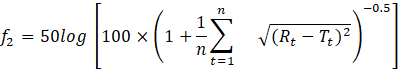
where Rt

and Tt

represent reference and test dissolution values at time t. Profiles are considered similar when f2 ranges between 50 and 100[31].
6.5 Dissolution Kinetics Modelling
Dissolution data are fitted to standard models (zero-order, first-order, Higuchi, Hixon-Crowell, Korsmeyer-Peppas) to determine drug release kinetics and mechanism of release.
6.6 In Vivo Bioavailability Studies
In vivo studies evaluate pharmacokinetic parameters:
These parameters are compared between optimised pulsatile formulation and marketed reference formulation using appropriate bioanalytical methods.
7. ADVANTAGES AND LIMITATIONS OF PULSATILE DRUG DELIVERY SYSTEMS
Therapeutic Efficacy: PDDS synchronise drug availability with disease chronobiology, maximising therapeutic benefit during periods of greatest pathophysiological activity.
Side Effect Reduction: By delivering drugs during specific time windows, cumulative toxicity and adverse effects are minimised compared to continuous-exposure formulations.
Biological Tolerance Prevention: Pulsatile delivery with intervening lag periods prevents tachyphylaxis and maintains long-term drug efficacy, particularly important for beta-2 agonists and corticosteroids.
Reduced First-Pass Metabolism: Delivery to distal GIT locations bypasses or reduces hepatic first-pass metabolism, improving bioavailability of extensively metabolised drugs.
Improved Patient Compliance: Single bedtime dose with programmed morning release improves compliance compared to multiple daily dosing schedules.
Site-Specific Delivery: Colonic-targeted delivery benefits patients with ulcerative colitis, irritable bowel syndrome, and Crohn's disease.
Protection from Gastric Environment: Enteric-coated PDDS protect acid-labile drugs (peptides, antibiotics) from gastric degradation.
7.2 Limitations and Challenges
Manufacturing Complexity: Multiple processing steps, especially for multi-particulate systems, increase production complexity and cost.
Variable GIT Transit Time: Individual variations in gastric emptying and intestinal transit can result in variable lag times and inconsistent drug release patterns.
Drug Loading Limitations: Some PDDS approaches (multi-particulate systems) require substantial excipient quantities, limiting drug loading capacity to <50% w/w.
Incomplete Drug Release: Some formulations show incomplete drug release (85-95%) rather than true pulse release, reducing therapeutic predictability.
Manufacturing Scale-Up Challenges: Compression-coating techniques and osmotic systems require specialised equipment for commercial scale-up.
Cost Implications: More expensive manufacturing processes and sophisticated equipment increase formulation costs 2-3-fold compared to conventional formulations.
Regulatory Complexity: PDDS require demonstration of bioequivalence, mechanistic validation of lag time, and in vivo proof of concept, increasing regulatory requirements.
Paediatric Dose Adjustment Limitations: Fixed-dose pulsatile systems cannot be easily modified for paediatric or elderly patients requiring dose adjustments.
8. CLINICAL APPLICATIONS AND THERAPEUTIC BENEFITS
The chronotherapeutic applications of PDDS extend across multiple disease states:
Cardiovascular Disease Management: Chronotherapeutic delivery of antihypertensive agents (verapamil, diltiazem) during early morning hours addresses the circadian blood pressure surge and early morning cardiovascular event peak.
Respiratory Disease Management: Bedtime delivery of beta-2 agonists and corticosteroids with morning peak release alleviates nocturnal asthma symptoms occurring between 3:00-5:00 AM.
Rheumatoid Arthritis: Prednisone delivery via Geoclock® technology achieving peak levels at 4:00 AM coincides with maximum inflammatory activity and morning stiffness severity, improving therapeutic efficacy while reducing daily corticosteroid requirements.
Gastroenterological Disease Management: Chronotherapeutic acid suppression with proton pump inhibitors or H?-receptor antagonists during nocturnal acid surge periods provides superior ulcer healing and symptom control compared to conventional dosing.
Diabetes Management: Glucose-responsive insulin delivery systems represent a paradigm shift toward "artificial pancreas" technology, enabling semi-automatic insulin administration based on real-time glycemic status [19].
Pain Management: Chronotherapeutic analgesic delivery (oxymorphone via Opana ER) addresses time-dependent pain symptom patterns in chronic pain conditions.
9. FUTURE PERSPECTIVES AND EMERGING TECHNOLOGIES
9.1 Advanced Formulation Approaches
Nanotechnology Integration: Incorporation of nanoparticles (liposomes, niosomes, polymeric nanoparticles) into PDDS enables enhanced permeability, targeted delivery, and reduced systemic exposure.
3D Printing Technology: Three-dimensional printing enables precise fabrication of pulsatile devices with customisable geometry, internal architecture, and drug-loaded compartments, facilitating personalised medicine approaches.
Combination Drug Delivery: Multi-API pulsatile systems enable coordinated delivery of two or more drugs with different pharmacological targets but complementary chronobiological requirements.
9.2 Bioresponsive and Smart Systems
Microbiota-Responsive Systems: Leveraging colonic microbiota-derived enzymes for colonic-specific drug delivery and pulsatile release without exogenous lag-time materials.
Dual-Responsive Hybrid Systems: Integration of multiple stimuli (pH + temperature, glucose + pH) for enhanced specificity and reduced off-target release.
Injectable Pulsatile Systems: Development of subcutaneously or intramuscularly injectable pulsatile delivery systems for long-acting formulations.
9.3 Personalised Chronotherapy
Circadian Phenotyping: Individual assessment of circadian rhythmicity parameters enables personalised chronotherapy timing optimisation based on patient-specific circadian characteristics.
Genetic Polymorphism Consideration: Pharmacogenetics variations in clock genes (CLOCK, BMAL1) influence optimal drug delivery timing; future PDDS may incorporate genetic profiling for personalised dosing schedules.
Digital Health Integration: Wearable biosensors and artificial intelligence enable real-time circadian rhythm monitoring and dynamic dosing schedule optimisation.
Pulsatile drug delivery systems represent a sophisticated advancement in pharmaceutical technology, offering distinct advantages over conventional immediate-release and sustained-release formulations by enabling chronotherapeutic drug delivery synchronised with the body's biological rhythms and disease-specific temporal patterns. The scientific rationale for PDDS is robust, grounded in established chronobiological principles demonstrating time-dependent variations in disease pathophysiology and drug pharmacokinetics. Multiple technological approaches time-controlled (capsular, osmotic, compression-coated), stimuli-responsive (temperature, pH, glucose-sensitive), and externally regulated (magnetic, ultrasonic, electrical) systems provide flexible platforms for achieving pulsatile release patterns tailored to specific therapeutic requirements.
The clinical efficacy of marketed PDDS formulations (Pulsincap™, CODAS®, Geoclock®, OROS®, IPDAS®, TIMERx®) demonstrates the feasibility of implementing chronotherapeutic drug delivery in clinical practice, with documented therapeutic benefits including improved disease control, reduced side effects, minimised biological tolerance, and enhanced patient compliance across diverse therapeutic domains (cardiovascular, respiratory, rheumatologic, gastrointestinal, and endocrine diseases).
While current PDDS technologies face challenges including manufacturing complexity, variable GIT transit times, incomplete drug release in some systems, and higher production costs, ongoing technological innovations nanotechnology integration, 3D printing, microbiota-responsive systems, and digital health integration promise to overcome existing limitations and enable next-generation chronotherapeutic platforms. The convergence of chronobiology, pharmaceutical technology, and precision medicine creates unprecedented opportunities for personalised chronotherapy approaches optimised for individual patient circadian phenotypes and genetic profiles.
In conclusion, pulsatile drug delivery systems represent a mature and clinically validated technology with significant therapeutic potential for chronopharmacotherapy. Future development in PDDS, particularly incorporating artificial intelligence, digital health monitoring, and personalised medicine paradigms, promises to establish chronotherapy as a cornerstone of precision medicine approaches for diseases exhibiting time-dependent pathophysiology. The field remains dynamic and innovative, with substantial potential to improve clinical outcomes and patient quality of life across diverse chronic disease populations.


Anju A. V., Ellappan D., Priya S., Yogeshwaran M. D., Vignesh G., Ramkumar B., Pulsatile Drug Delivery Systems: A Comprehensive Review of Chronotherapeutic Technologies and Clinical Applications, Int. J. of Pharm. Sci., 2026, Vol 4, Issue 3, 2590-2600. https://doi.org/10.5281/zenodo.19182132
 10.5281/zenodo.19182132
