B. pharmacy, shri. Sambhaji college of pharmacy khadkut nanded.
Made of the same substance as a cell membrane, a liposome is a tiny bubble, or vesicle. Drugs for cancer and other illnesses can be delivered using liposomes that have been loaded with medication. At the Babraham Institute in Cambridge, British haematologist Dr. Alec D. Bangham FRS initially described liposomes in 1961 (published 1964). They were found when Bangham and R. W. Horne added negative stain to dry phospholipids in order to test the institute's new electron microscope. The microscope images provided the first concrete proof that the cell membrane is a bilayer lipid structure, and the likeness to the plasmalemma was evident. The Greek terms "Lipos," which means fat, and "Soma," which means body, are the origin of the term "liposome."(7) This bibliographic study provides a broad overview of the history and evolution of liposomes, emphasizing both traditional (ethanol injection, reverse-phase evaporation, thin film hydration) and innovative scalable methods of manufacture. Liposomes' use in the food, cosmetics, and pharmaceutical industries as prospective new discoveries and products were also discussed, as well as the present growth and expansion of interest in them across numerous scientific fields. The effectiveness of liposomes in a variety of applications is finally discussed, with opinions ranging from unjustified pessimism to unsupported optimism. (1).
Lipid molecules, including phospholipids, scattered in an aqueous media, produce the enclosed vesicles known as liposomes. The inner and outer water phases are separated by one or more bilayers that resemble the cell membrane in structure. (7) There are several different liposome preparation techniques accessible today, and each one has unique benefits and characteristics. New liposomal formulations can be made quickly and simply if the right approaches are chosen and matching new procedures are applied. As a result of current developments, an increasing number of innovative technologies have been used to create the amazing vesicles in recent years. However, the vast array of new technologies might occasionally present challenges for liposome researchers. To assist in choosing appropriate preparation techniques to satisfy various objectives, we have created a methodical overview of the latest technology. Liposomes have grown in popularity as an experimental and commercial drug delivery system due to their biocompatibility, biodegradability, low toxicity, and ability to trap hydrophilic and lipophilic drugs [9] and streamline site-specific drug delivery to tumor tissues. Liposomes have been the subject of numerous investigations aimed at reducing medication toxicity and/or focusing on particular cells. (4)
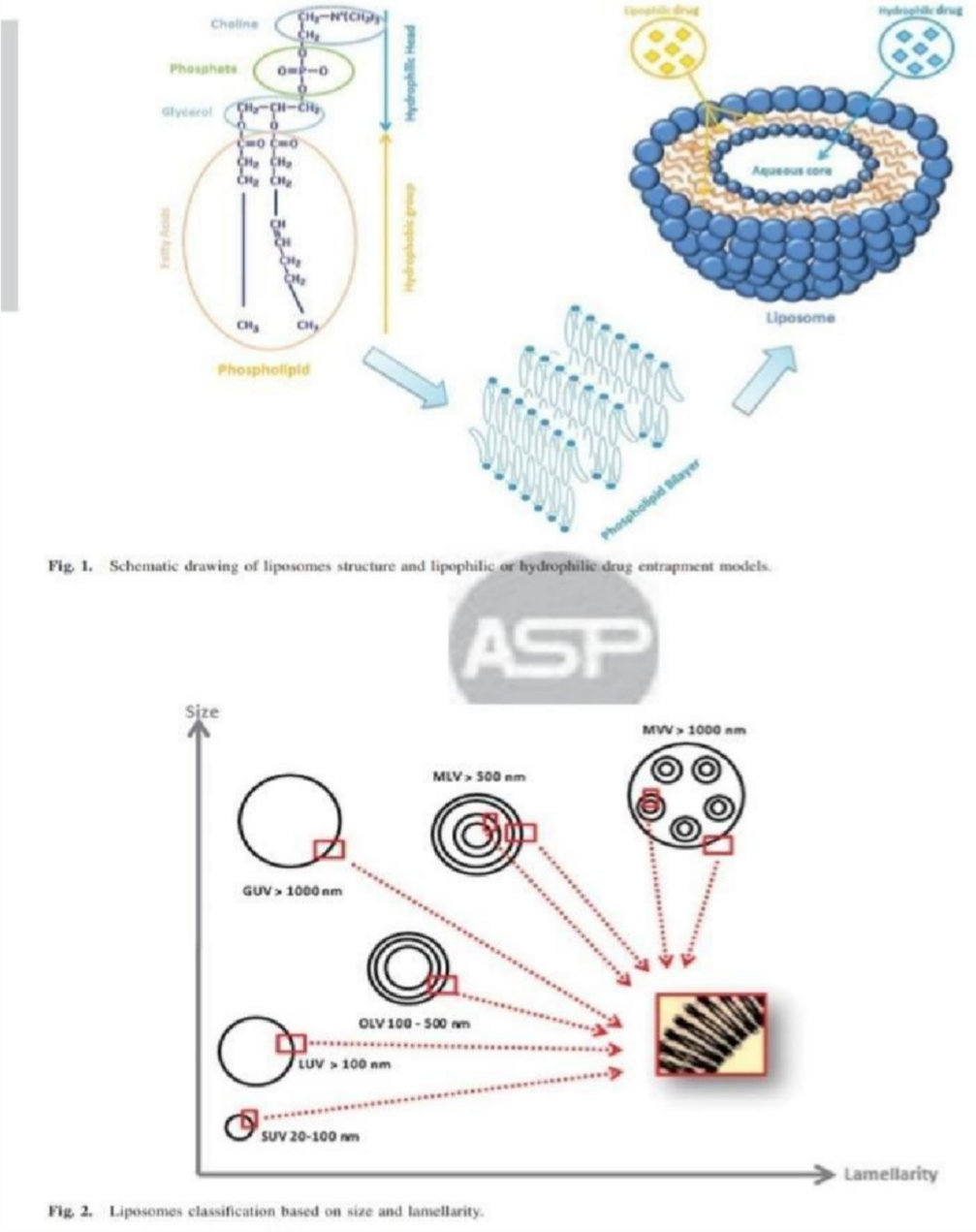
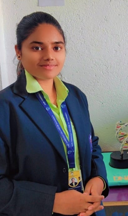
Liposomes are most frequently classified on the basis of their size (small, large and giant vesicles), number of bilayers (uni-, oligo- and multi-lamellar) (Vemuri and Rhodes, 1995, Vuillemard, 1991) and phospholipid charge (neutral, anionic or cationic) (Storm and Crommelin, 1998). Recently, liposomes have also been categorized with respect to their function such as conventional, stealth, ligand-targeted, long-release, and triggered-release (Sharma and Sharma, 1997, Storm and Crommelin, 1998). Multi-functional liposomes possessing a combination of these features have also been reported (Kale and Torchilin, 2010, Perche and Torchilin, 2013, Xiang et al., 2013). Even though liposomes were first reported close to half a century ago (Bangham et al., 1965), it was three decades after their discovery that the first liposomal drug Ambisome® entered the market (Davidson et al., 1994, Hann and Prentice, 2001). Since then, the list of liposomal drugs has continued to increase (Allen and Cullis, 2013). (22)
Literature Review:
liposomes have been categorized with respect to their function such as conventional, stealth, ligand-targeted, long-release, and triggered-release.
The preparation methods have been classified based on mean size, polydispersity and lamellarity of liposomes obtained, because control over these parameters remains a challenge with almost all preparation methods. This problem is exacerbated when moving from the laboratory to industrial scale.
Reduction of size and lamellarity of liposomes is typically carried out by subjecting them to homogenization, sonication, extrusion or freeze–thaw cycles.
Exclusively scalable techniques and focuses on strengths, respectively, limitations in respect to industrial applicability and regulatory requirements concerning liposomal drug formulations based on FDA and EMEA documents.
Liposomes were first described by British haematologist Dr Alec D Bangham FRS in 1961 (published 1964), at the Babraham Institute, in Cambridge. They were discovered when Bangham and R. W. Horne were testing the institute's new electron microscope by adding negative stain to dry phospholipids. The resemblance to the plasmalemma was obvious, and the microscope pictures served as the first real evidence for the cell membrane being a bilayer lipid structure
Liposomes are spherical vesicles having an aqueous core enclosed by one or more phospholipid bilayers or lamellae. Liposomes are most frequently classified on the basis of their size (small, large and giant vesicles), number of bilayers (uni-, oligo- and multi- lamellar)
Aim:
Aim: To conduct a comprehensive review of the preparation, and application of liposomes.
Objective:
Definition:
Liposomes were defined as an artificial microscopic vesicle consisting of a central aqueous compartment sur- rounded by one or more concentric phospholipid layers (lamellas). Furthermore, hydrophilic (in the aqueous cavity), hydrophobic (within lipidic membrane) and amphiphilic substances are able to be incorporated within these vesicles developing large potential applications. Numerous researchers have worked with these structures since Bangham’s discovery, making of liposomes the most popular nanocarrier systems (5)
Classification:
Five types of liposomes can be distinguished based on their composition and intracellular delivery method.
The amount of drug encapsulation in the liposomes is influenced by both the size and number of bilayers, and the vesicle size is a critical factor in determining the circulation half-life of liposomes. The following categories can also be used to group liposomes according on their size and number of bilayers: liposomes
can also be classified into following types-
Preparation Method
Classical Technique
There are four classical methods of liposome manufacture. The difference between the various methods is the way in which lipids are drying down from organic solvents and then redispersed in aqueous media. These steps are per- formed individually or are mostly combined
Bangham Method. This was the first technique that was applied to the manufacturing of liposomes. A blend of cholesterol and phospholipids was dissolved in an organic solvent. Next, evaporation—using a Rotar Evaporator at a lower pressure was used to evaporate the organic solvent. Lastly, an aqueous buffer solution was added to the dry lipidic layer that had formed on the flask wall while being stirred at a temperature higher than the lipid transition temperature. Although this technique is widely used and simple to use, the population of multilamellar liposomes (MLVs) produced by distributed phospholipids in aqueous buffer is diverse in terms of size and shape (1–5 m diameter). Therefore, liposome size reduction methods such extrusion through polycarbonate filters or sonication for SUV formation. In order to create a smaller and more uniformly sized population of vesicles, liposome size reduction procedures such sonication for SUV formation or extrusion through polycarbonate filters generating LUVs25–26 were helpful. (1)
A two-phase solution including phospholipids in an organic solvent (diethylether, isopropylether, or a combination of isopropyl ether and chloroform) and an aqueous buffer is briefly sonicated to create the first water-in-oil emulsion. A thick gel is created as a result of the organic solvents being eliminated at lower pressure. When the remaining solvent is eliminated by continuous rotating evaporation at lowered pressure, liposomes are created. This technique can achieve a high encapsulation effectiveness of up to 65% in a medium with a low ionic strength, such as 0.01M NaCl. Both large and tiny macromolecules have been encapsulated using this technique. The method's primary drawback is that the materials to be encapsulated are exposed to organic solvents and brief sonication periods. (9)
The solvent injection methods involve the dissolution of the lipid into an organic phase (ethanol or ether), followed by the injection of the lipid solution into aqueous media, forming liposomes (10)
An aqueous solution of the material to be encapsulated is slowly injected with a solution of lipids dissolved in diethyl ether or an ether-methanol mixture at 55°C to 65°C or with decreased pressure. Liposomes are produced as ether is then removed under vacuum. The technique's primary drawbacks are that the population is heterogeneous (ranging from 70 to 200 nm) and that the chemicals to be encapsulated are exposed to organic solvents at high temperatures. (11,12)
A large amount of buffer is quickly mixed with a lipid solution of ethanol. The MLVs are created instantly. The method's drawbacks include the population's heterogeneity (30–110 nm), the dilution of liposomes, the difficulty of removing all ethanol due to its formation of an azeotrope with water, and the high likelihood that the different physiologically active macromolecules will inactivate even in the presence of trace amounts of ethanol. (13)
Liposomes are defined mixed micelles that range in size from 40 to 180 nm and are created when lipids are dissolved with detergent. Following the removal of the detergent through controlled dialysis, phospholipids form uniform unilamellar vesicles with a sizable enclosed capacity. Other procedures, including calcium-induced fusion, nanoprecipitation, and emulsion processes, have already been employed to create liposomes. These traditional methods, however, call for significant amounts of organic solvent, which are bad for the environment and human health and necessitate the total elimination of any remaining organic solvent. Additionally, conventional procedures need a lot of energy and include numerous processes for size homogenization, making them unsuitable for mass production of liposomes. (1)
New Large-Scale Liposome Technique
Since industrial scale production of liposomes has become reality, the range of liposome preparation methods has been extended by a number of techniques such as Heat- ing Method, Spray drying, Freeze Drying, Super Critical Reverse Phase Evaporation (SCRPE), and several modified ethanol injection techniques which are increasingly attractive.
Mozafari (2005) suggested the heating approach, which produced liposomes on a huge scale without the use of any dangerous chemicals or procedures. This process involves heating the lipid mixture to 120 degrees Celsius after hydrating it with an aqueous phase that contains 3% (v/v) glycerol. By stopping vesicles from coagulating and sedimenting, glycerol can improve the stability of liposomes. Furthermore, because it is a water-soluble, nontoxic, and biocompatible chemical, it does not need to be separated from the final liposomal product. Thin layer chromatography shows that at the aforementioned temperature, there is no lipid breakdown. There are three ways to load bioactive materials into the liposomes during this process: (1) initially, in combination with glycerol and liposomal ingredients; (2) when the operating temperature is higher than the lipids transition temperature (Tg); and (3) at room temperature following the formation of the vesicles. Mortazavi et al. (2007) used calcium ions to successfully insert plasmid DNA (pCMV-GFP) with a high EE (81%) in their study. (14)
This method has been regarded as a quick, one-step process for producing liposomes on a wide scale (Kikuchi et al. 1991, Meure et al. 2008). This technique was employed by Skalko-Basnet et al. (2000) to create liposomes containing cyclodextrin (CD) and a medication. This approach involved dissolving lipids and mannitol in an organic solvent, like chloroform, and then using a spray drier to dry the suspension. Because of its highly amorphous character, the obtained spray-dried product was easily hydrated with an aqueous solution, and the agitation spontaneously generated liposomes. In this case, the amount of aqueous phase used to hydrate the spray-dried product affected the vesicles' size (300–500 nm). Furthermore, mannitol increased the lipid mixture's surface area, allowing the spray-dried material to be fully hydrated. Additionally, liposomes produced by immediately hydrating spray-dried powder or by hydrating dried powder (storage at 4 °C) after a year did not differ significantly in size or EE (Skalko-Basnet et al. 2000). The scientists also noted that metronidazole, a medication that is poorly soluble in both hydrophilic and lipophilic environments, had a comparatively high EE (around 47%). (14)
Lyophilization, often known as freeze-drying, is the process of removing water from frozen materials under very low pressure. Products that are thermolabile and would be damaged by heat- drying are often dried using this method. When it comes to liposomal stability, the technology holds a lot of promise as a long-term stability solution. It is known that confined materials may leak throughout the freeze-drying and reconstitution processes. It was recently demonstrated that liposomes could retain up to 100% of their original contents when freeze-dried in the presence of sufficient levels of trehalose, a carbohydrate that is frequently present in organisms at high concentrations. Trehalose is demonstrated to be a superior cryoprotectant (freeze-protectant) for liposomes. Pharmaceutical Equipment Suppliers sell freeze-driers in a variety of sizes, from tiny lab versions to massive industrial equipment. (15,16)
Three components make up the apparatus: a variable volume viewing cell, an HPLC pump that feeds aqueous solution into the viewing cell, and a high-pressure pump that controls CO2 and pressure by changing the viewing cell's piston. An electronic balance is used to quantify the ethanol solution of fatty components before it is introduced into the cell. Following the placement of the lipid components, the viewing cell is sealed, and gaseous CO2 is added while stirring inside the cell with a magnetic tip. After that, the temperature is increased to a selected level that may reach the supercritical temperature of carbon dioxide as well as the phase transition temperature of the phospholipids. Also, the pressure is maintained. Additionally, the pressure is maintained above the supercritical point. The HPLC pump gradually introduces an aqueous solution of the model drug into the cell until an adequate volume of solution is obtained after a few seconds for equilibration. Ultimately, a homogenous liposomal dispersion is created when the pressure is lowered to release CO2. (17)
There have been recent reports of novel methods for producing liposomes that are based on the idea of the ethanol injection technique, including the membrane contactor method, the crossflow-injection technique, and the microfluidic channel method. (1)
Jahn et al. created liposomes by infusing the water phase and the lipid phase into a microchannel using a microfluidic hydrodynamic focusing (MHF) device. Because of the narrow channel size and relatively modest flow rates, microfluidic flow is generally laminar. When several flow streams are introduced into a microchannel, interfacial diffusion subsequently produces well-defined mixing. Changing the flow rate was the primary method of controlling the liposomes' size. (1)
Membrane contactors are an effective way to combine two materials, and they can be utilized to create a variety of dispersion systems. According to reports, membrane contactors have already been used in the production of precipitates, emulsions. The organic phase is positioned in the pressurized tank, and a pump forces the aqueous phase into the module. The oil phase may be forced into the system by the nitrogen in the container. After that, the nitrogen bottle's connecting valve is opened, and the gas pressure is adjusted to a certain amount. The membrane contactor module is then pumped with the aqueous phase. The valve that connects the pressured vessel to the filtrate side of the membrane device opens when water reaches the hollow fiber module's input, allowing the organic phase to enter through the hollow fibers' pores. Liposomes are created spontaneously when the lipid and aqueous phases converge. The pressurized tank is deemed empty when air bubbles begin to form in the tube connecting it to the membrane module, signaling the end of the experiment. Magnetic stirring is then used to stabilize the liposomal suspension. The final preparation is finished by removing the ethanol using rotary evaporation. (18)
Application
Argo food industry
One of the basic components of practically every diet is a lipid molecule, including fats and polar lipids. Lecithin and a few other polar lipids, for example, are frequently derived from foods like soy beans and egg yolks. Polar lipids have historically been used to enhance the dispersion of different instant powders in water or to stabilize water-in-oil and oil-in-water emulsions and creams. However, liposomes have gained popularity since the development of microencapsulation technology because they are made solely of substances that are safe for consumption. Due to the fungicides, herbicides, or pesticides' extended presence and decreased harm to other living things, liposome-encapsulated biocides have demonstrated improved action. To keep liposomes on the leaves longer and prevent them from washing into the ground, their surface might be made sticky. These applications make advantage of low-cost liposomes made from synthetic lipids. Shellfish farms are experimenting with the same liposomes. Numerous parasite illnesses can affect these animals. They pump a lot of water through their bodies and are filter feeders. This appears to counteract the pool's high liposome dilutions, allowing for the delivery of medication molecules and some vital nutrients in ppm to ppb concentrations. liposomes are being tried in the preser-vation of cheeses. Addition of nitrates to cheese milk to suppress the growth of spore-forming bacteria is now being questioned due to health concerns and natural alternatives are under study. (19,20)
Cosmetic
The delivery of substances in cosmetics can likewise make use of liposomes' similar qualities. Furthermore, liposomes as a carrier itself have benefits as lipids are hydrated and can lessen skin dryness, which is a major contributor to skin aging. Additionally, liposomes can serve as a source that restores lipids and, crucially, linolenic acid. Since the introduction of Capture (C. Dior) and Niosomes (L'Ordal) in 1987, several hundred cosmetic items have become commercially available, and regulations governing the application of topical drugs and the delivery of other chemicals are often less strict than those governing parenteral administration. They range from straightforward liposome pastes that can be used in place of creams, gels, and ointments for DIY cosmetics to complex products with recombinant proteins for wound or sunburn healing and formulations with different extracts, moisturizers, and antibiotics. Anti- aging skin creams make up the majority of the goods. Long-lasting fragrances, hair conditioners, aftershaves, sunscreens that cannot be rinsed off, and other like goods are also gaining significant market share. More than 10% of the $10 billion market is currently accounted for by liposomal skin creams. (10)
Bioengineering
Modem genetic engineering and gene recombinant technology is based on the delivery of genetic material, i.e. fragments of DNA, into various cells and microorganisms in order to alter their genetic code and force them to produce particular proteins or polypeptides. The traditional method involves encasing the genetic material in liposomes, which function as an enhancer of endocytosis. More recently, liposomes have been used to mimic the precipitation of phosphate or DEAE. The size of the complex and its adsorption on the cell surface catalyze endocytosis or, potentially, fusion in these situations when the nucleic acid combines with several cationic liposomes to form a complex. Using fusogenic liposomes or causing fusion when the liposome adsorbs on the cell surface would be the third, as of yet unknown, method. However, transfection has recently been accomplished with success utilizing tiny unilamellar vesicles composed of lipids that are positively charged. Dioleoyl- propyl-trimethylammonium (DOTMA), a cationic lipid, was employed in the initial investigations. Subsequent research employing some of the commercially available cationic lipids demonstrated improved transfection efficiency. Liposomes with positively charged cholesterol were shown to have better transfection efficiency at lower toxicity. To enhance transfection, numerous new cationic lipids are being created. (19)
Topical application
Topical medication administration Drug distribution into the skin has been demonstrated to be successful when liposomes are applied to the skin's surface. Liposomes boost the skin's permeability for a variety of encapsulated medications while also reducing their negative effects because smaller dosages are now needed. (21) Similar to topical distribution in medical applications, there is disagreement among industry professionals regarding the mode of action. Others assert that liposomes are a water-based matrix for the active chemicals that is noninteractive, non-irritating to the skin, and free of alcohols, detergents, oils, and other artificial solubilizers, but some claim increased penetration into the skin. (19)
Drug targeting
The strategy for drug targeting via liposomes involves the use of ligands (e.g., antibodies, sugar residues, apoproteins or hormones), which are tagged on the lipid vesicles. Lipid vesicles concentrate at particular target sites because the ligand recognizes these receptor sites. This method prevents liposomes from preferentially distributing into the reticuloendothelial system (RES), which includes the liver, spleen, and bone marrow.
In conclusion, it seems that liposomes established themselves as an important model system in several different basic sciences and as a viable alternative in several applications. Despite over a 1- billion-dollar cosmetic liposome industry, I dare to say that the real future of liposomes is in anticancer and possibly other chemotherapies, gene therapy as well as some other medical applications such as artificial blood. Liposome based formulation have not entered the market in great numbers because of some problems limiting their development. Even that batch to batch reproducibility, low drug entrapment, particle size control, and short circulation half-life of vesicles seem to have been resolved, some other problems are still limiting the widespread use of liposomes, among them the stability issues, sterilization method and production of large batch sizes. Some of the stability problems may be overcome by lyophilization. The final product is freeze-dried liposome mixed with a suitable cryoprotectant that are particularly stable and have to be reconstituted immediately prior to administration. Another challenge is the identification of a suitable method for sterilization of liposome formulations as phospholipids are thermolabile and sensitive substance to procedures involving the use of heat, radiation and/ or chemical sterilizing agents. The alternative technique of liposome sterilization is filtration through sterile mem- branes (0.22 m). However, this method is limited by liposome size and is not suitable for large vesicles (>0-22 m). Finally, the major challenge for liposome is the largescale production method. Pharmaceutically acceptable procedures are those that can be easily scaled to larger batch sizes and economically feasible. However, unlike the classical pharmaceutical dosage forms (tablets, capsules, suppository---) which are produced in large batch sizes, liposome-based drugs even those already in the market are produced in small size batches and thus are costly for the manufacturers. Scale-up process to larger size batches is often a monumental task for the process development scientists. However, the accumulation of many novel experiences studying the practical aspects of liposomes, added to new developments in basic research, will bring the field of lipo- some biotechnology to the place it deserves in the future. An encouraging sign is the increasing number of clinical trials involving liposomes.
REFERENCES


Suryawanshi Yogita*, Waghmare Vyankatesh, Swami bhujayya durgadas, Shaikh shirin fatema maulana, Wangawar pallavi, Preparation And Application of Liposome, Int. J. of Pharm. Sci., 2025, Vol 3, Issue 3, 2615-2624. https://doi.org/10.5281/zenodo.15091173
 10.5281/zenodo.15091173
