Faculty of IES College of Pharmacy Bhopal M.P. India.
Pluchea indica is a reputed ever green tree belonging to the family Asteraceae; commonly known as Indian Fleabane. The plant is distributed tropical regions and coastal forest in India. It is well known and all the parts are used in traditional system of medicine. The plant has been used as astringent, antibacterial, hepatoprotective, haemostatic, anti-diarroheal and anti-inflammatory. The scientific parameter is necessary to identify the exact plant material and to find its quality and purity. The present study deals with preliminary phytochemical screening of various successive extracts were carried out and the parameters were reported. These studies indicated the possible information for correct identification and standardization of this plant material The preliminary phytochemical studies for different extracts of Pluchea indica show the presence of alkaloids, flavonoids, carbohydrates, phytosterols, tannins, saponins, proteins and aminoacids, terpenes, phenols.
Pluchea indica Linn commonly called as Indian Fleabane belongs to the Family: Asteraceae. Pluchea indicais an evergreen tree. The Leaves are alternate, simple, with petioles of length 5-10cm long. The flowers Hibiscus like single at upper leaf axils, corolla yellow with a red center. The Fruits are Globose. The Seeds are Black, hairy. The main chemical constituents are Kaempferol, Quercetin and its glycosides, herbacetin and its glucoside, populneol, populnin, populnetin, rutin, gossipetin, gossypol, lupeol, sesquiterpenoidal quinones viz ; thespeson, thespone, mansonones C,D,E and F, amino acids and carbohydrates. The main uses of Pluchea indicaare cutaneous infections, skin and liver diseases. Fruit juices are used on rheumatism sprains, scabies, swellings, insect bites and warts. Pulps of fresh fruits were applied for relief of migrane. Unripe fruit juice was used to cure piles. Decoction of bark was given to treat diarrhoea and arthritis. Root and leaf used in treatment of anxiety Epilepsy Bark was used for the treatment of hemorrhoids and chronic dysentery. Leaf used as an anti-inflammatory Ayurveda is a medical system primarily practised in India that has been known for nearly5000 years. It includes diet and herbal remedies, while emphasizing the body, mind and spirit in disease prevention and treatment.
MATERIALS AND METHOD
Selection of plant: - The plant selection on their availability and folk usage o the plant. The plant was chosen.
Collection of Plant Material: The Plant material of Pluchea indicawas collected from Ratibad Bhopal (M.P.), during the month of july 2024.
Authentication of plant: - The plant was identified And authenticated by Dr. Zia ul Hasan H.O.D. Department of Botany, Saifia Sciences College Bhopal (M.P.) and stored in the herbarium of the Institute and a specimen voucher no.814/Bot./Saf. /24 was assigned.
Defatting of plant material: - The shade-dried plant materials are coarsely powdered and fats and oil removed by soxhlation process with petroleum ether. The extraction proceeded until the substance was defatted.
Extraction by Soxhlation Process:- Accurately weight 60 gram of dried powdered of aerial portion (leaf) of Pluchea indica were extracted with Hydroalcohalic solvent using a48-hour soxhlation procedure, filtered and dried with vaccum evaporator at 400C,and prepared extract was also subjected to colour, odour and consistency.
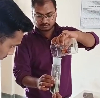
Fig. Solvent Prepration
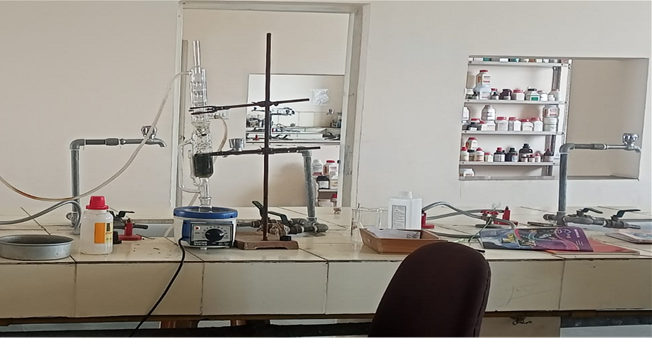
Fig. Soxhlate Assembly
Determination of percentage yield of the extract: - The crude extract after the soxhalation extraction process,extract was further on vaccum evaporater dried extract of aerial part of Pluchea indicawas done by using solvent Hydroalcohalic (ethanol:water, 70:30 v/v). The percentage yield of extract were calculated 4.7 gm.
Qualitative Analysis
Following standard protocols were used for qualitative analysis of samples to check for the presence of Alkaloids, glycosides, Flavonoids, Steroids , carbohydrates, Protein... etc
1.Test for alkaloids
1- Wagner's reagent (Iodine-potassium Iodide solution solution)
A solution of 1.3 g iodine and 2 g of potassium iodide in 100 ml of water. It gives reddish brown ppt. with most of the alkaloids even the purine bases.
2- Dragendorff's reagent (potassium bismuth Iodide).
Bismuth nitrate, nitric acid, pot. Iodide and water. It gives an orange ppt.
3- Mayer's reagent (potassium mercuric iodide).
HgCl2 (1.36 g) + KI (S g) + H2O (100 mI)
It gives white ppt. It is the most generally used of the alkaloidal reagents. The solution should be added to distinctly acidic solution of the alkaloid, only few drops of the reagent should be used and the solution should not contain acetic acid or alcohol.
2.Test for Glycosides
I. Keller Killiani test: 1ml of the extracts were dissolved in 1ml of glacial acetic acid and cooled, after cooling, 2-3 drops of ferric chloride was added. To this solution 2ml of conc. sulphuric acid was added carefully along the walls of the test tube. Appearance of reddish brown colored ring at the junction of two layers indicates the presence of glycosides.
II. Conc. sulphuric acid test: To 1ml of the extracts, 1ml of conc. sulphuric acid was added and allowed to stand for 2 min. a reddish color precipitate indicates the presence of glycosides.
III. Molish’s test: 2-3 drops of molisch reagent was added to the extracts and mixed well. To this, a few drops of conc. sulphuric acid was added carefully. Formation of reddish-purple colored ring at the junction of two layers indicates the presence of glycosides.
3.Test for phyotsterol
Solkowski Test
Solkowski test was done with the plant extracts. 2 ml extract taken in a test tube. 2 ml Chloroform and 2 ml conc.Sulphuric acid was added in it; brown or red colored ring on the sulphuric acid layer given the confirmatory test.
Libermann Burchard’s Test
Libermann and Burchurd’s test was done after the extraction and reflux of the plant material. 2 ml extract taken in a test tube. 2 ml Chloroform, 2 ml Acetic Anhydride and 2 ml conc. Sulphuric acid was added in it; translucent green colour given the confirmatory test.
4. Test for Carbohydrates
Extracts were dissolved individually in 5 mL distilled water and filtered. The filtrates were used for the detection of carbohydrates.
(a) Molisch Test
To 2.0 mL of the extract, 2 drops of Molisch reagent was added and mixed. 2.0 mL of concentrated sulphuric acid was added to this solution. Formation of the red violet ring at the junction of the solution and its disappearance on addition of excess alkali solution indicates the presence of carbohydrates.
(b) Benedict's Test
Few drops of Benedict’s reagent was added to the test solution and boiled on water bath. Formation of reddish brown precipitate indicates the presence of sugars. Depending on the concentration of the reducing sugar, the amount and colour of the precipitate produced varied. A positive Benedict’s test appears green, yellow, orange, or red.
(c) Fehling’s test
To 1 mL of the extract, 1 mL of Fehling’s A and 1 mL of Fehling’s B solutions were added in a test tube and heated in a water bath for 10 minutes. Formation of red precipitate indicates the presence of a reducing sugar. The filtrate was treated with 1 mL of Fehling’s A and B, and heated in a boiling water bath for 5-10 min. Appearance of reddish orange precipitate shows the presence of carbohydrates.
5. Test for phenolic compounds
(a) Ferric chloride test
A little extract was dissolved in distilled water. To this, 2 mL of 5% ferric chloride solution was added. Formation of blue, green or violet colour indicates the presence of phenolic compounds.
(b) Lead acetate test
A little extract was dissolved in distilled water. To this, a few drops of lead acetate solution was added. Formation of white precipitate indicates presence of phenolic compounds.
(c) Dilute iodine solution test
To 2-3 mL of extract, a few drops of dilute iodine solution was added. Formation of transient red colour indicates the presence of phenolic compounds.
6. Test for Flavonoids
(a) Ammonia test
5 mL of dilute ammonia solution were added to a portion of the crude extract followed by addition of concentrated H2SO4. Formation of a yellow colouration in the extract indicates the presence of flavonoids. The yellow colouration disappears after some time.
(b) Shinoda’s test
The extracts were dissolved in 5 mL of (95%) ethanol. To this, a piece of magnesium followed by concentrated hydrochloric acid was added drop wise and heated.
Appearance of magenta colour shows the presence of flavonoids.
(c) Zinc–hydrochloride test
To the extract, a pinch of zinc dust was added followed by addition of concentrated hydrochloric acid along the sides of the test tubes. Appearance of magenta color indicates the presence of flavonoids.
(d) Lead acetate test
The extract was treated with a few drops of lead acetate solution. Formation of yellow precipitate indicates the presence of flavonoids. Orange to crimson colour shows the presence of flavonones.
(e) Alkaline reagent test
The extract was treated with a few drops of sodium hydroxide. Formation of intense yellow colour, which becomes colour less on addition of few drops of dilute acid, indicates the presence of flavonoids.
(g) Ferric chloride test
To the extract, a few drops of neutral ferric chloride solution was added, a blackish red colour forms, indicating the presence of flavonoids.
7.test for protein
a)Ninhydrin Test
To 1ml of extract few drops of Ninhydrin reagent was added and heated in a boiling water bath. A purple blue colour indicates the presence of proteins
b) Biuret Test
To 1ml of extract, equal volume of 5% NaOH solution and copper sulphate solution added. A blue colour indicates the presence of proteins.
Quantitative phytochemical analysis
1.Estimation of Total polyphenol content (TPC)
The total polyphenol content of the extract was estimated using the Folin Ciocalteau reagent based assay as previously described by Singleton and Rossi [30]. 25-400 µg/ml methanolic gallic acid solutions were used as standards and methanol was used as a blank. The absorbance of the developed colour was recorded at 765 nm using a UV-Vis spectrophotometer (Jasco V-550). All determinations, for gallic acid as well as the plant extract, were carried out in triplicate. Data are represented as an average of the three determinations. Using these readings, a calibrated gallic acid standard curve was made. Based on the measured absorbance of the plant extract, the concentration of phenolics was estimated (µg/ml) from the calibration line. The content of polyphenols in the extract was calculated and expressed in terms of gallic acid equivalent (mg of GAE/g of dry weight material).
2.Total Flavonol Content
Flavones and flavonols contents were analyzed by the colorimetric method. 9.8 ml of the prepared extract was mixed with a 10% solution of aluminum chloride (200 μl). After 30 min, absorption was measured at a 425 nm wavelength. The amount was calculated using quercetin calibration curve. The results were expressed as the quercetin equivalent (QE) mg per 100 ml of the sample. Accurately weighed 100 mg of quercetin was dissolved in 100 ml of distilled water which gives the concentration of 1000 µg/ml. 10 ml of this solution was taken and made up to 100 ml with quercetin which contains the concentration of 100 µg/ml. Further 10 ml of this solution was taken and made up to 100 ml with quercetin which contains the concentration of 10 µg/ml. 1 to 10 ml were taken from this solution and made up to 10 ml to get the concentration ranges of 1 to 10 µg/ml. Calibration curve was plotted by mixing 9.8 ml aliquots of quercetin solutions with a 10% solution of aluminum chloride (200 μl). The absorbance was measured 30 min at 425 nm using UV spectrophotometer, against blank solution.
3.Separation of Phyotsterol
Dried Part of Plant

Hot Maceration

Extracted With Petroleum Ether

Filter

Volume Reduction

Extracted Lipid Refluxed With 5 %Alcoholic koH for 4 hrs

Filtrate Extracted With di-ethyl Ether

Dried And yield calculated

Positive Test for Steroids
RESULTS AND DISCUSSION:
The hydroalcoholic extract of Pluchea indicashow the presence of steroid, tannins and phenolic compounds, alkaloids, glycoside, carbohydrate. The results are shown intable .The Percentage yield of hydroalcoholic extract was (4.7gm).The plant was found to be rich in steroidal and flavonoid content. The phytoconstituents which are responsible for many pharmacological activities
Table 1: Qualitative analysis of Pluchea indica hydroalchoholic extract of presence of different phytoconstituents
|
S. No. |
Test |
Observation |
Inference |
|
1 |
Alkaloid |
|
|
|
|
Wagner's reagent |
Reddishbrown PPT |
+ve |
|
|
Dragendorff's reagent |
Reddishbrown PPT |
+ve |
|
|
Mayer'sreagent |
Creamcolour PPT |
+ve |
|
2 |
Glycoside |
|
|
|
|
Keller Killiani test. |
Appearance of reddish-brown colored ring at the junction of two layers |
+ve |
|
|
Conc.sulphuricacid test |
Reddish color precipitate |
+ve |
|
|
Molish’stest |
Formation of reddish-purple colored Ring at the junction of two layers. |
+ve |
|
3 |
Steroid |
|
|
|
|
SolkowskiTest |
Brown or red colored ring on the sulphuric acid layer given the confirmatory test. |
+ve |
|
|
Libermann Burchard’sTest |
translucentgreencolourgiventhe confirmatory test. |
+ve |
|
4 |
Carbohydrates |
|
|
|
|
MolischTest |
Formation of the red violet ring at the junction of the solution and its disappearance on addition of excess alkali solution indicates the presence of carbohydrates. |
+ve |
|
|
Benedict'sTest |
|
+ve |
|
|
|
Depending on the concentration of the reducing sugar, the amount and colour of the precipitate produced varied. A positive Benedict’s test appears green, yellow, orange, or red. |
|
|
5 |
Phenolic Compounds |
|
|
|
|
Ferric chloride test |
Formation of blue, green or violet colour indicates the presence of phenolic compounds. |
+ve |
|
|
|
Formation of white precipitate indicates presence of phenolic |
+ve |
|
|
Lead acetate test |
|
|
|
|
Dilute iodine solution test |
Formation of transient red colour indicates the presence of phenolic compounds |
+ve |
The total phenolic content for aqueous, hydro alcoholic extract was estimated by Folin Ciocalteu’s method using gallicacid as standard. The gallic acid solution of concentration(10-100ppm) conformed to Beer’s Law at 750 nm with a regression co-efficient (R2) = 0.997. The plot has a slope (m) = 0.028 and intercept = 0.003. The equation of standard curve is y = 0.028x + 0.003 (Fig. 1). The total flavonoid content for hydroalcoholic extract was measured with the aluminium chloride colorimetric assay using quercetin as standard. The quercetin solution of concentration (5-25 ppm) conformed to Beer’s Law at 510 nm with are gression co-efficient(R2)=0.999.The plot a slope(m)=0.043 and intercept =0.013. The equation of standard curveisy=0.043x+0.013(Fig. 2).
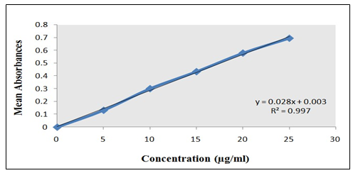
Fig.1: Total Phenolic Content for Standard Gallic Acid.
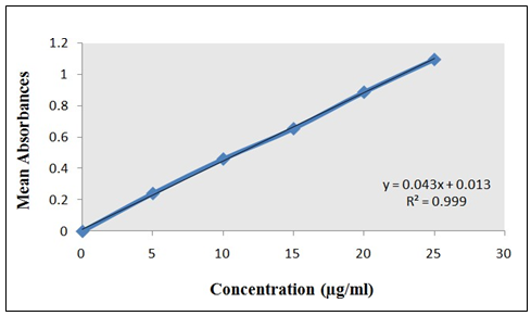
Fig.2: Total Flavonoid Content for Standard Quercetin
CONCLUSION
The preliminary phytochemical studies for different extracts of Pluchea indicashow the presence of alkaloids, flavonoids, carbohydrates, phytosterols, tannins, saponins, proteins and aminoacids, terpenes, phenols, gums and mucilage’s.
REFRENCES


Jitendra Jaiswal*, Mohit Lokhande, Abhishek Choudhary, Krish Upadhyay, Shivam Singh, Shumaila Khan, Ali Hussain Ansari, Phytochemical Analysis of Hydroalcoholic Extract of Pluchea Indica Leaves, Int. J. of Pharm. Sci., 2025, Vol 3, Issue 3, 1595-1602. https://doi.org/10.5281/zenodo.15043278
 10.5281/zenodo.15043278
