School of pharmacy, Abhilashi university, Chail Chowk, Mandi (Himachal Pradesh) India 175028
Diabetes mellitus is a chronic metabolic disorder currently a highly concerning health topic due to increased prevalence and lack of complete cure. conventional therapies such as oral hypoglycaemic agents and insulin injections are effective to combat increased insulin levels but suffer from poor bioavailability and side effects. hence in recent years nanotechnology-based drug delivery systems have gained a lot of attention as potential tool for overcoming the limitations of traditional therapies. niosomes are one among the nanotechnology-based approaches that can act as promising carrier for various therapeutic agents. Niosomes are non-ionic surfactant vesicles that can encapsulate and deliver drugs, offering improved bioavailability and targeted delivery for managing diabetes. Niosomal systems not only enhance the pharmacokinetic profile of drugs like metformin, Glibenclamide, and insulin but also allow for alternative administration routes such as transdermal and oral, reducing the need for invasive injections. Furthermore, co-encapsulation of antioxidants or anti-inflammatory agents with antidiabetic drugs may provide synergistic effects beneficial for long-term management of diabetes and its complications. This review explores the recent advancements in niosomal drug delivery systems for diabetic management, discussing formulation strategies, therapeutic outcomes, challenges, and future perspectives in the development of niosome-based antidiabetic therapies.
The chronic metabolic disorder of diabetes mellitus is a group of physiological dysfunctions characterized by hyper-glycemia resulting directly from insulin resistance, inadequate insulin secretion, or excessive glucagon secretion (1,2)
Diabetes mellitus (DM) is probably one of the oldest diseases known to man. It was first reported in Egyptian manuscript about 3000 years ago Diabetes was first documented by the Egyptians and is characterized by weight loss and polyuria. However, it was the Greek physician Aertaeus who coined the term diabetes mellitus (OM). In Greek, diabetes means "to pass through" and mellitus is the Latin word for honey (referring to sweetness) Type 2 diabetes mellitus (T2DM), formerly known as non-insulin-dependent diabetes mellitus or adult-onset diabetes is a metabolic disorder that is characterized by high levels of blood glucose as a result of insulin resistance and relative insulin deficiency.(3) Type 2 diabetes is a worldwide health crisis. Type 2 diabetes mellitus (T2DM) affects about 3% of the population or 100 million people worldwide and is the most common form of diabetes mellitus. It is predicted that the prevalence of DM in adults of which type 2 DM is becoming prominent will increase in the next two decades and much of the increase will occur in developing countries where the majority of patients are aged between 45 and 64 years (4) It is estimated that in 20I0 there were globally 285 million people (approximately 6.4% of the adult population) suffering from this disease. This number is estimated to increase to 430 million in the absence of better control or cure. An ageing population and obesity are two main reasons for the increase. Furthermore, it has been shown that almost 50% of the putative diabetics are not diagnosed until 10 years after onset of the disease, hence the real prevalence of global diabetes must be astronomically high. It is estimated that 366 million people had DM in 2011; by 2030 this would have risen to 552 million. The number of people with type 2 DM is increasing in every country with 80% of people with DM living in low- and middle-income countries. DM caused 4.6 million deaths in 2011 Type 2 diabetes mellitus (T2DM) affects about 3% of the population or 100 million people worldwide. The prevalence is higher in the United States, affecting 6–7% of the population and is increasing at an astounding rate. According to the National Institutes of Health (NIH) and Centers for Disease Control (CDC), over 30% of individuals with diabetes are undiagnosed. Furthermore, a third of children born in the United States in the year 2000 will develop diabetes, an estimated 28 million diabetic individuals within the next 50 years. About 422 million people worldwide have diabetes, the majority living in low-and middle-income countries, and 1.5 million deaths are directly attributed to diabetes each year. Both the number of cases and the prevalence of diabetes have been steadily increasing over the past few decades. According to the International Diabetes Federation (IDF), approximately 415 million adults between the ages of 20 to 79 years had diabetes mellitus in 2015. DM is proving to be a global public health burden as this number is expected to rise to another 200 million by 2040 It is predicted that the prevalence of DM in adults of which type 2 DM is becoming prominent will increase in the next two decades and much of the increase will occur in developing countries where the majority of patients are aged between 45 and 64 years. Type 2 diabetes is a disease with a complex multifactorial pathogenesis. The main pathogenetic mechanisms are considered to be impaired insulin secretion and insulin resistance, however, the number of new defects causing chronic hyperglycemia in type 2 diabetes is constantly increasing:
Niosomes
Niosomes or non-ionic surfactant vesicles are microscopic lamellar structures formed on admixture of non-ionic surfactant of the alkyl or di alkyl polyglycerol ether class and cholesterol with subsequent hydration in aqueous media. The first niosome formulations were developed and patented by L’Oreal in 1975. Niosomes are a novel drug delivery system, which entrapped the hydrophilic drug in the core cavity and hydrophobic drugs in the non-polar region present within the bilayer hence both hydrophilic and hydrophobic drugs can be incorporated into niosomes. The niosomes are amphiphilic in nature, in which the medication is encapsulated in a vesicle which is made by non-ionic surfactant and hence the name niosomes. Several factors affect the structure of niosomes, such as: the nature and type of surfactants, amount of cholesterol, the critical packing parameter (CPP), and the used drug. They are vesicular systems similar to liposomes that can be used as carriers of amphiphilic and lipophilic drugs.(6) Niosomes are mostly studied as an alternative to liposomes because they alleviate the disadvantages associated with liposomes. Niosomes overcome the disadvantages associated with liposomes such as chemical instability. Chemical instability of liposomes is due to their predisposition to oxidative degradation and variable purity of phospholipids. Non-ionic surfactants provide a few advantages over the phospholipids because they are more economical and are chemically more stable as they are not easily hydrolyzed or oxidized during storage. The vesicular structure can be modified to provide sustained or controlled drug delivery thus enhancing efficacy of system for prolonged periods. Thus, the main purpose of developing niosomal system is chemical stability, biodegradability, biocompatibility, chemical stability, low production cost, easy storage and handling and low toxicity. The twofold structure of surfactant molecules encourages them to orient in bilayer architecture. In this two-layered structure, hydrophilic and hydrophobic ends of surfactant tend to arrange into external and internal sites, respectively. This lamellar morphology allows us to entrap two kinds of drugs in a unique carrier simultaneously. Thus, niosomes are promising vehicle for drug delivery and being non-ionic, it is less toxic and improves the therapeutic index of drug by restricting its action to target cells. Niosomes can be administrated through various routes such as oral, parenteral, topical. Niosomes are used as a carrier to deliver different types of drugs such as synthetic and herbal, antigens, hormones and other bioactive compounds. They remain in the bloodstream for a reasonable time, which is useful for targeted delivery of drug.
Structure of niosomes –
The main components of niosomes are non-ionic surfactants, hydration medium and lipids such as cholesterol. The self-assembly of non-ionic surfactants in aqueous media results in closed bilayer structures. A high interfacial tension between water and the hydrophobic tails of the amphiphile causes them to associate. The steric and hydrophilic repulsion between the head groups of non-ionic surfactants ensure that hydrophilic termini point outwards and are in contact with water. The assembly into closed bilayers usually requires some input of energy such as mechanical or heat.(8) To form the closed bilayer structure, energy such as heat or physical agitation is required. Various forces inside the vesicles were found to play an important role in maintaining the vesicular structure, for example, van der Waals and repulsive forces that exist among the surfactant molecules. Varying the vesicle’s components (including type, composition, and concentration), size, surface charge, or volume will likely modify the properties of resultant niosome. The bilayer is formed by non-ionic surfactants, with or without cholesterol and a charge inducer. Different types of surfactants at variable combinations and molar ratios are used to form niosomes Examples of surfactants include alkyl ethers, alkyl glyceryl ethers, sorbitan fatty acid esters, and polyoxyethylene fatty acid esters. Addition of cholesterol maintains the rigidity of the bilayer, resulting in less leaky niosomes. Meanwhile, charge inducers provide charge to the vesicles and increase vesicle size, increasing drug entrapment efficiency. Negative charge inducers, including dicetyl phosphate, dihexadecyl phosphate, and lipoamino acid, and positive charge inducers, including stearylamine and cetylpyridinium chloride, help to stabilize the vesicles Non-ionic surfactants in niosomes tend to orient themselves in such a way that hydrophilic end faces outward (toward the aqueous phase), whereas the hydrophobic end faces inward to each other to form a closed bilayer structure, which encloses solutes in an aqueous solution As a result, the closed bilayer structure of niosomes has hydrophilic inner and outer surfaces, with a sandwiched lipophilic area in between.
Advantages of niosomes –
Niosomes combine several advantages with respect to other nanocarriers: (10,11)
Disadvantages of niosomes –
METHOD OF PREPARATION
Thin film hydration method – The mixture of vesicles forming ingredients like surfactant and cholesterol are dissolved in a volatile organic solvent (diethyl either, chloroform or methanol) in a round bottom flask. The organic solvent is removed at room temperature (20°C) using rotary evaporator leaving a thin layer of solid mixture deposited on the wall of the flask. The dried surfactant film can be rehydrated with aqueous phase at 60°C with gentle agitation. This process forms typical multilamellar niosomes.
Micro fluidization method-
Micro fluidization is a recent technique used to prepare unilamellar vesicles of defined size distribution. This method is based on submerged jet principle in which two fluidized streams interact at ultra-high velocities, in precisely defined micro channels within the interaction chamber. The impingement of thin liquid sheet along a common front is arranged such that the energy supplied to the system remains within the area of niosomes formation. The result is a greater uniformity, smaller size and better reproducibility of niosomes formed
Sonication method-
A typical method of production of the vesicles is by sonication of solution as described by Cable. In this method an aliquot of drug solution in buffer is added to the surfactant/cholesterol mixture in a 10-ml glass vial. The mixture is probe sonicated at 60°C for 3 minutes using a sonicator with a titanium probe to yield niosomes. The resulting vesicles are small and unilamellar. In the case of niosomes the resulting vesicle sizes are in general larger than liposomes, niosomes being no smaller than 100 nm in diameter. (13)
Reverse phase evaporation technique-
Cholesterol and surfactant (1:1) are dissolved in a mixture of ether and chloroform. An aqueous phase containing drug is added to this and the resulting two phases are sonicated at 485°C. The clear gel formed is further sonicated after the addition of a small amount of phosphate buffered saline (PBS). The organic phase is removed at 40°C under low pressure. The resulting viscous noisome suspension is diluted with PBS and heated on a water bath at 60°C for 10 min to yield niosomes
Multiple membrane extrusion method-
Mixture of surfactant, cholesterol and dicetyl phosphate in chloroform is made into thin film by evaporation. The film is hydrated with aqueous drug polycarbonate membranes, solution and the resultant suspension extruded through which are placed in series for up to 8 passages. It is a good method for controlling noisome size
Extrusion method-
In this method, a mixture of cholesterol and diacetyl phosphate is prepared and then solvent is evaporated using rotary vacuum evaporator to leave a thin film. The film is then hydrated with aqueous drug solution and the suspension thus obtained is extruded through the polycarbonate membrane (mean pore size 0.1 mm) and then placed in series up to eight passages to obtain uniform size niosomes
Bubble method –
It is novel technique for the one step preparation of liposomes and niosomes without the use of organic solvents. The bubbling unit consists of round8bottomed flask with three necks positioned in water bath to control the temperature. Water cooled reflux and thermometer is positioned in the first and second neck and nitrogen supply through the third neck. Cholesterol and surfactant are dispersed together in this buffer (PH 7.4) at 70°C, the dispersion mixed for 15 seconds with high shear homogenizer and immediately afterwards “bubbled” at 70°C using nitrogen gas.
Trans membranes pH gradient (inside acidic) Drug Uptake Process or Remote Loading Technique
Surfactant and cholesterol are dissolved in chloroform. The solvent is then evaporated under reduced pressure to get a thin film on the wall of the round bottom flask. The fim is hydrated with 300mM citric acid (PH 4.00 by vertex mixing. The multilamellar vesicles are frozen and shared 3 times and later sonicated. To this niosomal suspension. Aqueous solution containing 10 mg ml of drug is added and vortexes. The PH of the sample is then raised to 7.087.2 with 1M disodium phosphate. This mixture is later heated at 60°c for 10 minutes so give niosomes.
Ether injection method- This method provides a means of making niosomes by slowly introducing a solution of surfactant dissolved in diethyl ether into warm water maintained at 60°C. The surfactant mixture in ether is injected through 148-gauge needle into an aqueous solution of material. Vaporization of ether leads to formation of single layered vesicles. Depending upon the conditions used the diameter of the vesicle range from 50 to 1000 nm.(14)
Ethanol Injection Method
Ethanol injection is a fast and simple technique for the preparation of small unilamellar vesicles. The technique consists of the injection of an ethanolic solution of lipids into an aqueous phase, where spontaneous vesicle formation takes place. Co-solvents, such as isopropanol, can be used with ethanol in order to optimize lipid solubility and enhance encapsulation efficiency. Furthermore, the speed of injection and temperature can be changed in order to modify vesicle size and prevent aggregation, which makes this method even more versatile. The method is inexpensive, does not require sophisticated equipment, has the possibility of scaling up, and is easy to set up . The size of the niosomes obtained by this method is smaller compared to the thin film hydration and microfluidics methods(15)
Factors governing niosomal formulation –
Amount and type of surfactant-
As the HLB value of surfactants like span 85 (HLB 1.8) to span 20 (HLB 8.6) increased, the mean size of niosomes also increases proportionally. It is due to the fact that surface free energy decreases with increase in hydrophilicity of surfactant. Alkyl chain is present in well-ordered structure in gel state, while in the liquid state the structure of bilayer is more disordered. The gel–liquid phase transition temperature (TC) is used for characterization of surfactant and lipids. Entrapment efficiency is also affected by phase transition temperature i.e. span 60 having higher TC, provide better entrapment efficiency. Entrapment efficiency of the niosomes is affected by the HLB value for e.g. niosomes have high entrapment efficiency at HLB value 8.6 but HLB value 14 to 17 is not suitable for niosomes formulation
Cholesterol content and charge-
Hydrodynamic diameter and entrapment efficiency were found to be increased due to cholesterol content in the niosomal bilayer. Cholesterol can act by two ways. First, it can increase the chain order of liquidated bilayer and second; by decreasing the chain order of the gel state bilayer, cholesterol affects the hydrodynamic diameter and entrapment efficiency. It has been reported that release rate of drug decreases and rigidity of bilayer increases due to high concentration of cholesterol.
CMC (Critical micelle concentration)
The surfactant has ability to reduce the surface tension between two phases. If the concentration of surfactant is not going above to critic micelle concentration, then formation of niosome are not takes place. Some surfactant is showing the micelle formation when concentration of surfactant goes above CMC, but some surfactant is showing the formation of circular bilayer structure and that are niosome
Nature of encapsulated drug
The drug entrapment in niosomes is affected by molecular weight, chemical structure, hydrophilicity, lipophilicity as well as the hydrophilic lipophilic balance (HLB) value of the drug.15) Vesicle size may increase due to entrapment of drug. Drug particle interacts with the surfactant head groups, which may increase charge on polymer and thus cause repulsion of the surfactant bilayer which leads to increase in vesicle size. Entrapment of drug in niosomes increases vesicle size, probably by interaction of solute with
surfactant head groups, increasing the charge and mutual repulsion of the surfactant bilayers, thereby increasing vesicle size. In poly oxyethylene glycol (PEG) coated vesicles, some drug is entrapped in the long PEG chains, thus reducing the tendency to increase the size. The hydrophilic lipophilic balance of the drug affects degree of entrapment (16)
Quantity of surfactant and lipid
Generally, the maximum quantity of surfactant and lipid used for preparing niosome is 1–2.5% w/w. Any change in surfactant: cholesterol ratio changes the amount of drug entrapped, and the viscosity of the system also changes. Any change in the above ratio during the hydration step may influence the properties of niosomes
Hydration time
Yeo et al. prepared niosomes of methylene blue using thin film hydration technique and probe sonicated the vesicles for size uniformity. They studied the effect of hydration time and hydration volume on entrapment efficiency and vesicle size. They found short hydration time produces vesicles with larger sizes and less drug entrapment. They suggested 60 min as optimum hydration time and 5 ml hydration volume for full hydration of span 60 and formation of vesicular size. Longer hydration time produces small-sized vesicles(17)
Method of preparation
Method of preparing niosome may affect its size and entrapment efficiency. Abdelkader et al. prepared niosomes of naltrexone, by freeze and thaw, dehydration-rehydration and reverse-phase evaporation method thin-film hydration technique. In the results, he reported that the entrapment efficiency was significantly depended upon the method of preparation. Abdelkader et al. prepared ethoniosomes (ethanol-based niosomes) of prednisolone using a thin film hydration method and ethanol injection method. He found that the thin-film hydration method produces niosomes with better entrapment efficiency than ethanol injection method, but ethanol injection method produces small size niosomes in comparison to the thin-film hydration method. Small size niosomes can also be produced from reverse-phase evaporation and micro fluidization method. Niosomes prepared by transmembrane pH method shows better entrapment efficiency
pH of the hydration medium
The pH of the hydration medium is another factor that can influence the entrapment efficiency of the drug. For example, flurbiprofen shows higher entrapment at acidic pH (maximum 94.6% at pH 5.5). The entrapment efficiency of flurbiprofen increases as the pH decreases from 8 to 5.5 and entrapment efficiency decreases significantly at pH 6.8 (18)
Structure of surfactants –
The geometry of the vesicles formed during the niosomal preparation also depends upon the critical packing parameter (CPP). According to CPP the geometry of the vesicles can be predicted. CPP can be calculated using following equation:
Critical packing parameterCPP= vlc×a0
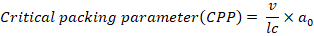
where v= hydrophobic group volume,
lc = the critical hydrophobic group length,
a0 = the area of hydrophilic head group
CPP is helpful in predicting the structure of niosome vesicles in following way;
spherical micelles formed if CPP<1/2
bilayer micelles are formed if ½<CPP<1
inverted micelles are formed if CPP>1.
Characterization of niosomes –
Transmission Electron Microscopy (TEM) and scanning electron microscopy (SEM)
TEM plays an important role in characterization of niosomes providing details about morphology, size and structure. TEM provide high resolution images that allow visualization of size and surface features of niosomes. TEM can also visualize lamellar structure of niosomes confirming their bilayer arrangement. Thus, single layered (unilamellar) and multilayered (mullilamellar) vesicles can be differentiated using transmission electron microscopy. (19) Scanning electron microscopy is an important technique used in characterization of niosomes focussing mainly on surface morphology and topographical features. SEM provides detailed images of surface of niosomes that helps to identify surface roughness, smoothness or irregularities along with three dimensional images of vesicles. It is also useful to monitor surface morphology changes during stability testing. (20)
Zeta Potential (ZP), and Polydispersity Index (PDI)
Zeta potential measures the electrical charge on surface of niosomes. The zeta potential of +-30mV is considered optimum. It suggests strong electrostatic repulsion preventing aggregation and stability. Lower vales indicate low electrostatic repulsion which may lead of aggregation and fusion of vesicles. It is determined using zeta potential analyzers. (19) Polydispersity index (PDI) indicates uniformity of particle size distribution. A PDI value between 0.1 to 0.3 is considered optimum for niosomal formulation.
Encapsulation Efficiency (EE)
Centrifugation was used to measure the %EE of all the niosomal formulations. For this purpose, a niosomal suspension was poured into 1.5 mL Eppendorf and centrifuged for 1 h at 16,000 rpm at 4 ?C using a cooling centrifuge. The supernatant was discarded, and the separated niosomes were washed with PBS before being centrifuged an additional two times under the same conditions. The amount of the entrapped drug was determined by adding 1 mL of isopropanol to 0.1 mL of the separated niosomes, which was then diluted with PBS up to 20 mL and then sonicated and centrifuged again at 14,000 rpm for 15 min at 25 ?C to obtain a clear solution to be analysed.
The EE (%) was determined using
EE%= Amount of entrapped drugTotal amount of drug ×100
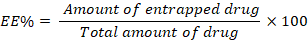
where the Amount of entrapped drug is the actual amount of the drug successfully encapsulated in the vesicles and the total CLR amount refers to the entire quantity of CLR utilized during preparation.(19)
Both the entrapment efficiency (EE) and yield of niosome depend on the method of preparation as well as physicochemical properties of drug. The number of double layers, vesicle size and its distribution, entrapment efficiency of the aqueous phase, and the permeability of vesicle membranes are influenced by the methodology used for formulation as well as the addition of cholesterol as they make the niosomes less leaky
Optical Microscopy Technique
This technique is also used for observation of niosome size and shape. Nearly 100 niosome are used for particle size determination. In this method size of stage micrometre coinciding with the eye piece micrometre is recorded and size of niosome is then calculated
In Vitro Release Studies
In vitro drug release can be done by
Stability study
Stability studies are done by storing niosome at two different conditions, usually 1 °C and 2 °C. Formulation size, shape and number of vesicles per cubic mm can be assessed before and after storing for 30 d. After 15 and 30 d, residual drug can also be measured. Light microscope is used for determination of size of vesicles and the numbers of vesicles per cubic mm is measured by hemacytometer
Niosomes in diabetes treatment-
Oral therapy-
In a study titled "Pharmacokinetic study of niosomes loaded transdermal patch," streptozotocin-induced diabetic rats were orally administered niosomes, and the resulting hypoglycaemia and elevation of serum insulin levels were compared to those achieved with subcutaneous insulin injections. Oral administration of niosome-encapsulated insulin (100 IU/kg) resulted in decreased blood glucose levels and elevated serum insulin. (23) Separately, lycopene, a principal carotenoid, was extracted from ripe tomatoes and used to formulate lycopene-loaded nano niosomes. Diabetes was induced in Wistar rats using alloxan monohydrate. An antidiabetic study demonstrated a marked decrease in blood glucose levels and improved biochemical parameters in diabetic rats treated with the lycopene formulation at doses of 100 & 200mg/kg for 14 days, compared to the antidiabetic drug Glibenclamide (5 mg/kg), indicating the formulation's significant antidiabetic potential. (24) Metformin hydrochloride loaded niosomes were formulated to enhance bioavailability and reduce lactic acidosis, a common metabolic complication during diabetes treatment. The niosomes were prepared using the reverse phase evaporation technique. Pharmacokinetic data of the MH-loaded niosomal preparation demonstrated a significant prolongation and increased intensity of the hypoglycaemic effect compared to the free MH solution. These results suggest that MH-loaded niosomes are a promising extended-release preparation with improved hypoglycaemic efficiency. Canagliflozin (CFZ) is widely used for managing type 2 diabetes mellitus. However, its low oral bioavailability, stemming from poor solubility and restricted membrane permeability, presents a challenge. Encapsulating CFZ in niosomes significantly sustained drug release compared to the aqueous drug dispersion. Oral administration of these niosomal formulations significantly enhanced the oral antidiabetic effect of CFZ. This study indicates that niosomes are promising carriers for improving the oral anti-diabetic activity of CFZ. (26) ß-sitosterol loaded niosomes were designed and evaluated for its anti-diabetic properties. In vivo experiments demonstrated significant improvements in body weight and blood glucose levels in diabetic rats. The optimized formulation exhibited controlled release and substantial antidiabetic activity, suggesting its potential as an effective treatment for type 2 diabetes mellitus.(27) Recombinant human insulin niosomes were prepared using polyoxyethylene alkyl ether surfactants via the classic film hydration method. Diabetes was induced by IP injection of streptozotocin (65 mg/kg) in male Wistar rats. Animals treated with oral niosomes (Brij 52 and 92)-encapsulated insulin (100 IU/kg) showed decreased blood glucose levels and elevated serum insulin; the hypoglycaemic effect of Brij 92 niosomes was particularly significant.(28) Insulin-loaded niosomes were optimized using response surface methodology. This study evaluated the effects of cholesterol and surfactant concentration, as well as sonication duration, on the zeta-potential, polydispersity index (PDI), and entrapment efficiency (EE%) of the insulin-loaded niosomal vesicles. Optimum conditions were assessed based on cytocompatibility and in vitro drug release. The resulting optimum insulin-loaded niosomal vesicles, characterized across different aspects, displayed a spherical morphology with minimal aggregation. Furthermore, they exhibited good biocompatibility with Caco-2 cells and sustained release in simulated intestinal fluid. Glimepiride-loaded niosomes were prepared to enhance its oral bioavailability and hypoglycemic efficacy. An in-vitro release study was performed using dialysis. In-vivo pharmacodynamic studies and pharmacokinetic evaluation were conducted on alloxan-induced diabetic rats. In-vivo studies demonstrated the superiority of GLM niosomes in lowering blood glucose levels (BGL) and maintaining therapeutic GLM levels for a longer duration compared to the free drug and the marketed product. While there was no significant difference in the mean plasma AUC0-48 hr between GLM-loaded niosomes and the marketed product, GLM-loaded niosomes exhibited a seven-fold enhancement in relative bioavailability compared to the free drug. (30) Sustained-release metformin niosomes were formulated to decrease side effects and reduce dosing frequency. Non-ionic surfactant vesicles using different surfactants were prepared via thin-film hydration and characterized for morphology, entrapment efficiency, in-vitro release, TEM (transmission electron microscopy), and physical stability. The optimized formulation was further studied to determine the effect of surfactant concentration, DCP (Dicetyl phosphate), surfactant: cholesterol ratio, and hydration volume. Release data were analysed using release kinetics models. Metformin-loaded niosomes effectively sustained drug release, potentially decreasing side effects and increasing patient compliance. The effect of niosomal encapsulation on intestinal absorption and oral bioavailability of nateglinide was studied by Amal A. Sultan in 2017. The in-situ rabbit intestinal absorption of nateglinide was monitored using both its aqueous solution and niosomes. Streptozotocin-induced diabetic albino rats were used to assess the hypoglycaemic effect of nateglinide following oral administration of both aqueous dispersion and niosomal formulations. Niosomes demonstrated a significant improvement in both the rate and extent of the hypoglycaemic effect compared to the unprocessed drug. In conclusion, niosomes can enhance the oral bioavailability of nateglinide, potentially through a nontraditional absorption pathway.(32) Formulation of a Repaglinide deformable lipo-niosomal (LNH) was achieved using reverse ethanol injection. In vitro characterization tests and an in vivo study performed on the optimal RPG-loaded LNHs demonstrated a significant increase in entrapment efficiency and permeability. The RPG-loaded LNHs exhibited a superior hypoglycaemic effect compared to free RPG, likely due to enhanced intestinal absorption of RPG upon encapsulation within the LNHs. (33) The study aimed to investigate the synergistic effect of trans-activator of transcription (Tat) and niosomes to enhance the hypoglycaemic activity of orally delivered human insulin. For the in vitro release, insulin loaded in elastic anionic niosomes (T10 = 4 h) exhibited a slower release rate compared to insulin in the mixture loaded in niosomes (T10 = 3 h). Oral administration of the mixture loaded in elastic anionic niosomes to alloxan-induced diabetic mice, at insulin doses of 25, 50, and 100 IU/kg body weight, demonstrated significant hypoglycaemic activity. Specifically, the percentage reduction in fasting blood glucose was 1.95, 2.10, and 2.10 times greater than that achieved with subcutaneous insulin injection at 12 h, respectively. Repaglinide (RPG) is a monotherapy insulin secretagogue used to treat type II diabetes; however, because of hepatic first pass metabolism, it has variable bioavailability (about 50%) and poor water solubility. For diabetes patients with dysphagia, the formation of chewable tablets with RPG-loaded proniosomes offers a promising new oral drug administration method. (13) Another study looked at a new proniosomal formulation of pioglitazone with regulated drug delivery for the treatment of type 2 diabetes. A 32-factorial design was used to examine the effects of independent factors such as the kind of surfactant and the ratio of surfactants to cholesterol. The provesicular powders' shape, vesicle size, in vitro drug release, and encapsulation efficiency were all evaluated. Stability tests and in vivo performance evaluation were performed on the ideal provesicular powder that was discovered. The results demonstrated that it had a greater in vivo hypoglycaemic effect on both normal, healthy, and STZ-induced diabetic albino rats. (14) To increase the bioavailability of linagliptin, a BCS class-III drug, by improving its permeability, linagliptin-loaded non-ionic surfactant vesicles (Niosomes) were formulated and evaluated using statistical optimization. Niosomes were prepared by both the thin film hydration method (TFHM) and the modified ether injection method (MEIM). The relationship between dependent and independent variables was determined using mathematical equations and response surface methodology (RSM). Statistical analysis was performed using ANOVA. In conclusion, all in vitro and ex vivo experiments demonstrated promising results for treating type II diabetes mellitus with linagliptin-loaded non-ionic surfactant vesicles. Glibenclamide (GB), an oral antidiabetic sulfonylurea, is commonly used to manage type 2 diabetes mellitus. However, its low water solubility leads to poor bioavailability. This study aimed to enhance GB dissolution, and consequently its pharmacological effect, by formulating it as a proniosomal powder. GB proniosomes were prepared using a slurry method with sucrose as a carrier. The resulting formulations were characterized for particle size, zeta potential, entrapment efficiency, powder flow properties, and in vitro dissolution. Furthermore, the pharmacological effect was assessed by measuring fasting blood glucose levels (BGL) before and after treatment. A significant 73% reduction in fasting blood glucose levels was observed in animals treated with the proniosomal formulation, with no evidence of liver damage. Pharmacodynamic results demonstrated a significantly greater reduction in fasting blood glucose levels in animals treated with proniosomes compared to a 17.6% reduction after treatment with the pure drug. Histopathological analysis confirmed the absence of liver damage following proniosomal treatment.(36)
Transdermal delivery
The coacervation phase separation process was used to create the metformin proniosomal gel. By encapsulating the medication in multiple proniosomal gel formulations made of varying ratios of Span 60/Span 40, cholesterol, and lecithin, the potential of proniosomes as a transdermal drug delivery method was assessed. The systems that were prepared were described. Metformin proniosomal gel has a relatively excellent stability profile and shown a promising extended drug delivery strategy. Pioglitazone (PZ) encapsulated in a Carbopol-based trans gel system (proniosomes/niosomes) in order to provide a transdermal medication delivery method that promotes skin penetration. Using the quality by design (QbD) technique, the created formulations were optimized, and the transdermal flux, particle size, and entrapment % were calculated. High encapsulation and improved flux value were shown to be more effective delivery carriers, and the proposed formulation considerably boosted PZ penetration into the skin. When compared to tablet format, the bioavailability of Carbopol trans gel was significantly higher (2.26 times), according to an in vivo pharmacokinetic investigation. Our findings imply that Carbopol-based trans gels are an effective vehicle for pioglitazone administration via the skin because they also demonstrated superior antidiabetic action when compared to commercially available tablets.(38)
In one research, Glibenclamide was encapsulated in niosomes and then incorporated into an aqueous gel foundation to provide a regulated release profile. Span 20/Span 80 and cholesterol were used in a modified ether injection approach to create Glibenclamide-incorporated niosomes. By using FT-IR, morphology, vesicle dimension, encapsulation effectiveness, in-vitro diffusion, and drug release kinetics, the produced niosomes were assessed for chemical incompatibility. The improved niosomes were added to a gel basis that included Carbopol 934 to create niosomal gels, which were then assessed for viscosity, in-vitro diffusion, and in-vivo pharmacodynamic activity. Comparing Glibenclamide gel to niosomal gel, the former displayed the highest percentage of drug release. Pharmacodynamic action and indications of a steady reduction in blood glucose levels suggest the well-controlled release of Glibenclamide from the niosomal gel.(39)
Vaginal therapy
In a study to investigate the potential of the niosomes vaginal delivery system for systemic treatment of insulin, two kinds of vesicles with Span 40 and Span 60 were prepared by lipid phase evaporation methods with sonication. The niosomal entrapment efficiency was determined by column chromatography. The particle size and morphology of the vesicles also were evaluated. The hypoglycaemic effects, and insulin concentrations after vaginal administration of insulin vesicles to rats were investigated. Compared with subcutaneous administration of insulin solution, the relative pharmacological bioavailability and the relative bioavailability of vaginal administration of insulin vesicles were determined. The results indicate insulin-Span 60, Span 40 niosomes had an enhancing effect on vaginal delivery of insulin. (40)
Inhalation therapy
The invention and optimization of nano-niosomes for the inhaler dosage form of Glibenclamide (Gbn), a hypoglycaemic medication, was the goal of this study. Results obtained in vivo demonstrated that the inhalation canister effectively delivered nano-niosomes, with a mass median aerodynamic diameter of 1.4 micron. The blood glucose level of hyperglycaemic rats was reduced by 51.42 ± 5.2%± after 180 minutes by the inhaled nanoniosomal dispersion loaded with Gbn, which was almost twice as much as when Gbn was taken orally. An innovative and efficient dosage form for the treatment of diabetes mellitus might be the Glibenclamide nano-niosomes inhaler.(41)
Combination therapy
Samed (2018) investigated the encapsulation of two or more drugs in a single niosomal carrier. The primary formulation problem was that the two medications had differing release patterns. In a niosomal formulation, metformin HCl and glipizide were encapsulated, with metformin accumulating at the core and glipizide accumulating at the surface. The formulation is appropriate for combinatorial sustained release treatment of antidiabetic medications, as evidenced by the dual drug loaded niosomes' prolonged release activity for up to 14 hours. For the oral treatment of type 2 diabetes mellitus, calcium alginate microspheres encapsulating metformin hydrochloride niosomes and chitosomes were developed. Oral administration of metformin as chitosomal and even more as niosomal dispersion entrapped in alginate beads significantly improved the medication's hypoglycaemic impact in rats used in in vivo investigations. This benefit was shown not only for the drug itself but also for the alginate beads containing the plain drug.(43) Using the ether injection approach, metformin/gliclazide niosomes were created with span 60. Drug entrapment, vesicle size measurement, and in vitro drug release were used to characterize the prepared niosomes. Since niosomes serve as a reservoir mechanism for continuous drug delivery, they demonstrated a sustained drug release pattern. The medication was released by the prepared niosomes over a period of 6–8 hours and had acceptable physicochemical characteristics. Nanotransferosomal formulations containing pioglitazone and eprosartan mesylate were developed as a combined therapy for concurrent conditions such as diabetes and hypertension. The Box-Behnken Design was used to improve the nanotransferosomal formulations. Comparing the ONTF to oral and drug-loaded NT formulations, pharmacodynamic tests demonstrated better and longer-lasting control of diabetes and hypertension in Wistar rats. The current study's findings imply that the creation of such a combinational delivery system may lead to a logical treatment plan for the successful management of patients with diabetes and hypertension who have coexisting medical problems.(45)
CONCLUSION
The development of niosomal drug delivery systems has gained significant attention in recent years as a novel strategy to enhance the therapeutic potential of oral and transdermal treatments for diabetes mellitus. Unlike conventional dosage forms, niosomes—vesicular systems formed by non-ionic surfactants and cholesterol—offer multiple advantages, including controlled drug release, improved bioavailability, and better permeation across biological membranes. These characteristics are especially valuable for chronic conditions like type 2 diabetes, where consistent glycaemic control is essential. Numerous studies have investigated the niosomal encapsulation of commonly used oral antidiabetic agents such as metformin, Glibenclamide, pioglitazone, and repaglinide. These studies report significant improvements in pharmacokinetic parameters, including prolonged half-life, enhanced area under the curve (AUC), and improved oral bioavailability. For instance, metformin-loaded niosomes have demonstrated sustained release and reduced gastrointestinal side effects, while Glibenclamide- and pioglitazone-loaded niosomes have shown enhanced permeation and better blood glucose regulation in animal models. The formulation variables critically influence the efficacy of the niosomal systems. Factors such as the type and concentration of surfactants (e.g., Span 60, Tween 80), cholesterol-to-surfactant ratios, hydration media, and preparation techniques (thin-film hydration, reverse-phase evaporation) all determine the vesicle size, entrapment efficiency, and drug release profile. Incorporation of penetration enhancers like oleic acid or ethanol and bio adhesive agents such as Carbopol or hydroxypropyl methylcellulose (HPMC) have further optimized transdermal niosomal formulations. Despite promising preclinical outcomes, the transition of niosomal formulations into clinical use remains limited. Challenges including scale-up production, long-term stability, regulatory hurdles, and variability in skin permeability among patients must be addressed through rigorous studies and standardization. Moreover, detailed toxicological assessments and well-designed clinical trials are essential to validate the safety and efficacy of these formulations in human subjects. In summary, niosomal drug delivery systems present a promising platform for enhancing the efficacy of non-insulin antidiabetic therapies. Their ability to modulate drug release, improve absorption, and reduce systemic side effects positions them as a strong candidate for next-generation diabetes management. With continued research and technological advancement, niosomes may soon offer practical and superior alternatives to conventional dosage forms in the long-term treatment of diabetes.
REFERENCES


Anchal Sharma*, Dr. Dev Prakash Dahiya, Anchal Sankhyan, Richa Kumari, Sachin Thakur, Niosomal Carriers for Diabetic Management: A Novel Approach, Int. J. of Pharm. Sci., 2025, Vol 3, Issue 5, 2577-2593. https://doi.org/10.5281/zenodo.15436489
 10.5281/zenodo.15436489
