Abstract
Nanotechnology in medicine represents a transformative approach to diagnosing, treating, and preventing diseases at the molecular and cellular levels. With the ability to manipulate materials at the nanoscale, nanotechnology enables the development of novel medical devices, drug delivery systems, diagnostic tools, and therapeutic interventions. Nanoscale materials, such as nanoparticles, nanotubes, and nanostructured surfaces, exhibit unique physical, chemical, and biological properties that can be exploited for targeted drug delivery, early disease detection, and regenerative medicine. For instance, nanoparticles can be engineered to deliver drugs directly to diseased cells, minimizing side effects and improving therapeutic efficacy. Additionally, nanosensors can be used for highly sensitive detection of biomarkers, enabling early diagnosis of diseases such as cancer, cardiovascular conditions, and neurological disorders. While promising, challenges such as biocompatibility, toxicity, regulatory issues, and manufacturing scalability remain, necessitating continued research and development. This review highlights the current advancements in nanomedicine, its potential applications, and the hurdles that must be overcome for widespread clinical adoption.
Keywords
Nanotechnology, drug delivery, diagnostics, nanoparticles, nanomedicine, disease detection, therapeutic applications, biocompatibility, Nanotechnology incancer, Nanotechnology in diabetics.
Introduction
Using nanotechnology to improve human health and well-being is known as nanomedicine. The application of nanotechnology in a variety of therapeutic domains has transformed the medical industry, as nanoparticles with sizes ranging from 1 to 100 nm are created and employed for biomedical research tools, diagnostics, and therapeutics1. With the aid of these instruments, it is now feasible to treat diseases and aid in the investigation of their pathogenesis by offering therapy at the molecular level. Conventional medications have significant drawbacks, such as non-specificity of action and ineffectiveness due to incorrect or inefficient dosage formulation (e.g., cancer chemotherapy and antidiabetic medicines).
Drugs having a higher degree of cell specificity are more effective and have fewer side effects. More sensitive diagnostic techniques can identify the disease early and offer a better prognosis. Targeted medication therapy, diagnostics, tissue regeneration, cell culture, biosensors, and other molecular biology tools are all made possible by the widespread use of nanotechnology. Fullerenes, nanotubes, quantum dots, nanopores, dendrimers, liposomes, magnetic nanoprobes, and radio-controlled nanoparticles are among the many nanotechnology platforms under development.[1]
According to the late Nobel physicist Richard P. Faynman's 1959 dinner talk, "there is plenty of room at the bottom." He suggested using machine tools to create smaller machine tools, which would then be used to create still smaller machine tools, and so on, all the way down to the atomic level, stating that this is "a development which i think cannot be avoided." He proposed that in the end, nanomachines, nanorobots, and nanodevices may be utilized to create a variety of automatically accurate tiny instruments and manufacturing tools, as well as to manufacture enormous amounts of ultrasmall computers and different nanoscale microscale robots.Feynman's concept was generally ignored until the middle of the 1980s, when engineer K. Eric Drexler, who had a degree from Mit, wrote a book called "Engines of Creation" to raise awareness of the possibilities of molecular nanotechnology.[2]
•The use of nanotechnology In a 1974 study [34], Norio Taniguchi of Tokyo Science University proposed the following definition of "nanotechnology": "'nano-technology' primarily consists of the processing, separation, consolidation, and deformation of materials by one atom or one molecule. The early 1980s saw two significant advancements in nanotechnology and nanoscience: the development of cluster science and the creation of the scanning tunneling microscope (STM). Fullerenes were discovered in 1985 as a result of this progress.
Three different nanotechnologies are used at Rice University:
• "Wet" nanotechnology is the study of biological systems that are mainly found in aquatic environments. Here, membranes, enzymes, genetic material, and other biological constituents are the functional nanometer-scale structures of interest. The existence of living things whose form, function, and evolution are controlled by the interactions of nanometer-scale structures provides ample evidence of the success of this nanotechnology.11
• "Dry" nanotechnology, which comes from physical chemistry and surface science, is concerned with creating structures out of silicon, carbon (such as fullerenes and nanotubes), and other inorganic materials. In contrast to "wet" technology, "dry" approaches allow for the use of semiconductors and metals. These materials' active conduction electrons make them too reactive to function in a "wet" environment, but they also give "dry" nanostructures their promising physical characteristics for use in electrical, magnetic, and optical devices. Creating "dry" structures with some of the same characteristics of self-assembly as the wet ones is another goal.
• computational nanotechnology enables the simulation and modeling of intricate structures at the nanoscale. Success in nanotechnology depends on the predictive and analytical capabilities of computation. For example, it took nature several hundred million years to evolve a functional "wet" nanotechnology. With the help of computation, we should be able to shorten the time it takes to develop a working "dry" nanotechnology to a few decades, and it will also have a significant impact on the "wet" side. There is a strong interdependence among these three nanotechnologies. The main developments in each have frequently resulted from the adaptation of knowledge or the use of methods from one or both of the others.[3]
• Nanotechnology's History
Beginning in 1958, the field of nanotechnology had several stages of development, which are compiled in the table.1.

Table 1: periodical development in nanotechnology
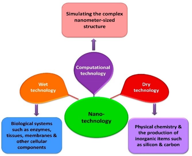
Fig 1. Different Nanotechnology at Rice Universit
• Applications of nanotechnology:-
The different fields that find potential applications of nanotechnology are as follows:
A. Health and medicine.
B. Medical use of nano materials.
C. Drug delivery.
D. Applications in ophthalmology.
E. Application of nanotechnology in modified medicated textiles.
G. Nanotechnology in cancer.
H. Application progress of nanotechnology in diabetic retinopathy prevention.
A. Nanotechnology in health and medicine: -Even today, a number of serious and complex illnesses that pose a significant challenge to humanity include diabetes, cancer, Parkinson's disease, Alzheimer's disease, cardiovascular disease, multiple sclerosis, and various serious inflammatory or infectious diseases (like HIV). One use of nanotechnology in the medical and health fields is called "nanomedicine." Nanomaterials and nanoelectronic biosensors are used in nanomedicine. Molecular nanotechnology will profit from nanomedicine in the future.All human races could profit from the numerous anticipated advantages of the medical application of nanotechnology. Early detection and prevention, better diagnosis, appropriate therapy, and illness follow-up are all made feasible by nanomedicine. The use of specific nanoparticles as tags and labels allows for faster biological testing, increased sensitivity, and increased flexibility. The development of nanodevices such as gold nanoparticles, which can identify genetic sequence in a sample when tagged with brief DNA segments, has increased the efficiency of gene sequencing. Nanotechnology can be used to replicate or mend damaged tissue. The use of these so-called artificially stimulated cells in tissue engineering has the potential to transform artificial implants or organ transplantation.
B. Medical use of nano materials The science and technology of nanomedicine is still in its infancy. Nanotechnology expands research and application by interacting with biological molecules at the nanoscale. It is possible to comprehend how nanodevices interact with biomolecules both inside human cells and in the extracellular media. Utilizing physical characteristics that differ from those seen at the microscale, including the volume/surface ratio, is made possible by operating at the nanoscale. The use of gold nanoshells to aid in the diagnosis and treatment of cancer, as well as the use of liposomes as adjuvants for vaccines and as drug delivery vehicles, are two types of nanomedicine that have previously undergone testing in mice and are awaiting human trials.Likewise, another use of nanomedicine that has been effective in rats is drug detoxification. Smaller, less intrusive devices that may be implanted inside the body and have significantly faster biochemical reaction times are used in medical technologies. Nanodevices are quicker and more sensitive than conventional medication delivery methods.
C. Delivery of drugs:-Nanoparticles are employed in nanotechnology to deliver drugs to precise sites. Because the active substance is exclusively deposited in the morbid zone, this procedure uses the necessary dosage of the medicine and greatly reduces adverse effects. This extremely selective method can lessen suffering and expenses. To the sufferers.Consequently, a range of nanoparticles, including dendrimers and nanoporous materials, find use.Drug encapsulation uses micelles made from block co-polymers. They deliver tiny medication molecules to the intended spot. Likewise, active release of medications is achieved by the use of nano electromechanical systems. Gold shells or iron nanoparticles are being used extensively in the treatment of cancer. A tailored medication lowers drug use and treatment costs, resulting in more economical patient care. Drug bioavailability can be increased by using nanomedicines, which are composed of molecules or particles at the nanoscale.Nano-engineered technologies, like nano robots, target molecules to maximize bioavailability over time and at specific locations in the body.The molecules are targeted, and drugs are delivered precisely to the cells. Nanotools and devices for in vivo imaging are also being developed in this field. Nanoparticles are employed as contrast in images, such as those from mris and ultrasounds. Materials with nanoengineering are being created to treat diseases and ailments including cancer. Self-assembled biocompatible nanodevices that can identify malignant cells, autonomously assess the condition, treat it, and provide reports can be developed thanks to the development of nanotechnology.
D. Ophthalmology applications
With the aid of nanostructures and devices that function massively in parallel at the unit cell level, nanomedicine seeks to monitor, control, build, repair, defend, and enhance human biological systems at the molecular level in order to produce medicinal benefits. Nanotechnology concepts like biomimicry and pseudointelligence are used in nanomedicine. Treatment of oxidative stress, intraocular pressure measurement, the ragnostics, the use of nanoparticles to treat choroidal new vessels, prevent scarring following glaucoma surgery, and treat retinal degenerative disease through gene therapy, prosthetics, and regenerative nanomedicine are some of the ways that nanotechnology is being applied in ophthalmology. In addition to helping with many unresolved issues including sight-restoring therapy for patients with retinal degenerative disease, nanotechnology will transform the current therapeutic hurdles in drug delivery and postoperative scarring [35]. This developing field is anticipated to provide treatments for ocular illnesses. For the treatment of severe evaporative dry eye, a new nanoscale dispersed eye ointment (ndeo) has been effectively created [36]. Medium-chain triglycerides (mct) were combined with petrolatum and lanolin, which are semisolid lipids used in traditional eye ointment, to create a liquid lipid. The two phases were then dispersed in polyvinyl pyrrolidone solution to create a nanodispersion. The ointment matrix was confined in the mct nano emulsion, which had a mean particle size of roughly 100 nm, according to a transmission electron microscopy. When compared to commercial polymer-based artificial tears (tears natural® forte), the improved formulation of ndeo showed no cytotoxicity to human corneal epithelial cells and remained stable after six months of storage at 4°C. When compared to a commercial product, the therapeutic effects of ndeo were assessed and showed therapeutic improvement, showing a trend of positive correlation with higher concentrations of ointment matrix in the ndeo formulations. According to histological analysis, the ndeo is safe for use in ophthalmology and restored the normal morphology of the cornea and conjunctiva. Current studies demonstrates how different nanoparticulate systems, such as microemulsions, nanosuspensions, nanoparticles, liposomes, niosomes, dendrimers, and cyclodextrins, can be used in the field of ocular drug delivery. It also illustrates how the frontiers of ocular drug delivery and therapy can be explored through the use of various emerging nanotechnology, such as nanodiagnostics, nanoimaging, and nanomedicine.
E. Application of nanotechnology in modified medicated textiles:-
Newer antimicrobial cotton has been created and used for antibacterial textiles using nanotechnology. Nanotechnology has been used in the development of novel improved antimicrobial textiles. There have already been reports of the use of traditional antibacterial chemicals on fabrics. Focusing on inorganic nanostructured materials that exhibit strong antibacterial properties and applying them to textiles has improved this technology. [4]
F. Nanotechnology in cancer:-

Figure 2: nanotechnology improve cancer detection and dignosis
Utilizing nanotechnology to identify extracellular cancer biomarkers Indicating the presence of cancer in the body, a cancer biomarker is a detectable biological molecule present in blood and other bodily fluids, including urine and saliva .When cancer is present, the body or cancer cells may release proteins (secreted proteins or cell surface proteins) ,carbohydrates , or nucleic acids (circulating tumor dna, mirna, etc.) As cancer biomarkers .Measuring specific amounts of cancer biomarkers helps track the effectiveness of treatment and allows for early detection of cancer or tumor recurrence.However, a number of obstacles have hindered the use of biomarkers, such as low concentrations of biomarkers in bodily fluids, variations in the timing and quantity of biomarkers within patients, and the challenge of conducting prospective studies High sensitivity and selectivity as well as the capacity to assess several targets at once are provided by nanotechnology. Nanoparticles and nanomaterials can enhance biosensors by offering targeted specificity Furthermore, using nanoparticles increases the surface-to-volume ratio, which improves the sensitivity of biosensors in meeting the requirements of particular biomolecular diagnostics.The quantum dots Three popular nanoparticle probes for cancer diagnosis are polymer dots (pds), gold nanoparticles (aunps), and quantum dots (qds).
F.1 Protein detection:-The FDA has approved several proteins for use in cancer detection, including as ca-125 (ovarian cancer), afp (liver cancer), psa (prostate cancer), and cea (colorectal cancer). The identification of these characteristics may be aided by particular interactions with aptamers, antibody fragments, or antibodies. After that, the interaction event will be transformed into a measurable signal.QD-based biosensors have been employed in recent research to find cancer biomarkers. High resistance to photobleaching, exceptional resistance to degradation, extensive absorption with narrow, high-efficiency stokes shifts, and a high quantum yield and molar extinction coefficient are the distinctive characteristics of Qds. A sandwich-type test, which consists of a biomarker, a capture antibody, a second capture antibody, and a secondary antibody that binds to the capture antibody, is a popular method for finding protein biomarkers. Staining and fluorescence are two techniques that can be used to visualize the secondary antibody.This approach used two qd-conjugated antibodies against carcinoembryonic antigen (cea) and neuron-specific enolase (nse) to detect two biomarkers, with a limit of detection (lod) of 1.0 ng/ml for each.A sandwich immunoassay based on zinc oxide (zno) qd was created for zno nanowire substrates because of its enormous surface area, which presents several binding sites for detection.The most commonly researched cancer biomarker, cea, has been used to predict tumor recurrence after surgical resection in individuals with late-stage cancer and to track anticancer treatment. The enzyme Nse, which catalyzes the transformation of 2-phosphoglycerate into phosphoenolpyruvate, has been linked to islet cell cancers, carcinoids, and small cell lung cancer. They each had a lod of 1.0 ng/ml and were detectable at concentrations greater than 15 ng/ml after secretion. Zno qds coated with antibody against carbohydrate antigen 19-9 (ca 19-9, a biomarker for pancreatic ductal adenocarcinoma) is another example of a qds-based immunosensor. This is a significant application in the detection of ca 19-9.Due to the high isoelectric point of zinc, electrostatic adsorption mostly facilitated the immobilization process, while the sandwich shape was created by the immunological response to the ca 19-9 antigens and antibodies. The detection of amplified signals by square wave stripping voltammetry (swv) and the intrinsic photoluminescence (pl) displayed by the labeled qds were transformed from Ca 19-9 immunological recognition. The optical spectral detection showed a dynamic range of 1–180 u/ml and a lod of 0.25 u/ml, whereas the electrochemical assay's dynamic range was 0.1–180 u/ml and the lod was 0.04 u/ml [30]. In vivo, peptides are commonly used to actively target malignant areas. The arg-gly-asp (rgd) peptide motif has been used to target tumor tissue in vivo for diagnostics because it is recognized by a cell surface receptor (integrin ?v?3) linked to angiogenesis and cancer spread. In one work, the tumor-homing penetration peptide irgd was simply co-administered with a legumain-responsive aggregable gold nanoparticle to create an irgd (crgdkgpdc)-mediated and enzyme-induced precise targeting gold nanoparticle system (irgd/aunps-a&c).In four t1 mammary tumors, Irgd/aunps-a&c demonstrated significant penetration and accumulation .Nanoparticles can also be coupled to aptamers, which are single-stranded DNA (ssdna) or rna sequences that can be extracted via exponential enrichment (selex), which depends on ligand systematic evolution.For their specific targets, which include ions, bacteria, peptides, viruses, phospholipids, and even entire cells, aptamers exhibit high affinity and remarkable binding specificity. Cy5-containing polymeric nanoparticles coupled with the A10 rna aptamer have the ability to attach to the prostate-specific membrane antigen (psma). Only lncap cells and canine prostate cancer cells, which are positive for psma, could bind to Cy5-pla/aptamer nps; pc3 cells, which are negative for psma, could not. When used in Balb/c mice, Cy5-pla nps show exceptional signals with little background fluorescence in various organs.Ucnps, or rare-earth upconverting nanophosphors, hold promise as a biological luminescent label of the future. By using the upconversion process following multiple-photon absorption, Ucnps may absorb near-infrared (nir) light and convert it into visible light. Prostate cancer cell growth has been linked to the overexpression of secreted phospholipase a2 (spla-2),an enzyme that catalyzes phospholipid hydrolysis. Phosphate surfactants, like phospholipids, are helpful in identifying and cleaving the spla-2 enzyme. Since the enzyme hydrolyzes an ester group between the fatty acid and ethylene glycol, the nanoparticles are immediately released from the prostate cancer surface, making this special feature potentially very helpful in controlling ucnp release.
F.2 Ctdna detection:-
Tumor-derived DNA fragments, roughly 100–200 base pairs long, are known as circulating tumor DNA (ctdna) and are found in the bloodstream .Primary tumors or circulating tumor cells (ctcs) can release CTDNA, which can be used to identify cancer by looking for genetic abnormalities unique to the disease. Even before any symptoms of cancer appear, the identification of genetic abnormalities in the CTDNA can aid in the detection of cancer. Genetic abnormalities linked to cancer can be found by highly specific hybridization with nucleic acid probes that have complementary sequences.To identify a single exon in the brca1 gene in breast cancer, a DNA silver nanocluster (nc) fluorescent probe was created [42]. This probe raised the lod to 6.4 × 10⁻¹¹ m under ideal circumstances. Based on nanocluster fluorescence following recognition-induced hybridization, large deletion mutations in brca1 were identified. The brca1 deletion was distinguished by the efficient enhancement of the agnc fluorescence, which came in different intensities, through specific hybridization of the dna-templated silver nc fluorescent probe to target dnas. The presence of the deletion type increased in a dynamic range of 1.0 × 10⁻¹⁰ to 2.4 × 10⁻⁶ m, and the lod reached 6.4 × 10⁻¹¹ m. Under ideal conditions, the fluorescence intensity displayed by dna-agncs at emission peaks near 440 nm (excitation at 350 nm) rose. The normal kind of this sensing system usually exhibits modest fluorescence, however the deletion type produced increased fluorescence emission.
F.3Microrna detection:- Microrna is linked to the diagnosis of cancer. Mir-141 is a promising biomarker for prostate cancer, and Jou af described a two-step sensing method for its sensitive detection. The sensing platform's initial phase involved using cdse/zns qds modified with fret quencher-functionalized nucleic acids, which included a recognition sequence for the mir-141 recognition sequence as well as a telomerase primer sequence. The nucleic acid-functionalized cdse/zns qds showed covalent binding to the fret quencher. A duplex was created when the probe and mir-141 hybridized, and duplex-specific nuclease (dsn) would cleave it. The cleavage triggered the qds' fluorescence and liberated the quencher unit. The telomerase primer sequence was also made visible as a result of this cleavage. In the second phase, telomerase/dntps-stimulated primer unit elongation, hemin incorporation, and chemiluminescence produced with luminol/h2o2 were all involved. This platform distinguished between healthy people and those who carried prostate cancer and assisted in detecting mir-141 in a serum sample.
F.4DNA methylation detection :- According to recent reports, the genome methylation landscape, or methylscape, is shared by the majority of cancer types and may serve as a common biomarker for cancer .The authors of this study used DNA-gold affinity and DNA solvation to identify changes between cancer and normal genomes. They then created straightforward, rapid, sensitive, and selective electrochemical or colorimetric one-step assays to identify cancer.
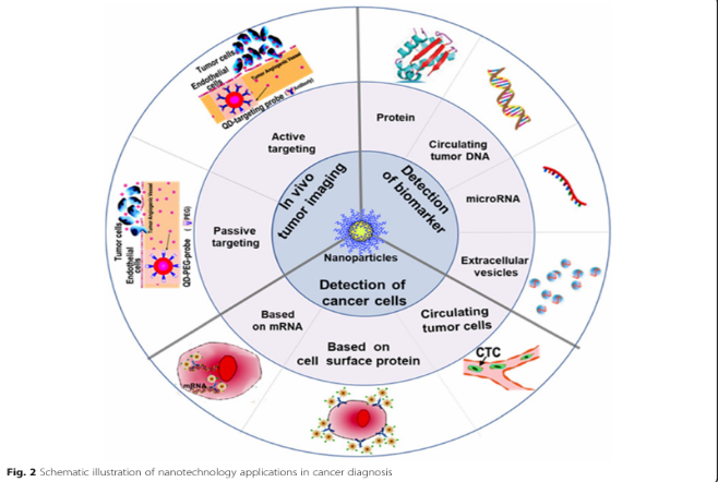
[5] Fig .3 schematic illustration of nanotechnology application in cancer dignosis
• Application progress of nanotechnology in diabetic retinopathy prevention :- In addition to avoiding intense exercise and managing blood pressure and sugar levels, routine fundus examinations are essential for detecting dr early. The gold standard for examining the fundus is fluorescein angiography, which first appeared in the 1960s. The fluorescent dye used in the technique is sodium fluorescein, which is rapidly injected into the antebrachial vein. Angiograms are created by taking pictures of the brilliant green light that the dye emits as the blood vessels return to the eye after the agent has flowed into the retinal vessels with blood. By using the acquired angiograms, the microstructure and microcirculation alterations of fundus vessels can be seen, as well as the dynamic cycling of blood within the retina. The use of polyethyleneimine-modified fluorescein sodium (pei-nhac-fs) nanoparticles (nps) in fundus fluorescein angiography and its safety were investigated by Qian et al.Pei-nhac-fs nps may be used to inspect the fundus and detect the onset of dr. Since the study showed that it can effectively decrease the body's retention of fluorescein sodium and create retinal blood vessels. Pei-nhac-fs nps are also distinguished by their rapid metabolic rate, stable characteristics, strong biocompatibility, and easy manufacturing procedures. But like other fundus angiography methods, it invariably makes patients feel more invasive. According to Vadanasundari Vedarethinam, vanadium core-shell nanorods can be utilized to monitor the severity of dr, identify metabolic changes in the condition, and enable early diagnosis and detection. Motivated by this idea, Vadanasundari et al. Created a range of vanadium core-shells with various elements and structural parameters using vanadium oxide loaded on silica nanorods in an effort to support laser desorption ionization mass spectrometry and create plasma metabolic fingerprints of diabetic retinopathy.Blood testing for diabetic patients will be required in the future in order to detect the disease sooner, which will greatly aid in its prevention and treatment. Additionally, the technique created by Vadanasundari et al. Has notable performance in disease grading and can be utilized to differentiate between the severity levels of non-proliferative diabetic retinopathy. As a result, it offers Dr. A new, trustworthy, and effective blood testing method.40 The results of this most recent study have been used in clinical trials.
![the role of nanotechnology in the pathogenesis of diabetic retinopathy.[6].png](https://www.ijpsjournal.com/uploads/createUrl/createUrl-20241202130453-3.png)
Fig. 4 the role of nanotechnology in the pathogenesis of diabetic retinopathy.[6]
• Advantages of nanotechnology in medicine :-
1]Targeted drug delivery:- By delivering medications specifically to particular cells or tissues, nanoparticles can reduce side effects and increase the effectiveness of treatment.
2]Improved imaging:-By improving imaging methods, nanomaterials enable earlier and more precise diagnosis of conditions like cancer.
3] Enhanced therapies:- By employing nanocarriers to increase the solubility and bioavailability of poorly soluble medications, for example, nanotechnology can increase the efficacy of treatments.
4]Diagnostics:-Early diagnosis and improved health status monitoring are made possible by nanosensors' ability to identify diseases at extremely low concentrations.
5]Vaccines: By enhancing the immune response and stability of vaccine formulations, nanoparticles can be utilized to provide more potent vaccinations.
6]Regenerative medicine: Through the creation of scaffolds that encourage cell growth and healing, nanotechnology supports tissue engineering and regenerative medicine.
7]Reduced dosage: Nanotechnology can lower the total dosage needed by improving the delivery and efficacy of drugs, hence lowering possible toxicity.[7]
•Disadvantage of nano technology in medicine:- one significant disadvantage of nanotechnology in medicine is the potential for toxicity and biocompatibility issues. Nanoparticles can interact with biological systems in unpredictable ways, leading to adverse effects on cells and tissues. For example, certain nanoparticles may provoke immune responses or cause oxidative stress, which can harm healthy cells.[8]
• Manufacturing of nanotechnology
A] Nanomaterials for the 21st century
This chapter discusses a number of novel or improved nanomaterials that are expected to significantly influence technical applications generally and medical technology specifically. Carbon and inorganic nanoparticles are the two types of nanomaterials. Because the physical interactions between biological structures and materials at the nanoscale interface are essential for the biological response, it should be underlined that not only nanoparticles but also nano-structured bulk materials are studied.
•Carbon nanomaterials
1.Introduction: carbon bonds and structures Some of the most intriguing nanostructures are formed as a result of the bonds that exist between carbon atoms. Diamond and graphite are the two traditional allotropes of solid carbon at normal temperature. In a tetrahedral lattice structure, carbon atoms in diamond are joined to four other carbon atoms to form a three-dimensional network. The hardest mineral in the world, diamond is also a great electrical insulator. Hexagon-shaped carbon atoms are firmly bound together to form parallel planar sheets in graphite. Because the Van der Waals forces holding the sheets together are significantly weaker, graphite can be utilized as a base for some lubricants and as a material for pencils.Graphite conducts electricity, unlike diamond. The physical characteristics of pure carbon compounds can differ significantly, as these classical examples demonstrate.
2.C60 A significant advance in the field of carbon chemistry was made in 1985 when it was discovered that there was a third and new carbon allotrope with sixty precisely symmetrically distributed carbon atoms (C60) (Kroto et al., 1985). In honor of the American inventor and architect R. Buckminster Fuller (1895–1983), who created geodesic domes resembling the structure of C60, the C60 molecule was first and officially known as buckminsterfullerene. Since the spherical shape of C60 resembles a football, scientists immediately dubbed it a "buckyball." There are sixty vertices and thirty-two faces in the geometric configuration, twelve of which are pentagonal and twenty of which are hexagonal.The facets are arranged symmetrically to create a molecular ball that is around 1.0 nm in diameter. Numerous more fullerene molecules with different shapes and structures, including C70, C76, C80, and C84, were synthesized shortly following the discovery of C60 (Dresselhaus et al., 1996). The higher fullerenes are intriguing as well and have a number of potential benefits on their own.
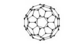
Fig.5 Representation of a C60 molecule.
3.Carbon nanotubes:- One of the amazing things that science occasionally finds and that will probably transform 21st-century technology advancements are carbon nanotubes. Carbon nanotubes are molecules made entirely of carbon atoms and are the fourth allotrope of carbon. They are elongated fullerenes, if you will. Only a small number of fullerene uses have made it to the market, despite the high expectations around their discovery. Nonetheless, since carbon nanotubes' mechanical, electrical, thermal, and optical characteristics outperform those of widely used materials, optimistic forecasts are being made about them. The existence of concentric multi-walled carbon nanotubes (mwcnts) as byproducts of fullerene production was reported by Sumio Iijima in 1991 (Iijima, 1991).Two years later, the true breakthrough came when two research groups independently found single-walled carbon nanotubes (swcnts), which are made up of a single seamless cylindrical wall of carbon atoms. This discovery was made by Bethune et al. (1993) and Iijima and Ichihashi (1993). Similar to cylinders inside cylinders, mwcnts are a group of concentric swcnts with varying sizes. Swcnts were extremely novel nanoobjects with distinct characteristics and behaviors. It has recently been possible to create pure double-walled carbon nanotubes (dwcnts) (Endo et al., 2005b). In a number of applications, these intermediate structures may be used in place of mwcnts or swcnts due to their potentially higher material qualities.
Structure:- In essence, carbon nanotubes are highly organized, rolled-up sheets of hexagonal carbon honeycomb that can be either closed or open at the end.(Figure 6).
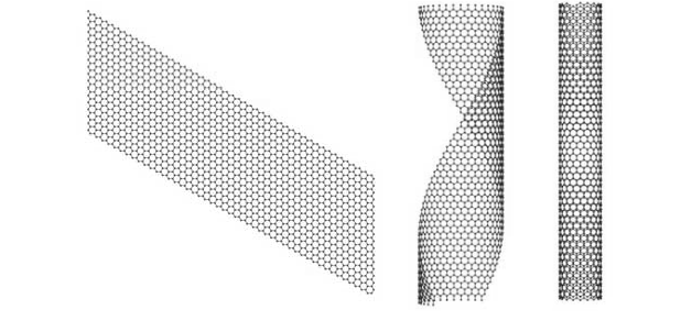
Fig. 6 Illustration of a flat carbon sheet (left) rolled-up into a partially rolledup sheet (middle) and carbon nanotube.
4.Other carbon nanotube-based materials
4.1 Doped carbon nanotubes:- It is possible to replace the carbon atoms in all kinds of nanotubes with another element, such nitrogen or boron. The synthesis of these carbon nanotubes doped with B and/or N was initially documented in 1994 (Stephan et al., 1994). Besides partial replacement, carbon can be totally replaced. One remarkable outcome of these efforts is a sandwich-like structure composed of stacked concentric nanotubes, the coaxial tubes of which are composed of either boron nitride graphenes or carbon graphenes (Suenaga et al., 1997). Arc discharge, laser ablation, and chemical vapour deposition are synthesis methods that are comparable to those used to create pure carbon nanotubes. See, for example, Ma et al. (2004) for a summary.
4.2 Endohedral carbon nanotubes Other atoms, molecules, compounds, or crystals can partially or completely occupy the interior cavity of scents or mwcnts. These hybrid nanotubes are referred to as X@SWCNT or X@MWCNT, where X is the atom, molecule, etc. involved, much like fullerenes. By thermally annealing C60 powders over swcnts at >600 °C under vacuum, swcnts were first filled with C60 in 1998 (Smith et al., 1998). Within the SWCNT, the C60 molecules form a self-assembled chain that resembles a nanoscopic peapod. Additionally, endohedral fullerenes have been added to SWCNTS, and more intricate hybrid materials based on nanotubes, including Gd@C82@swcnts, have been created (Suenaga et al., 2000). Hybrid materials based on nanotubes may find use in micro-electromechanical and electrical systems.
However, due in part to their great chemical stability, extreme hydrophobicity, and likely poor biocompatibility, fullerenes, carbon nanotubes, and peapods have not yet found widespread (bio)medical applications. However, these characteristics could be altered by carefully functionalizing these carbon nanostructures (Sayes et al., 2004).
4.3 Functionalised carbon nanotubes:- Carbon nanotube functionalization has significant effects on the materials' characteristics and uses. Functionalization by non-covalent adsorption of (biological) molecules is simpler than covalent attachment of molecules on the sidewalls of nanotubes. According to Chen et al. (2001), non-covalently functionalized SWCNTS offer locations for selective binding while maintaining the sp2 hybrid bonds (no bonds are broken) and, consequently, the electronic structure of carbon nanotubes. When combined with biomolecular recognition skills (such as antibodies), the special qualities of carbon nanotubes may result in tiny electronic devices, such as sensors and probes. A recent assessment of the properties and uses of functionalized carbon nanotubes was conducted by Sun et al. (2002).
•Inorganic nanomaterials
1.Inorganic fullerene-like molecules:- Although there hasn't been any experimental proof, theoretical research indicates that totally inorganic fullerene-like compounds made up of group 13/15 (such as boron-nitrogen) and C60 analogues with silicon atoms should be achievable. There have been recent reports of the synthesis and structural characterization of soluble, completely inorganic, spherical molecules that resemble fullerenes and contain Cu, Cl, Fe, C, P, and N (Bai et al., 2004). There are currently no anticipated uses for these compounds; they are primarily of scientific interest.
2.Inorganic nanotubes:-
Synthesis:- In recent years, reports on the synthesis of several inorganic nanotubes have been published (see Tenne and Rao (2004) for a current overview). The structure of carbon nanotubes is similar to that of inorganic nanotubes. Chalcogenides, like Mos2 (Chhowalla and Amaratunga, 2000), oxides, like Tio2 (Kasuga et al., 1998), nitrides, like BN (Gleize et al., 1994), halides, like Nicl2 (Hacohen et al., 1998), and metals, like Ni (Bao et al., 2001), are a few of the significant inorganic nanotubes that are manufactured. Similar synthesis methods to those for carbon nanotubes include laser ablation (Parilla et al., 2004) and arc discharge (Chhowalla and Amaratunga, 2000).Furthermore, the most effective methods are suitable chemical reactions, like sol-gel chemistry (Kasuga et al., 1998).One flexible method for creating inorganic compounds is sol-gel chemistry. In this process, a molecular precursor is hydrolyzed and then heated, usually in air.
3.Dendrimers First created in the early 1980s, dendrimers are spherical, complex, and synthetic molecules with highly distinct chemical structures (Newkome et al., 1985; Tomalia et al., 1985). The Greek word "dendron," which means tree, is where the word "dendrimers" comes from. The term "dendrimer" has replaced other words including "arborols," which comes from the Latin word "arbor," which also means tree, and "cascade molecule." Dendrimers are almost perfect monodisperse macromolecules having a regular and highly branching three-dimensional, or fractal, architecture from the perspective of polymer chemistry. The core, branches, and terminal groups at the periphery are their three main architectural components (Figure 7). The components of the macromolecule radiate forth from the central core in branching forms, forming an interior cavity and a sphere of end groups that can be shaped to meet specific needs..
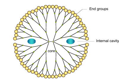
Fig. 7 Schematic representation of a dendrimer.
An initiator core serves as the foundation for the dendrimer's successive shells, or generations (the fourth generation is shown). Quest molecules are contained within the "dendritic box's" interior cavity.[9]
CONCLUSION: -
Nanotechnology in medicine represents a transformative frontier with the potential to revolutionize diagnostics, therapeutics, and drug delivery systems. By manipulating materials at the nanoscale, researchers can enhance the efficacy of treatments, improve targeted delivery to specific cells or tissues, and minimize side effects. Applications include advanced imaging techniques, cancer treatment, and regenerative medicine.However, while the promise of nanotechnology is significant, challenges remain. These include concerns about biocompatibility, potential toxicity, and the need for comprehensive regulatory frameworks to ensure safety. Ongoing research is critical to address these issues and to translate laboratory findings into clinical practice.In conclusion, as we continue to explore and harness the capabilities of nanotechnology, its integration into medicine could lead to more personalized, effective, and safer healthcare solutions, ultimately improving patient outcomes and transforming the landscape of medical treatment.
REFERENCES
-
-
-
- Novel applications of nanotechnology in medicine, A. Surendiran, S. Sandhiya, Pradhan & c. Adithan, Indian j med res, december 2009,http://journals.lww.com/ijmr by bhdmf5ephkav1zeoum1tqfn4a+kjlhezgbsiho4xmi0hcywcx1awnyqp/ilqrhd3i3d0odryi7tvsfl4cf3vc4/oavpdda8k2+Ya6H515kE= on 11/02/202,689
- Mallanagouda Patil1, Dhoom Singh Mehta2, Sowjanya Guvva3, Future impact of nanotechnology on medicine and dentistry, Journal of Indian Society of Periodontology - Vol 12, Issue 2, May-Aug 2008, http://journals.lww.com/jisp by bhdmf5ephkav1zeoum1tqfn4a+kjlhezgbsiho4xmi0hcywcx1aw nyqp/ilqrhd3i3d0odryi7tvsfl4cf3vc1y0abggqzxdggj2mwlzlei= on 11/02/2024, 34
- Mritunjai singh, Shinjini singh, s. Prasada, i. S. Gambhir, nanotechnology in medicine and antibacterial effect of silver nanoparticles, digest journal of nanomaterials and biostructures vol. 3, no.3, September 2008, p. 116 117.
- Anna Pratima Nikalje Nanotechnology and its Applications in Medicine, Med chem Volume 5(2): 081-089 (2015) - 81 ISSN: 2161-0444 Med chem, an open access journal p 81-88.
- Ye Zhang1†, Maoyu Li1,2†, Xiaomei Gao3, Yongheng Chen1 andting Liu2 Nanotechnology in cancer diagnosis: progress, challenges and opportunities zhang et al. Journal of Hematology & Oncology, https://doi.org/10.1186/s13045-019-0833-3, 1-13.
- Yuxin Liu & Na Wu, Progress of Nanotechnology in Diabetic Retinopathy Treatment, International Journal of Nanomedicine,1393-1396.
- Patel, r. S., & patel, m. M. (2017). "nanoparticle-based drug delivery systems: a review of their applications in cancer therapy." journal of drug delivery science and technology, 39, 30-41. Doi:10.1016/j.jddst.2017.08.001.
- S. S. G. S. Rao et al. (2013). "toxicological aspects of nanomedicine: an overview." nanomedicine: nanotechnology, biology, and medicine, 9(4), 505-511.
- B. Roszek, W.H. de Jong and R.E. Geertsma, Nanotechnology in medical applications: state-of-the-art in materials and devices, RIVM report 265001002, Bilthoven.,21-123.
- Abe M, Hiraoka M, Takahashi M, Egawa S, Matsuda C, Onoyama Y, Morita K, Kakehi M and Sugahara T (1986). Multi-institutional studies on hyperthermia using an 8-MHz radiofrequency capacitive heating device (Thermotron RF-8) in combination with radiation for cancer therapy. Cancer 58, 1589-1595.
- R. Holladay, W. Moeller, D. Mehta, J. Brooks, R. Roy, M. Mortenson, Application Number WO2005US47699 20051230 European Patent Office (2006).
- Zhang, L., et al. (2024). "Advanced Drug Delivery Systems: Current Status and Future Perspectives." Nature Nanotechnology, 19(1), 23-45.
- 2. Chen, X., & Wilson, J. (2023). "Nanostructured Materials for Tissue Engineering Applications." Advanced Materials, 35(15), 2200134.
- Smith, A.B., et al. (2024). "Smart Nanocarriers for Targeted Cancer Therapy." Journal of Controlled Release, 355, 125-147.
- Johnson, M.R., & Brown, P. (2023). "Neural Interfaces: Bridging Biology and Electronics." Nature Reviews Materials, 8(4), 341-362.
- Patel, S., & Roberts, M. (2024). "Nanomaterials for Brain Drug Delivery." Advanced Drug Delivery Reviews, 192, 114578.
- Thompson, D.A., et al. (2023). "Regulatory Framework for Nanomedicine." Nature Reviews Drug Discovery, 22(8), 623-645.
- Williams, E.M., et al. (2023). "Nanomedicine in Cancer Immunotherapy." Nature Cancer, 4(3), 278-295
- Hughes, R.A., et al. (2024). "Environmental Applications of Nanomaterials." Environmental Science: Nano, 11(2), 345-367.
- Lee, K.M., & Wang, X. (2023). "Next-Generation Nanomedicine." Science Translational Medicine, 15(723), eabc9876.
- Tomalia DA, Reyna LA, Svenson S. Dendrimers as multipurpose nanodevicesfor oncology drug delivery and diagnostic imaging. Biochem Soc Trans 2007; 35 : 61-7


 Sanjay Wagh*
 Shaikh Ansari F.
 Dr. Rajendra Kawade


![the role of nanotechnology in the pathogenesis of diabetic retinopathy.[6].png](https://www.ijpsjournal.com/uploads/createUrl/createUrl-20241202130453-3.png)


 10.5281/zenodo.14258034