1,2 Madhav University Sirohi, Pindwara Rajasthan -307026
3 Ideal College of Pharmacy, Kalyan (E), Mumbai
4 Mula Education Society Sharda college of pharmacy, Vadgaongupta, Ahmednagar
The present study focuses on the formulation and evaluation of an in situ gelling ophthalmic drug delivery system for Ofloxacin, a broad-spectrum fluoroquinolone antibiotic. The aim was to develop a formulation that undergoes sol-to-gel transition upon exposure to physiological conditions, thereby enhancing ocular residence time and drug bioavailability. Poloxamer 407 and 188 were employed as thermosensitive polymers, with Hydroxypropyl Methylcellulose (HPMC) and Carbopol 934 as viscosity enhancers and mucoadhesive agents. The gels were prepared using a cold method and evaluated for parameters such as pH, gelling temperature, gel strength, drug content, rheological behavior, clarity, sterility, and in vitro drug release. Among the developed formulations, F2 demonstrated optimal performance with satisfactory gelling temperature (37.3°C), high drug content (94.90%), appropriate gel strength, and excellent clarity and sterility. In vitro release studies indicated sustained drug release up to 8 hours, achieving 94.25% cumulative release. Stability studies confirmed the formulation's robustness over time. This in situ gel system presents a promising alternative to conventional eye drops by reducing dosing frequency and improving patient compliance and therapeutic efficacy in ocular infections.
In Situ Hydrogels
Hydrogels are polymeric networks that absorb large quantities of water while remaining insoluble in aqueous solutions due to chemical or physical crosslinking of individual polymer chains. They resemble natural living tissue more than any other class of synthetic biomaterials due to their high water content; furthermore, the high water content of the materials contributes to their biocompatibility. Hydrogels show minimal tendency to adsorb proteins from body fluids because of their low interfacial tension. Further, the ability of molecules of different sizes to diffuse into (drug loading) and out of (drug release) hydrogels allows the possible use of dry or swollen polymeric networks as drug delivery systems for oral, nasal, buccal, rectal, vaginal, ocular and parenteral routes of administration. These are polymers endowed with an ability to swell in water or aqueous solvents and induce a liquid-gel transition. Currently, two groups of hydrogels are distinguished, namely preformed and in situ forming gels. Preformed hydrogels can be defined as simple viscous solutions which do not undergo any modifications after administration. The use of preformed hydrogels still has drawbacks that can limit their interest for ophthalmic drug delivery or as tear substitutes. They do not allow accurate and reproducible administration of quantities of drugs and, after administration; they often produce blurred vision, crusting of eyelids, and lachrymation. Thus in situ hydrogels can be instilled as eye drops and undergo an immediate gelation when in contact with the eye In situ-forming hydrogels are liquid upon instillation and undergo phase transition in the ocular cul-de-sac to form viscoelastic gel and this provides a response to environmental changes. Three methods have been employed to cause phase transition on the surface: change in temperature, pH, and electrolyte composition. Increase in solution viscosity by using polymers improves retention of product on the corneal surface. More recently, the approach to improve precorneal retention is based on the use of mucoadhesive polymers. The principle for use of Bioadhesive vehicles relies on their ability to interact with the mucin-coating layer present at the eye surface. The polymers chosen to prepare ophthalmic hydrogels should meet some specific rheological characteristics. It is generally well accepted that the instillation of a formulation should influence tear behavior as little as possible. Because tears gave a pseudoplastic behavior, pseudoplastic vehicles would be more suitable as compare to Newtonian formulations, which have a constant viscosity independent of the shear rate, whereas pseudoplastic solution exhibit decreased viscosity with increasing shear rate, thereby offering lowered viscosity during blinking and stability of the tear film during fixation.
Drug release from hydrogels
As discussed in the previous sections, hydrogels have a unique combination of characteristics that make them useful in drug delivery applications. Due to their hydrophilicity, hydrogels can imbibe large amounts of water (N90 wt.%). Therefore, the molecule release mechanisms from hydrogels are very different from hydrophobic polymers. Both simple and sophisticated models have been previously developed to predict the release of an active agent from a hydrogel device as a function of time. These models are based on the rate limiting step for controlled release and are therefore categorized as diffusion, swelling & chemically controlled mechanism.
Smart Hydrogels
"Smart" hydrogels, or stimuli-sensitive hydrogels, are very different from inert hydrogels in that they can "sense" changes in environmental properties such as pH and temperature and respond by increasing or decreasing their degree of swelling. The volume-changing behavior of "smart" hydrogels is particularly useful in drug delivery applications as drug release can be triggered upon environmental changes. These "intelligent" or "smart" polymers play important role in drug delivery since they may dictate not only where a drug is delivered, but also when and with which interval it is released. Swelling of hydrogel increases as the external pH increases in the case of weakly acidic (anionic) groups, but decreases if polymer contains weakly basic (cationic) groups. Another mechanism of in situ hydrogel is ion induced gelation. In this, polymers may undergo phase transition in presence of various ions. Gellan gum commercially available as Gelrite® is an anionic polysaccharide that undergoes in situ gelling in the presence of mono- and divalent cations, including Ca2+, Mg2+, K+ and Na+. Gelation of the low-methoxy pectins can be caused by divalent cations, especially Ca2+
Sol (particles unconnected)
Gel (particles connected)

Fig No. 01: A Schematic diagram of Sol-gel mechanism
In Situ gel system
The use of preformed hydrogels still has drawbacks that can limit their interest for ophthalmic drug delivery or as tear substitutes. They do not allow accurate and reproducible administration of quantities of drugs and, after administration; they often produce blurred vision, crusting of eyelids, and lachrymation. A new approach is to try to combine advantages of both solutions and gels, such as accuracy and facility of administration of the former and prolonged residence time of the later. Thus, in situ hydrogels can be instilled as eye drops and undergo an immediate gelation when in contact with eye. The liquid to semisolid phase change can be triggered by increased temperature, increased pH and ionic strength of the tear film.
Based on different stimuli, in situ forming hydrogels can be classified as follow:
Ionically induced gelation
Gellan gum is an anionic exocellular polysaccharide by the bacterium pseudomonas elodea, having the characteristic property of cation-induced gelation. The acetylated form is commercially available as gelrite (Kelco division of Merck and Co, USA). The sol-gel transition process is induced by the presence of monovalent or divalent ions such as Na+ and Cat. Some other parameters influence the phase transition. e.g.: The concentration of polysaccharide, the temp of the preparation, and the nature and the concentration of cations. It was determined that divalent ions such as magnesium or calcium were superior to monovalent cations in promoting the gelation of the polysaccharide. However the concentration of sodium tears (2-6g/l) is quite sufficient to induce the gelation.
pH induced gelation
Pseudolatexes can be defined as artificial latexes prepared by the dispersion of a preexisting polymer in aqueous medium in situ gelling pseudo latexes for ophthalmic use can be described as aqueous colloidal dispersions of polymer, which become viscous gels after instillation in the conjunctival cul-de-sac due to modification of the pH. Pseudo latexes are obtained by dispersion of an organic solution of a preformed polymer in an aqueous medium, leading to an o/w emulsion. Two principal methods are commonly used to prepare ophthalmic pseudo latexes, the solvent evaporation process and the salting out process. Both methods allow the production of a lyophilized and easily re dispersible power. Thus, pseudo latexes have the advantage of the latex as well as the stability of active compounds such as pilocarpine, which is sensitive to aqueous media.
Thermo reversible hydro gels
These hydro gels are liquid at room temperature (20-250 C) and undergo gelation when in contact with body fluids (35-370 C), due to an increase in temperature. Different thermal settings gels have been described in this Review. For example acrylic acid copolymers and N-isopropylacrlamide derivatives ophthalmic administration such as tolerance have limited the choice of such polymers. Poloxamers, commercially available as pluronic (BASF-Wyandotte, USA), are the most commonly used thermal setting polymers in ophthalmology
Advantages of In situ gels:
Importance of In Situ Gelling System:
The major importance is the possibilities of administrating accurate & reproducible quantities compared to already formed gel.
In situ forming polymeric delivery system such as ease of administration & reduced frequency of administration improved patient compliance & comfort.
LITERATURE REVIEW:
1. Velpandian T., et. al., The present work describes the formulation and evaluation of an ocular delivery system of timolol maleate based on the concept of both temperature and pH-triggered in situ gelation. Pluronic F-127 (a thermosensitive polymer) in combination with chitosan (pH-sensitive polymer also acts as permeation enhancer) was used as gelling agent. The developed formulation was characterized for various in vitro parameters e.g.. clarity, gelation temperature and pH, isotonicity, sterility, theological behavior, drug release profile, transcorneal permeation profile, and ocular irritation. Developed formulation was clear, isotonic solution that converted into gel at temperatures above 35oC and pH 6.9-7.0. A significant higher drug transport across corneal membrane and increased ocular retention time was observed using the developed formulation. The developed system is a viable alternative to conventional eye drops for the treatment of glaucoma and various other ocular diseases.
2. Patel U. L., et. al., Formulation and in vitro evaluation of moxifloxacin hydrochloride ophthalmic inserts were carried out by using different polymers like hydroxypropyl methylcellulose (4-7%), methylcellulose (2-3.5%) and polyvinyl alcohol (4-7%) and by solvent casting method. The aim of this study was to increase the contact time, achieving controlled release of drug, reducing frequency of administration and lastly improving therapeutic efficacy. The prepared ophthalmic insert were then evaluated for uniformity of thickness, weight, drug content, %moisture absorption, %moisture loss, folding endurance and surface pH. In vitro drug release of formulated batches of ophthalmic insert was performed by studying the diffusion through artificial membrane. On the basis of all physicochemical parameters and in vitro drug release studies, the formulation (F12) with PVA (7%) was found to be promising and was selected as an optimized formulation. The result of in vitro diffusion study of selected formulation (F12) exhibited first order kinetic, which is non-fickian in nature. The higuchi plot revealed that the release might be diffusion controlled.
3. Stella M. R., et. al., Ocular Pharmacokinetics of Moxifloxacin after topical treatment of animals and humans was discussed in this study. The ocular penetration and pharmacokinetics of moxifloxacin in comparison to other fluoroquinolones (ofloxacin, ciprofloxacin, gatifloxacin, norfloxacin, levofloxacin, and lomefloxacin) have been determined by in vitro and ex vivo techniques, as well as in animal and human studies. This article reviewed the original pharmacokinetics work performed by Alcon and other studies reported in the ocular fluoroquinolone literature. The results consistently demonstrated higher maximum concentrations for moxifloxacin relative to the other fluoroquinolones in ocular tissues with levels well above its minimum inhibitory concentrations for relevant ocular pathogens. This superior performance is due to the unique structure of moxifloxacin that combines high lipophilicity for enhanced corneal penetration with high aqueous solubility at physiological pH.
4. Wei-San Pan et al 81 (2008) studied Pluronic F127-g-poly(acrylic acid) copolymers as in situ gelling vehicle for ophthalmic drug delivery system to prolong the precorncal resident time and improve ocular bioavailability of the drug. The rheological properties and in vitro drug release of Pluronic-g-PAA copolymer gels were investigated. The rheogram and in vitro drug release studies indicated that the drug release rates decreased as acrylic acid/Pluronic molar ratio and copolymer solution concentration increased. But the drug concentration had no obvious effect on drug release. The release rates of the drug from such copolymer gels were mainly dependent on the gel dissolution. In vivo resident experiments showed the drug resident time and the total resident amount in rabbit's conjunctiveal sac increased by 5.0 and 2.6 folds for in situ gel, compared with eye drops. The decreased loss angle at body temperature and prolonged precorneal resident time also indicated that the copolymer gels had bioadhesive properties. These in vivo experimental results, along with the rheological properties and in vitro drug release studies, demonstrated that in situ gels containing Pluronic-g-PAA copolymer may significantly prolong the drug resident time and thus improve bioavailability. Pluronic-gPAA copolymer can be a promising in situ gelling vehicle for ophthalmic drug delivery system.
5. Liu Z, et al 73 (2006) investigated that the poor bioavailability and therapeutic response exhibited by conventional ophthalmic solutions due to rapid pre-corneal elimination of the drug may be overcome by the use of in situ gel-forming systems that are instilled as drops into the eye and then undergo a sol-gel transition in the cul-de-sac. The present work describes the formulation and evaluation of an ophthalmic delivery system of an antibacterial agent, gatifloxacin, based on the concept of ion-activated in situ gelation. Alginate (Kelton®) was used as the gelling agent in combination with HPMC (Methocel E50Lv) which acted as a viscosity-enhancing agent. The rheological behaviors of all formulations were not affected by the incorporation of gatifloxacin. Both in vitro release studies and in vivo pre-corneal retention studies indicated that the alginate/HPMC solution retained the drug better than the alginate or HPMC ES0Lv solutions alone. These results demonstrate that the alginate/HPMC mixture can be used as an in situ gelling vehicle to enhance ocular bioavailability and patient compliance.
DRUG PROFILE:
|
Name |
Ofloxacin |
|
Structure: |

Fig No 03: Structure of Ofloxacin |
|
IUPAC name: |
7-fluoro-2-methyl-6-(4-methylpiperazin-1-yl)-10-oxo-4-oxa-1-azatricyclo[7.3.1.0]trideca-5(13),6,8,11-tetraene-11-carboxylic acid |
|
Molecular Weight: |
361.368 g/mol |
|
Molecular formula: |
C??H??FN?O? |
|
Solubility: |
It is slightly soluble in water, alcohol, dichloromethane, and methyl alcohol but sparingly soluble in chloroform. |
|
Melting Point: |
250-257°C |
|
Physicochemical Properties: |
Ofloxacin is a pale yellow or bright yellow crystalline powder, |
|
Mechanism of Action: |
Ofloxacin is a fluoroquinolone whose primary mechanism of action is inhibition of bacterial DNA gyrase. In vitro it has a broad spectrum of activity against aerobic Gram-negative and Gram-positive bacteria, although it is poorly active against anaerobes. |
|
Uses: |
Ofloxacin is used to treat certain bacterial infections in many different parts of the body. It may also be used for other problems as determined by your doctor. Ofloxacin may mask or delay the symptoms of syphilis. It is not effective against syphilis infections |
MATERIAL AND METHODOLOGY
Formulation Development
In situ gel was prepared by the cold method. A weighed amount of poloxamer 407 and poloxamer 188 (15-20% w/v) was slowly added to 15 mL water (at °C) in a beaker with continuous stirring using a magnetic stirrer at a speed of 500 rpm for 2 hrs. The temperature of water was maintained at °C throughout the preparation. This solution was kept overnight in refrigerator. HPMC K-100 (0.5% w/v), carbopol 934 (0.1%, 0.3%, and 0.5% w/v), and the preservatives (methyl and propyl paraben 0.1% and 0.01%, w/v resp.) were added to poloxamer dispersion with continuous stirring. The preservative solution was prepared by solubilizing it in hot water. It was mixed with above dispersion after cooling. The weighed amount of drug (2% w/v) was dissolved in the mixture of tween 80 and ethanol (1:2) or glycerin. The drug solution was then mixed in the above described poloxamer dispersion. The final volume was made up and pH of the poloxamer dispersion was adjusted to 7 using triethanolamine, whereas the dispersion containing carbopol was adjusted to pH 5.8.
Table No. 01 Formulation Table
|
Batch |
Ofloxacin (gm) |
Tween 80 (ml) |
Poloxamer 407 (gm) |
Poloxamer 188 (gm) |
Carbopol 934 (gm) |
Hydroxy Propyl Methyl Cellulose |
Methyl Paraben (gm) |
Propyl Paraben (gm) |
Distilled Water (ml) |
|
F1 |
6.66 |
6 |
6 |
- |
- |
0.15 |
0.03 |
0.003 |
30 |
|
F2 |
6.66 |
6 |
4 |
2 |
- |
0.15 |
0.03 |
0.003 |
30 |
|
F3 |
6.66 |
6 |
4.5 |
- |
0.09 |
0.15 |
0.03 |
0.003 |
30 |
|
F4 |
6.66 |
6 |
6 |
- |
0.03 |
0.15 |
0.03 |
0.003 |
30 |
|
F5 |
6.66 |
6 |
6 |
- |
0.09 |
0.15 |
0.03 |
0.003 |
30 |
|
F6 |
6.66 |
6 |
6 |
- |
0.15 |
0.15 |
0.03 |
0.003 |
30 |
RESULT AND DISCUSSION
Preformulation Study:
Table No. 02: Melting point of Ofloxacin
|
Sr. No. |
Sample |
Melting point |
Reported |
|
1 |
Ofloxacin |
250-257 °C |
250-257 °C |
lmax of Ofloxacin was found to be 269 nm in Distilled Water
Table No. 03: Solubility study of Ofloxacin
|
Solvent |
Solubility |
|
Distilled Water |
Slightly soluble |
|
Methanol |
Slightly soluble |
|
acetic acid |
Freely soluble |
Compatibility study determines stability of drug with excipients under experimental conditions. The IR spectra did not show any difference in wavelength from those obtained for their physical mixture with polymers as compared to drug.
These obtained results indicate that there was no interaction between Ofloxacin, polymers and the other excipients. Hence they are compatible with each other. Thus, Ofloxacin can be used in combination with excipients for the preparation thermoreversible in situ gel.
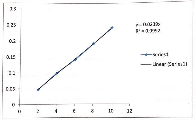
Fig No.03: Calibration curve in Distilled water
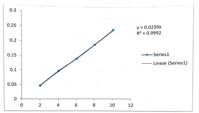
Fig No. 16: Calibration curve in STF
Evaluation of Formulations
Table No. 04: Determination of visual appearance
|
Sr. No. |
Formulation |
Appearance of solution |
Appearance of gel |
|
1 |
F1 |
Transparent |
Transparent |
|
2 |
F2 |
Transparent |
Transparent |
|
3 |
F3 |
Cloudy |
Cloudy |
|
4 |
F4 |
Transparent |
Transparent |
|
5 |
F5 |
Cloudy |
Transparent |
|
6 |
F6 |
Cloudy |
Cloudy |
Table No. 05: Determination of pH
|
Sr. No. |
Formulation |
pH |
|
|
F1 |
7.2 |
|
|
F2 |
7.4 |
|
|
F3 |
7.9 |
|
|
F4 |
7.0 |
|
|
F5 |
7.0 |
|
|
F6 |
6.9 |
Table No. 06: Determination of drug content
|
Sr. No |
Formulation |
Drug Content(%) |
|
1 |
F1 |
89.04 |
|
2 |
F2 |
94.90 |
|
3 |
F3 |
92.06 |
|
4 |
F4 |
91.10 |
|
5 |
F5 |
89.13 |
|
6 |
F6 |
90.67 |
Table No. 07: Gel strength of formulations
|
Sr. No |
Formulation |
Gel strength (S) |
|
|
F1 |
0.76 |
|
|
F2 |
0.59 |
|
|
F3 |
0.55 |
|
|
F4 |
0.51 |
|
|
F5 |
0.63 |
|
|
F6 |
0.77 |
Table No. 08: Determination of gelling temperature
|
Sr. No |
Formulation |
Gelling Temperature |
|
|
F1 |
27.1 |
|
|
F2 |
37.3 |
|
|
F3 |
24.8 |
|
|
F4 |
30.3 |
|
|
F5 |
41.6 |
|
|
F6 |
35.37 |
Table No. 09: Gelling Capacity
|
Sr. No |
Formulation |
Gelling Capacity (min) |
|
|
F1 |
70 |
|
|
F2 |
139 |
|
|
F3 |
181 |
|
|
F4 |
76 |
|
|
F5 |
284 |
|
|
F6 |
276 |
Table No.10: Rheological study
|
Sr. no |
Shear rate(1/s) |
Viscosity (CP) |
|||||
|
F1 |
F2 |
F3 |
F4 |
F5 |
F6 |
||
|
1 |
0.03 |
15.3 |
23.7 |
28.8 |
46.7 |
21.3 |
25.6 |
|
2 |
0.1 |
24.6 |
24.8 |
29.3 |
42.2 |
25.7 |
36.5 |
|
3 |
0.3 |
34.7 |
28.8 |
30.4 |
43.7 |
36.8 |
38.9 |
|
4 |
0.9 |
34.8 |
38.7 |
33.6 |
45.2 |
39.9 |
39.8 |
|
5 |
2.7 |
35.7 |
45.9 |
41.8 |
53.7 |
44.8 |
48.8 |
|
6 |
8.1 |
38.8 |
55.2 |
42.1 |
59.6 |
53.3 |
57.4 |
|
7 |
14.6 |
87.7 |
56.7 |
49.8 |
59.9 |
57.6 |
57.8 |
The prepared in-situ gelling systems were evaluated for the sterility. After 7 days of incubation the results showed that no microbial growth was found.
Table no. 11: Evaluation of sterility testing
|
Formulation |
Days of Incubation |
|||||
|
Day 1 |
Day 2 |
Day 3 |
Day 4 |
Day 5 |
Day 6 |
|
|
F2 |
- |
- |
- |
- |
- |
- |
Table No. 12: Drug release profile of optimized batch
|
Sr No. |
Time (Hr.) |
Percentage cumulative drug release |
|
1 |
0 |
0 |
|
2 |
15 |
30.86 |
|
3 |
30 |
31.74 |
|
4 |
60 |
65.63 |
|
5 |
120 |
73.45 |
|
6 |
180 |
82.45 |
|
7 |
240 |
83.92 |
|
8 |
360 |
85.72 |
|
9 |
480 |
94.25 |
Table No.13: Stability study of optimized batch at 4°C
|
Sr. No |
Time (day) |
Appearance |
PH |
Gelation Temp °C |
Drug Content (%) |
|
1 |
15 |
Transparent |
7.1 |
37 |
98.1 |
|
2 |
30 |
Transparent |
7.0 |
34 |
97.17 |
|
3 |
45 |
Transparent |
7.4 |
35 |
98.19 |
|
4 |
60 |
Transparent |
7.6 |
38 |
98.01 |
Table No. 14: Stability Study optimized batch at 40°C
|
Sr. No |
Time (day) |
Appearance |
PH |
Gelation Temp °C |
Drug Content (%) |
|
1 |
15 |
Transparent |
7.4 |
37 |
99.14 |
|
2 |
30 |
Transparent |
7.2 |
37.6 |
99.17 |
|
3 |
45 |
Transparent |
7.5 |
38 |
99.06 |
|
4 |
60 |
Transparent |
7.4 |
38 |
99.07 |
CONCLUSION
The formulated in situ gel of Ofloxacin using thermosensitive polymers like Poloxamer 407 and 188, along with mucoadhesive agents such as HPMC and Carbopol 934, successfully exhibited desirable physicochemical properties, sustained drug release, and good ocular retention. The optimized formulation (F2) showed excellent clarity, appropriate gelling temperature, and high drug content, with no microbial contamination. This study concludes that the developed in situ gel offers a promising, patient-friendly alternative to conventional ophthalmic solutions by enhancing bioavailability and reducing the frequency of administration.
ACKNOWLEDGMENT
The author expresses sincere gratitude to the guide and faculty members of the Department of Pharmaceutics for their valuable guidance and support throughout the project. Heartfelt thanks to the laboratory staff for their assistance during the experimental work.
REFERENCES


Babu Anmulwad, Asha Chopde, Preet Chavarkar, Salman Shaikh, Formulation and Development of In Situ Gel of Ofloxacin, Int. J. of Pharm. Sci., 2025, Vol 3, Issue 7, 3033-3043. https://doi.org/10.5281/zenodo.16312680
 10.5281/zenodo.16312680
