Abstract
Skin is the body's largest organ and the most exposed to the environment, making it more susceptible to mechanical, chemical, burn, and electrical injury. However, due to the numerous factors that impede wound healing, there is a need to develop a solution with rapid wound healing capacity. Skin tissue engineering aims to improve the potential and replace damaged tissue with healthy tissue by creating extracellular matrix-based bio-ink embedded with medicines and growth factors. Fish air sacs are a rich source of extracellular matrix, collagen, nutrients, and growth factors such as IGF and TGF ?-1. Fish air sacs are an effective material for wound healing. They contain numerous active forms of extracellular matrix that support angiogenesis, cell migration, and proliferation, providing growth factors involved in signaling tissue formation and assisting wound healing. It just not only promotes cell migration and differentiation but also restores the lost dynamic reciprocity between cells and the extracellular matrix, thereby accelerating wound healing. The initiative attempted to create bio-ink with an extracellular matrix obtained from xenogenic sources such as fish air sac. The fish air sac sample was incised and obtained from a local fish shop. It was preserved in D/W with 1% v/v of Antibiotic-Antimycotic solution liquid to avoid contamination, properly washed, and stored for collagen isolation. Once the fish air sac slurry was prepared, polymers were added and polymerized using mechanical and freeze-thaw cycles. The prepared bio-ink was characterized by wetting properties, hemocompatibility, biocompatibility, and in-ovo and in-vitro experiments to ensure its biocompatibility and hemocompatibility. In-ovo investigations confirmed normal angiogenesis and in-vitro investigations revealed a greater cell growth rate in comparison to the control group. This study shows that created extracellular matrix-based bio-inks are hemocompatible, biocompatible, angiogenic, and have minor immunogenic properties. investigations revealed a greater cell growth rate in comparison to the control group. This study shows that created extracellular matrix-based bio-inks are hemocompatible, biocompatible, angiogenic, and have minor immunogenic properties.
Keywords
Swim bladder, air bladder, wound healing, skin tissue engineering, topical cream, collagen.
Introduction
A wound is a physical, chemical, or electrical damage to the skin tissue which can be commonly caused by an injury, accident, or surgery. Skin is considered the first line of barrier to keep a distance from foreign bodies and microbial infections in the internal body. When the wound happens to the body, it can range from minor cuts to serious injuries. Wounds break the outer skin layer barrier, exposing the body to infection. The natural process of wound healing is divided into four stages: 1) Hemostasis 2) Inflammation 3) Proliferation & 4) Remodelling1. But when there is a presence of intrinsic and extrinsic factors influencing the healing process, the wound healing is hindered and causes microbial infection and a slow healing process. The intrinsic factors are increased age, sex, nutritional status, obesity, and multiple diseases or illness and extrinsic factors are moisture, infection, mechanical stress, chemical stress, and consumption of alcohol and narcotic drugs2.These materials can be utilized to produce scaffolds that are suited for reconstruction of cells and tissues and that are nearly identical to the Extracellular Matrix ECM, which speeds up the healing process of wounds3. Scar formation is a very common phenomenon when healing occurs. It occurs due to a lack of collagen deposition at the wound site, so there is a need of a rich collagen source that would be feasible and non-immunogenic for healing purposes. So, to eliminate collagen deficiency, fish air bladder is a best, reliable xenogenic alternative which is introduced which will help to provide abundant collagen and help in the healing process.
Materials:
A fish bladder was purchased from the local fish market. PVA Poly vinyl alcohol CAS No.9002-89-5 Molecular Weight: ~ 1,60,000 purchased from HImedia Laboratories, Chitosan CAS No. 9012-76-4, Gelatine type A CAS No. 9000-70-8 from HIMedia Laboratories. Millipore water was collected from Ramnarain Ruia Autonomous College, Mumbai.
Methodology:
Preparation of Fish bladder slurry.
The Fish bladder sample was incised and procured from the fish market, kept in D/W with 1% v/v of Antibiotic Antimycotic solution 100X liquid to avoid contamination. The collected samples are washed with Millipore water. Then, samples are gashed into small pieces and kept in 80% v/v alcohol, kept in an orbital shaker for 15 minutes for washing to remove any contamination. The samples were taken out from the alcohol solution and kept in millipore water to remove the alcohol. This process was followed by a single wash of 0.5% w/v Sodium lauryl sulphate wash and then it was followed by 3 - 4 wash of millipore water to remove Sodium lauryl sulphate traces. The samples were weighed on the weighing scale and kept in Millipore water below 55°c for 8 h. The dissolved collagen was strained out with cheese-cloth to obtain a pure collagen, the pH was checked and kept sealed in the refrigerator before use.
Formulation of Extracellular matrix-based Gelatin-PVA-Chitosan topical gel/ cream
Chitosan was dissolved in 2N acetic acid, pH was stabilized with a buffer solution. Poly vinyl alcohol [PVA] was measured at 8% w/v and dissolved on a magnetic stirrer for overnight. After the PVA was fully dissolved, 2% w/v gelatin was introduced into the solution and stirred at the same rpm until it was fully dissolved. Once the topical gel was prepared it was sealed and kept in the refrigerator.
Characterizations:
Spreadability test:
Spreadability refers to how easily a cream or gel spreads when applied to the skin or wound area. Spreadability is defined as the amount of time it takes for a gel to spread without friction between two glass slides after a specific force is applied. The spreadability test involved drawing a 2 cm radius in the middle of the posterior side of the petri plate and loading 200µl of gel. Another petri plate was placed exactly over the reversed petri plate. Now, the load was placed on top for 5 minutes. Once the gel has been dispersed, the weight is removed and the circumference of the gel spread is measured4.
The Spreadability was calculated by given formula,
S= (m×w)/t
Formula. 1
Hemolysis Test:
Hemolysis is defined as the destruction of red blood cells RBC. The samples were washed with 0.9% saline. The samples were submerged in 10ml of 0.9% saline solution in a centrifuge tube for 30 minutes at 37°C. The samples were added to the tube and incubated at 37°C for one hour. Finally, the samples were centrifuged. The supernatant was removed, and absorbance was measured at 541nm using UV-visible spectroscopy5.
The Hemolysis rate was calculated by the given formula,
Hemolysis rate %=(O.Ds-O.Dnc)/(O.Dpc-ODn.c)
Formula. 2
Organoleptic assay:
The developed gels were evaluated for physical appearance, color, texture, phase separation, and homogeneity. The aforementioned features were examined using visual observations, and the findings were recorded. For initial skin feel, the emulsion or cream was placed on the thumb and rubbed against it with an index finger to assess whether the prepared gel was gritty, oily, smooth, or abrasive6.
Viscosity test:
The density of a gel or cream is examined for various in a variety of applications including film production, topical application, 3D printing, and others. The Brookfield Rheometer is a common device for measuring the viscosity of viscous gel. The plate design enables precise shear rate control of absolute viscosity. 0.5-1.0 mL of gel is poured into the center of the stage, and the cone spindle is adjusted as needed. The Rheo3000 software controls the time and program, which are set according to standards, and the viscosity is measured and graphed over a particular time period7.
CAM Chorio-allantoic membrane Assay:
We bought fertilized eggs from the Hatchery. The eggs were carefully delivered, cleaned, and incubated for three days at 37°C in an egg incubator. The assay was run on day four. To prevent contamination, the egg shell was cleaned with alcohol, and a syringe was used to remove any extra albumen. The CAM area window was left open for operation, and the upper section of the shell was fractured. The window was sealed using adhesive tape once the work was completed. Every two days, the infected eggs were monitored to assess the toxicity on the embryonic survival rate8.
FT-IR Test:
To determine the sample's physical and chemical makeup, Fourier transform infrared analysis was performed on the created gel. It's a method that works with IR spectroscopy and doesn't require extra preparation for both liquid and solid materials. Raw data is converted into a spectrum using a mathematical process that also aids in identifying the components and bonds that have been formed. The frequency range of infrared absorption is 600–4000 cm–1. Using spectrum data, the molecular groups in the sample will be examined9.
Adhesion test:
This is done to evaluate the prepared hydrogel and determine whether or not it has sticky characteristics. After removing two fresh pieces of meat that are the same weight and size, the hydrogel layer is applied to the flesh's upper surface, and another and the adhesion is examined10.
Histological studies:
Histological studies are employed to examine the cellular and tissue microstructure. They support the identification of anatomical and functional details, illness diagnosis, and the understanding of pathological changes at the cellular level. In medical research and diagnostics, hematoxylin and eosin H&E stains are essential for analyzing cellular characteristics and tissue architecture. Nuclei blue and cytoplasm pink are highlighted by the H&E staining, which facilitates the identification of tissue abnormalities, assessment of disease progression, and comprehension of tissue architecture in both healthy and pathological settings11.
In-vitro studies
MTT Assay:
This assay is known as MTT 3-4, 5-dimethylthiazolyl-2-2, 5-diphenyltetrazolium bromide. It is sensitive and shows the viability and metabolic activities of the cell. Similar to ATP, it also measures the end point. The assay is based on the reduction of MTT yellow tetrazolium dye, where mitochondrial dehydrogenases generate purple formazan crystals. After the product is dissolved in DMSO, it is measured at 550 nm12.
RESULTS:
Spreadability test:

Fig.1 Spreadability test of prepared topical cream
Table no.1

The prepared topical cream has spreading properties, as evidenced by the measured 10.5 g.cm/sec. These properties indicate the presence of rheological properties, which give the topical cream its gritty, thick, firm texture and aid in covering the applied area, all of which meet the requirements for topical applications.

Fig. 2 Hemolysis graph of topical cream
The hemolysis test aids in evaluating the loss of red blood cells, which ultimately reveals the sample's toxicity. According to the hemolysis test's graphical representation, the manufactured topical cream has 1% hemolysis compared to 100% for the positive control.The developed topical cream is described as non-hemolytic and suitable for use in additional research.
Organoleptic assay:
Table.2

The physical examination known as an organoleptic test was used to verify the various parameters. The prepared topical cream's soft texture and the immediate, gritty, moisturizing feeling on the skin were better understood when the gel drop was applied to the thumb and index finger and massaged. Topical cream had a hazy white color and a translucent physical aspect.
Viscosity test:

Fig.3 Viscosity graph of prepared topical cream
A viscosity test is critical for topical application purposes. The minimum viscosity is 1.6172 pascal, and the average viscosity of topical cream is 12.5992 pascal. It shows the rheological properties of the topical cream which makes the prepared topical cream eligible for using the formulation as a topical application and can be used for 3D printing purposes as well.
CAM assay:

Fig.4 Chorio-allantoic membrane assay of prepared topical cream
The chorio-allantoic membrane assay is an in-ovo study that helps in the evaluation of cell toxicity and angiogenesis. The sample was kept on the cam area on day 4 and was kept in an incubator for 48 hours. The control egg showed no abnormal angiogenesis or death because there was no sample in the control egg. The sample egg showed normal angiogenesis and no deformity, the color and structure of the cam area were also intact.
FT-IR analysis

Fig.5 FT-IR analysis of prepared topical cream
In FTIR spectroscopy, the 3279.68 cm??1; peak corresponds to O-H or N-H stretching, typically found in alcohols, phenols, or amines. The 2943 cm??1; peak indicates C-H stretching, characteristic of aliphatic hydrocarbons. The 1559 cm??1; peak can be assigned to C=C stretching, often found in aromatic rings or amides. Lastly, the 1406 cm??1; peak is linked to C-H bending deformation in alkanes or N-O symmetric stretching in nitro compounds. Together, these peaks suggest the presence of hydroxyl, alkyl, and aromatic groups in the sample. The FT-IR spectra of the scaffolds shows that both PVA, Gelatin, chitosan, and fish air sac were well dispersed within the scaffolds.
Adhesion test:
Fig.6 Adhesion test of topical cream
The topical gel was applied on the surface of the meat piece and the other meat piece was overlapped and kept for 30 minutes. After that the both of the meat pieces were stuck together which showed that the prepared topical cream can be adhere to the skin after it’s application.

Control Sample
Fig.7 H&E staining of topical cream
Fig.5 Hematoxylin & Eosin staining of prepared topical cream
Histological studies of CAM area was done. Hematoxylin and eosin staining was carried out to examine the nucleus availability and cell structure. In comparison to topical cream sample, the control sample has less amount of nucleus present which was confirmed by hematoxylin staining and the there was also less staining of eosin seen as compared to topical cream sample. It concludes that the prepared topical cream is biocompatible and supports angiogenesis.
MTT assay:
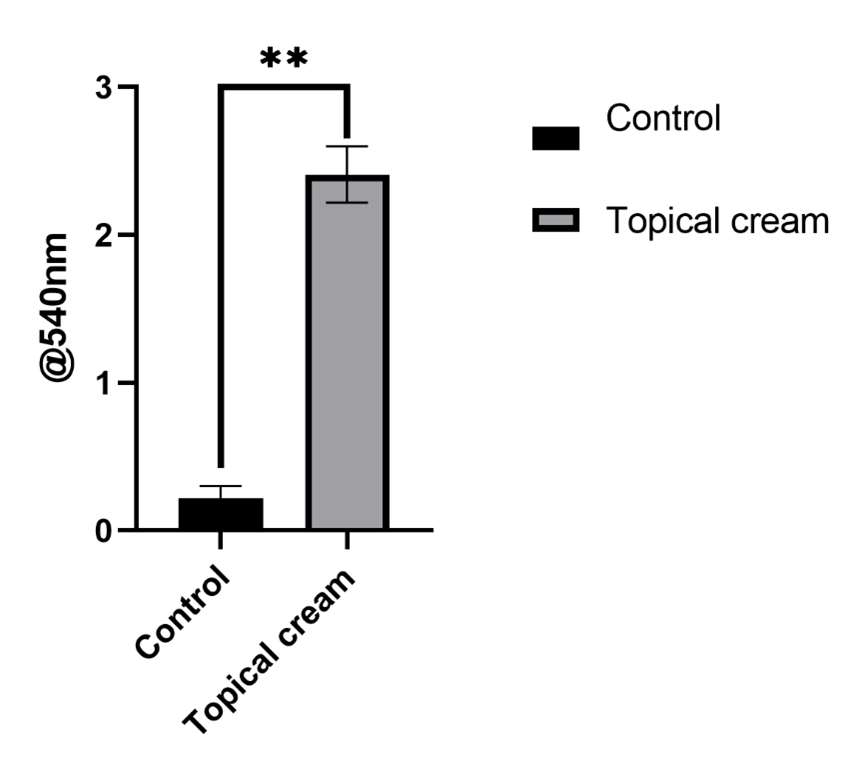
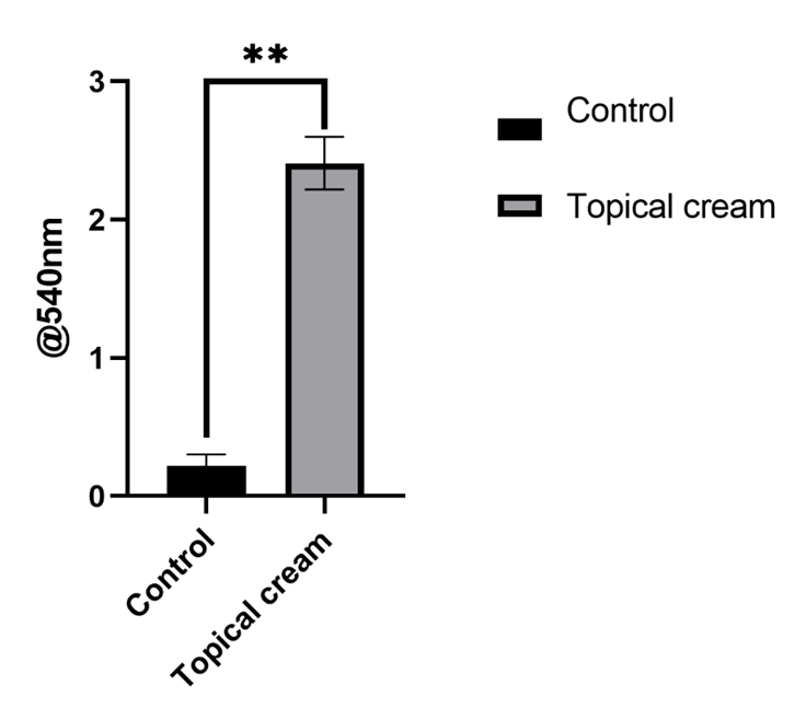
MTT is an colorimetric assay helps to understand the cell viability and cell metabolism. Higher the absorbance, higher is the viability. The absorbance of the topical cream sample is higher than the control which shows that the viability of the topical cream is higher than the control which would help to conclude that the prepared topical cream is non-cytotoxic, biocompatible and could be used for further investigation.
CONCLUSION:
The prepared topical cream is a multi-purpose hydrogel which could also be used for preparation of 3d printed skin and it is proposed like this because of it’s rheological property, the thickness of the topical cream makes it eligible for extrusion purposes. The Fourier transform spectroscopy helped to recognize the chemical composition of the topical cream. Hemolysis test confirmed the prepared topical cream is non-hemotoxic and high hemocompatible. The Spreadability test showed it has spreading properties like an emulsion which can easily spread and cover the larger area. CAM assay confirmed that the topical cream supports angiogenesis and it has no bad effect on the living cells. The histological studies also helped as a confirmatory test to the CAM assay and showed the cell structure and nucleus and no necrosis was seen. Adhesion test helped to understand the adhesive property of the prepared topical cream which critical for wound healing purpose. Collagen has the critical role in skin tissue engineering, the collagen bundle helps to form a healthy flawless skin without scar, this was the reason the fish air bladder was chosen. It is rich in collagen and growth factors. By performing several characterizations, it is confirmed that the prepared topical cream is high hemocompatible, biocompatible, supports angiogenesis, it is non-cytotoxic, supports cell metabolic activity and prepared product is also eligible to go for further investigation and in future it may be used as an effective alternative for wound healing purposes.
CONFLICT OF INTEREST:
The authors have no conflicts of interest regarding this investigation.
ACKNOWLEDGEMENT:
We acknowledge profound gratitude to the Department, Chemistry of Ramnarain Ruia Autonomous College and ASK Solutions analytical and research laboratories for providing facilities and technical assistance for research work.
REFERENCES:
- Guo S, DiPietro LA. Critical review in oral biology & medicine: Factors affecting wound healing. J Dent Res. 2010;893:219–29.
- Talekar YP, Apte KG, Paygude S V., Tondare PR, Parab PB. Studies on wound healing potential of polyherbal formulation using in vitro and in vivo assays. J Ayurveda Integr Med. 2017 Apr 1;82:73–81.
- Chocholata P, Kulda V, Babuska V. Fabrication of scaffolds for bone-tissue regeneration. Vol. 12, Materials. MDPI AG; 2019.
- Bakhrushina EO, Anurova MN, Zavalniy MS, Demina NB, Bardakov AI, Krasnyuk II. Dermatologic Gels Spreadability Measuring Methods Comparative Study. Int J Appl Pharm. 2022;141:164–8.
- Henkelman S, Rakhorst G, Blanton J, van Oeveren W. Standardization of incubation conditions for hemolysis testing of biomaterials. Mater Sci Eng C [Internet]. 2009;295:1650–4. Available from: http://dx.doi.org/10.1016/j.msec.2009.01.002
- S0926669022003880.
- Potanin A, Marron G. Rheological Characterization of Yield-Stress Fluids with Brookfield Viscometer. Appl Rheol. 2021;311:1–9.
- Zandvoort V, Spiegelaere D. Chorioallantoic Membrane Assay as Model for Angiogenesis in Tissue Engineering Chorioallantoic Membrane Assay as Model for Angiogenesis. 2024;2020.
- Torres-Rivero K, Bastos-Arrieta J, Fiol N, Florido A. Metal and metal oxide nanoparticles: An integrated perspective of the green synthesis methods by natural products and waste valorization: applications and challenges. Compr Anal Chem. 2021 Jan 1;94:433–69.
- Freedman BR, Uzun O, Luna NMM, Rock A, Clifford C, Stoler E, et al. Degradable and Removable Tough Adhesive Hydrogels. Adv Mater. 2021;3317.
- de Haan K, Zhang Y, Zuckerman JE, Liu T, Sisk AE, Diaz MFP, et al. Deep learning-based transformation of H&E stained tissues into special stains. Nat Commun. 2021;121:1–13.
- Intini C, Elviri L, Cabral J, Mros S, Bergonzi C, Bianchera A, et al. 3D-printed chitosan-based scaffolds: An in vitro study of human skin cell growth and an in-vivo wound healing evaluation in experimental diabetes in rats. Carbohydr Polym. 2018 Nov 1;199:593–602.


 V.V.Pongade*
 Iraa Gupta
 Vaishnavi Kamble


 10.5281/zenodo.13946802