Department of Pharmacology, Marri Laxman Reddy Institute of Pharmacy, Dundigal, Hyderabad-500043, Telangana, India
Objective: The present study was undertaken to evaluate the antiangiogenic activity of copper capped 4-aminopyridine which is a potassium channel modulator in chick embryo model and zebra fish assay and also to prepare and characterize copper capped nanoparticles of 4-aminpyridine. Methods: Biosynthesis and Characterization of 4-AP copper nanoparticles was done. For the purpose Zebra fish fin regeneration and embryo assay, chick CAM asssy was performed. In this study we have selected 4-AP as the objective drug to evaluate its effect on Zebra fish fin regeneration assay and embryo assay. Results: For that purpose eggs were used and the number of branching points on 7th day was evaluated per unit area and the results were presented where the formulation 4-APCuNP have shown significant reduction in branching points from the score of 4 to 1 which is a good sign for angiogenic activity. Conclusion: Angiogenesis is one of the important factors in many physiological and pathological processes. So, control of angiogenesis using drugs is essential for homeostasis in disease conditions such as wound healing and cancer. Based on the statistical analysis, we conclude that 4-APCuNPs is good anti-angiogenic and it is more effective in inhibiting angiogenesis
Formation of new blood vessels from the existing vessels is known as angiogenesis in other terms revascularization. Angiogenesis play an important role in maintenance of homeostasis in both normal and diseased states [1]. Regulation of angiogenesis is a balance between angiogenic and antiangiogenic factors. Various proangiogenic factors such as vascular endothelial growth factors (VEGF) fibroblasts growth factor (bFGF), Transforming growth factor (TGF), platelet derived growth factor (PDGF), TNF-α, IL-8 and angiopoietins stimulate neovascularization. Thrombospondin-1 and endostatin are natural anti-proangiogenic factors[2,3]. Various ion channels such as Na+, K+, Ca2+, Mg2+ and H+ and volume regulated ion channels are located on endothelial cell membrane7 Among them Cl-channel 4-aminopyridine (4-AP) and voltage gated K+ channel blocker have reported antiangiogenic activity. 4-AP inhibits fast voltage gated K+ channel in a dose dependant manner. Ion channel modulators are an extremely successful drugclass, second only drugs targettingG-protein couple receptors. There are three types of ion channels,i.e.,voltage- gated, extracellular ligand-gated, and intracellular ligand-gated along with two groups of miscellaneous ion channels. Nanoparticles have revolutionized drug and surgical therapies in recent times and found to be play a vital role in future drug therapies. Modern day applications such as formulation of drugs with some metal complexes where individual salts found to be toxic in therapeutic management.
Copper nano particles act as anti biotic, anti microbial,and antifungal agents when added to plastics, coatings and textiles. Nanoparticles of their size influence the properties of individual atoms or molecules when introduced into the biological system functions may vary with respect to distribution and biological responses. Copper is said to play dual role in cell growth based on the formulation either nanoparticles or in salt form[4]..
Copper is an essential micro nutrient that plays a vital role in normal and disease conditions[5].. It is one of the cofactor for many enzymes and serves as a catalyst that acts as a component of oxidative metabolism, clotting, iron acquisition and cellular immunity[6].. Role of Copper in angiogenesis have been reported in many studies either regulate or suppress the growth[7,8]. .Copper concentrations affect VEGF, FGF2 and TNF-α[9].. Copper activates HIF-1 that regulates VEGF[10,11].. Thus formulating drugs with copper nanoparticles gives the advantage of valuable therapeutic advantage when given individually is the importance of current investigation.
MATERIALS AND METHODS
Experimental animals:
Zebra fish was purchased from NIN Hyderabad. Chicken eggs were collected from the nearby poultry farms.
Housing of animals:
Zebra fish were placed under hygienic conditions and were exposure to 12 hours day and night cycle. Fish were fed with standard feed and fresh water is replaced daily. All the experimental methodology and conventions utilized in this investigation were approved by the Institutional Animal Ethics Committee (IAEC) of MLR Institute of pharmacy, Hyderabad, (1567/PO/RE/S/11/CPCSEA).
Chemicals:
Table 1: Chemicals used for experiments are listed below
|
S.No. |
Chemicals |
Quantity |
|
1. |
Lignocaine |
25ml |
|
2. |
Copper sulphate |
1mg |
|
3. |
Sodium hydrochlorite |
1mg |
|
4. |
Trypsin |
1gm |
Instruments:
Table 2: Various Instruments used for experiments are listed below
|
S.No. |
Instruments |
Model number |
|
|
Electronic balance |
CA 323 |
|
|
UV Visible spectrometer |
T-70 |
|
|
PH meter |
Microprolabmate, india |
|
|
Semi auto analyzer |
ES-100 |
|
|
Tissue homogenizer |
Rq-127/a |
|
|
Cooling centrifuge |
R-21 |
|
|
Probe sonicator |
ATP-750 |
Biosynthesis of copper Nanoparticle:
Firstly, we have to prepare 1mM CuSo4 solution by adding 0.02496g of CuSo4 in 100ml distilled water. To that add 2ml 4-AP solution dropwise. Then keep the solution on sonicator for 20 minutes by maintaining the temperature at 70°C and pH 5-5.5 and observed the reduction of Cu ions into copper nanoparticles (4-APCuNPs). The synthesized 4-APCuNPs were centrifuged at 12,000 rpm for 30 mins at 8°C. Pellet was collected by discarding supernatant and then washed with distilled water to remove impurities and 90% ethanol is used to wash the pellet to get pure 4-APCuNPs powder [12,13]..
Dose Selection:
Characterization of Prepared Nanoparticles:
Characterization and analytical techniques are methods used to identify ,isolate or quantify chemicals or materials, or to characterize their physical properties. The characteristics of synthesized CuNPs were studied by UV-Vis and IR spectrophotometric methods. Nano particles are characterized for various purposes, including nanotoxicology studies and exposure assesment in work places to assess their health and safety hazards. There is a wide range of instrumentation to these properties, including microscopy and spectroscopy.
ANTI -ANGIOGENIC ACTIVITY:
Zebra fish embryo assay:
Chosen sexually mature one male and two female zebra fish and put them all in tank for fertilization. Fertilized eggs were collected in the next morning. Fertilized eggs were transferred by pipette (point was cut off) to 0.5% sodium hypochlorite solution (prepared with embryo culture water), and then was shaken lightly to wash and sterilize embryos, transferred into embryo culture water after 2 mins. Then put it in a temperature control (28°C) illuminating incubator culture 24hr, for later use. When the development of zebra fish embryos reach to 24 h, chorionic villus of zebra fish embryos were digested and dissolved by 1mg/mL trypsin (prepared when needed). Ten minutes later, chorionic villus began fall off, and then wash with large amounts of water, repeated 5 times.
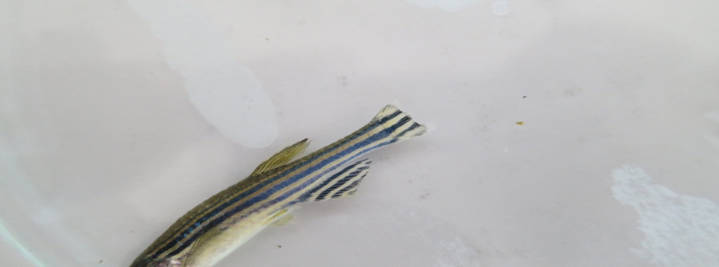
Zebra fish fin assay:
Adult fish obtained from NIN were kept in large tanks for acclimatization with continues supply of air. On the day of experiment, fish were taken out and they were placed in 150ml of marine water in 250ml beakers divided into 6-treatmentgroups 6 fishes in each group. They were anesthetized using 2% Lignocaine. Their fin was cut up to 50% by using sterilized straight razor blade and imaged under microscope. Pre and post amputation images were collected before transferring the fishes in recovery beaker containing fish water. Test drug, and standard drug were given to different groups, control group were maintained in similar conditions like test and standard. The fish water was changed on alternate day and dose was renewed. This was done for 30 days till full regeneration of fin is obtained normally. The images of amputed fin were collected on day 10, day 20, and day 30. The area of regenerated fin was calculated using ImageJ software. Percentage of regeneration was calculated and significance was obtained statistically[14]..
Chick chorioallantoic membrane (CAM) assay:
CAM assay was done by using chick eggs collected from local hatchery each group containing 6 eggs and were sterilized with 70% ethanol and were placed in incubator at 37°C in horizontal position. The eggs were placed in horizontal position on day 3 a fine small hole was done at narrow end to collect albumin 3 ml and then sealed with tape and again placed in incubator. On day 7 a small cut was made on the shell to place sterile gel foam of 3mmX3mm and 1 mm thickness was placed on the membrane.
On the third day of incubationa hole will be made on the narrow end of the egg to withdraw 3ml of albumin. This decreases pressure inside the egg and allows working on allontonic membrane. The hole will be sealed with surgical tape and the eggs are put back for incubation. On the 7th day of incubation a small window is cut opened on the shell and sterile gel foam (3mmX3mm and then 1mm piece is placed on the membrane.Then eggs are incubated undistured till day 14. On the 14th daythe cam tissues are removed out). The standard and 4-AP and 4-APCuNP 100μg/100ml and 200μg/100ml were placed on the gel foam and then placed in incubator. On day 14 CAM tissues were removed and placed in 10% formalin and then stained with hematoxylin and eosin. The tissue was examined in trinocular microscope for angiogenesis branching in a unit square region was considered for counting and were analyzed. Based on the branching points the angiogenetic score was given as 1-4 to each egg and shown as below [15].
|
Branching points |
Score |
|
≥35 |
4 |
|
25-34 |
3 |
|
15-24 |
2 |
|
≤15 |
1 |
Statistical Analysis:
Graphpad prism 5 was used to carry statistical analysis and to compare differences between the groups, one way ANOVA followed by post hoc dunnett’s test was performed and the values that are significant at p<0.05 were considered significant and were expressed as mean±SEM; n=6.
RESULT AND DISCUSSION
Biosynthesis of copper Nanoparticle:

Fig. 1: 4-Amino Pyridine Fig. 2: 4-AP Copper Nanoparticles
Characterization of Prepared Nanoparticles:
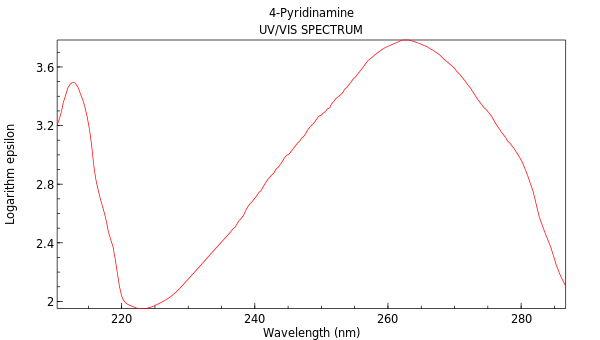
Fig. 3: 4-Pyridinamine

Fig. 4: 4-Aminopyridine
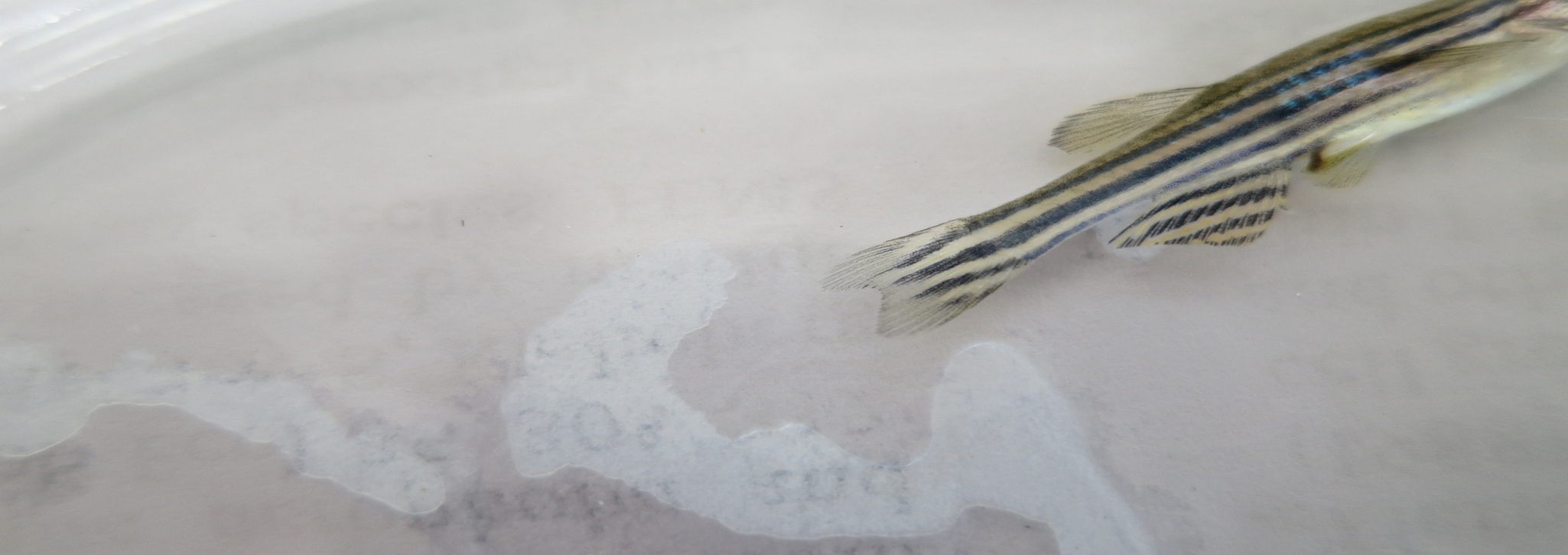
Zebra fish fin assay:

Control Standard
4-AP Low Dose 4-AP High dose


4-AP CuNP Low dose 4-AP CuNP high dose
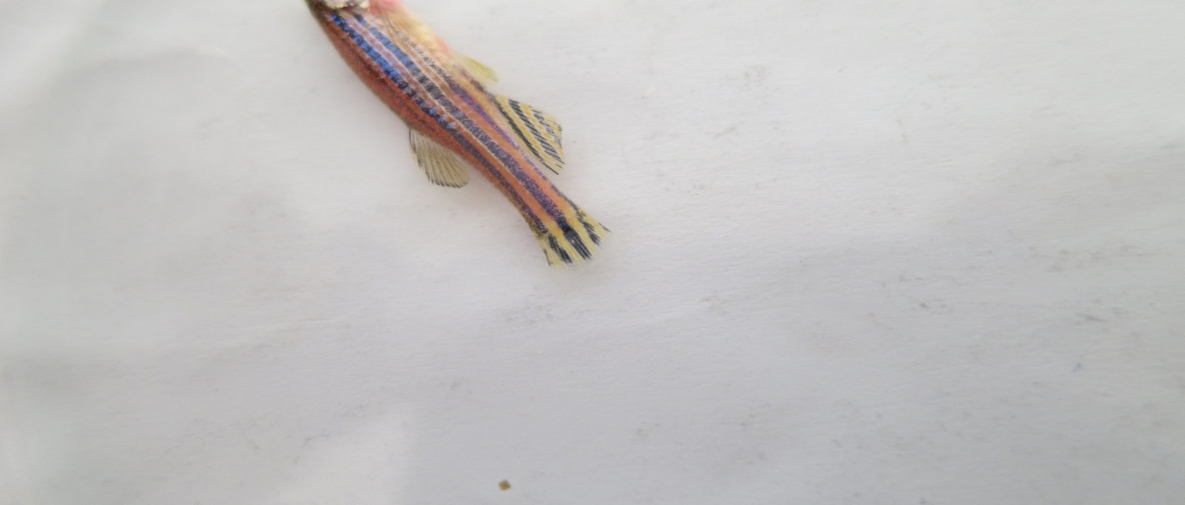
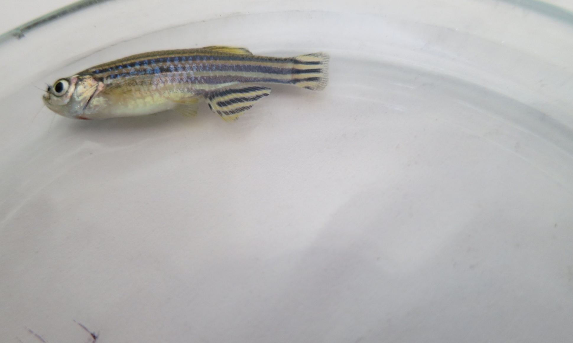
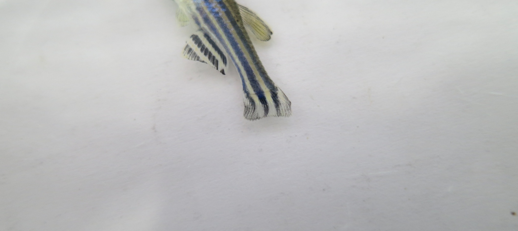
Fig. 5: Effect of control, standard and 4-APCuNP on angiogenesis in regeneration of
Zebrafish fin assay.

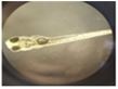
Fig. 6: Regeneration of Zebra fish Fin Assay
In zebra fish fin regeneration assay percentage fin regeneration was 19.2 in case of standard and the control was 62.3. when treated with 4-AP 100µl/100 ml fin regeneration was 22 and 4-AP 200µl/100 ml it was 28.2 which was significant when compared to control and standard. But when treated 4-AP formulated with copper capped nanoparticles 100µl/100ml and 200µl/100ml it was 18.2 and 16.4 respectively which indicates that when formulated with copper nanoparticles respectively.
Zebra fish embryo assay:


Day 1 All groups Control Day 3 4-AP 100ug


Day 3 4-AP 200ug Day 3 4-APCuNP 100ug Day 3 4-APCuNP 200ug
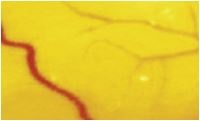
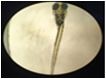
Fig. 7: Effect of 4-APCuNP on Zebra fish embryos.
Images of zebra fish embryos with decreased angiogenic vessel count which are treated with various drug solution.in zebra fish embryo model the percentage of angio genic vessels was taken as parameter to estimate the test drug to standard drug .
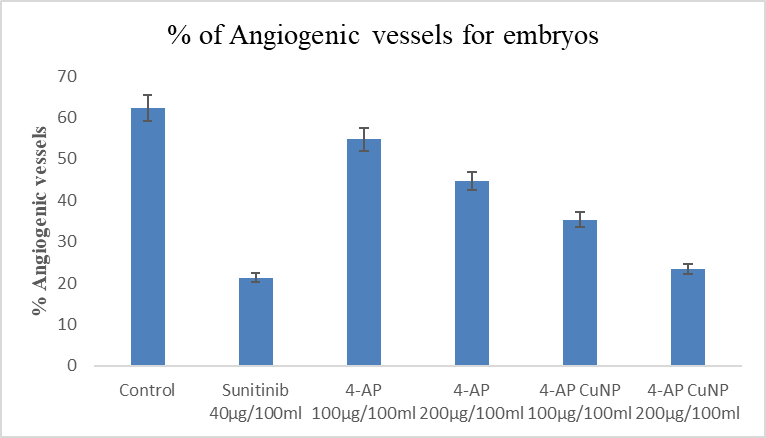
Fig. 7: Percentage of angiogenic vessels for Zebra fish embryo assay
The treated groups have shown significant, decrease in percentage of angiogenic vessels. In zebra fish model the percentage of angiogenic vessels was taken as parameter to estimate the test drug s 4-APCuNP.Sunitib is used as a standard drug.the treated group has shown significant decrease in percentage ofangiogenic vessel in the dose dependent manner. The low doses(100µg/100ml) treated group has not shown significant decrease inpercentage of angiogenic effect. Medium dose (200µg/100ml) treated groups have shown significant decrease in percentage of angiogenic vessels.
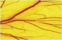
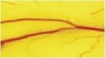
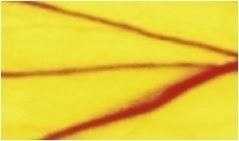
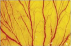
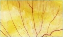
Chick chorioallantoic membrane (CAM) assay:
Points were counted under microscope. The numbers of vesseel branches points in a square region were counted and analyzed. Results have shown that significant reduction in Branching branching points at the given concentrations i.e., 100µg/100ml and 200µg/100ml of 4-AP and 4-APCuNP treated groups.

Control Sunitinib 40µg/100ml

4-AP 100µg/100ml 4-AP 200µg/100ml
4-APCuNP 100µg/100ml 4-APCuNP 100µg/100ml
Fig. 8: Effect of 4-AP and 4-APCuNP on angiogenesis in CAM assay
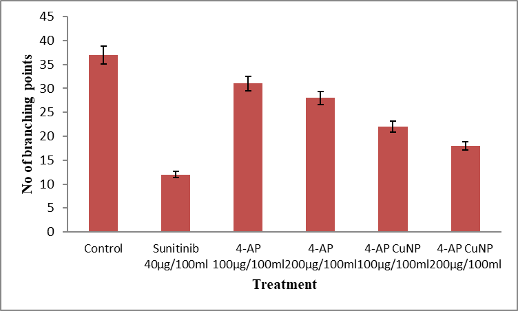
Fig. 9: Graph showing the effect of 4-APCuNP on the branching points in CAM assay
After treating with 4-AP 100µg/100ml and 4-AP 200µg/100ml the number of branching points per unit area were decreased to 28 and 24 and the scores was 3 which is relatively significant when compared control group.
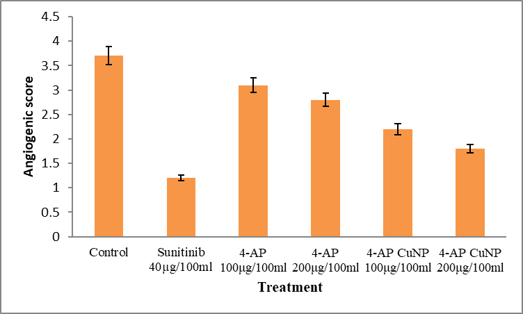
Fig. 10: Graph representing the effect of 4-APCuNP on angiogenesis score in CAM assay
After treatment with 4-APCuNP 100µg/100ml and 4-APCuNP 200µg/100ml the branching points were reduced to 22 and 18 respectively and score was 1 which is very significant at p<0.05. This shows that formulating 4-AP with copper nanoparticles have shown to be promising effect on angiogenesis in various models of evaluation.
CONCLUSION
Based on the statistical analysis, we conclude that 4-APCuNPs is good anti-angiogenic and it is more effective in inhibiting angiogenesis. Further research on volgate gated potassium channel blockers in proliferation and migration of endothelial cells may provide better rargets for the treatment of various diseases such as cancer, rheumatid arthritis, obesity, diabetic retinopathy, psoriasis, atherosclerosis, and endomerriosis in which angiogenesis is a part of pathology. Structural modifications of the drug may result in the development of cause specific molecules with lesser side effects.
ACKNOWLEDGEMENT
The Author Suvendu Saha is grateful to the Principal, MLR Institute of Pharmacy, Dundigal, Hyderabad for providing facilities to carry out the work.
REFERENCES
Formation of new blood vessels from the existing vessels is known as angiogenesis in other terms revascularization. Angiogenesis play an important role in maintenance of homeostasis in both normal and diseased states [1]. Regulation of angiogenesis is a balance between angiogenic and antiangiogenic factors. Various proangiogenic factors such as vascular endothelial growth factors (VEGF) fibroblasts growth factor (bFGF), Transforming growth factor (TGF), platelet derived growth factor (PDGF), TNF-α, IL-8 and angiopoietins stimulate neovascularization. Thrombospondin-1 and endostatin are natural anti-proangiogenic factors[2,3]. Various ion channels such as Na+, K+, Ca2+, Mg2+ and H+ and volume regulated ion channels are located on endothelial cell membrane7 Among them Cl-channel 4-aminopyridine (4-AP) and voltage gated K+ channel blocker have reported antiangiogenic activity. 4-AP inhibits fast voltage gated K+ channel in a dose dependant manner. Ion channel modulators are an extremely successful drugclass, second only drugs targettingG-protein couple receptors. There are three types of ion channels,i.e.,voltage- gated, extracellular ligand-gated, and intracellular ligand-gated along with two groups of miscellaneous ion channels. Nanoparticles have revolutionized drug and surgical therapies in recent times and found to be play a vital role in future drug therapies. Modern day applications such as formulation of drugs with some metal complexes where individual salts found to be toxic in therapeutic management.
Copper nano particles act as anti biotic, anti microbial,and antifungal agents when added to plastics, coatings and textiles. Nanoparticles of their size influence the properties of individual atoms or molecules when introduced into the biological system functions may vary with respect to distribution and biological responses. Copper is said to play dual role in cell growth based on the formulation either nanoparticles or in salt form[4]..
Copper is an essential micro nutrient that plays a vital role in normal and disease conditions[5].. It is one of the cofactor for many enzymes and serves as a catalyst that acts as a component of oxidative metabolism, clotting, iron acquisition and cellular immunity[6].. Role of Copper in angiogenesis have been reported in many studies either regulate or suppress the growth[7,8]. .Copper concentrations affect VEGF, FGF2 and TNF-α[9].. Copper activates HIF-1 that regulates VEGF[10,11].. Thus formulating drugs with copper nanoparticles gives the advantage of valuable therapeutic advantage when given individually is the importance of current investigation.
MATERIALS AND METHODS
Experimental animals:
Zebra fish was purchased from NIN Hyderabad. Chicken eggs were collected from the nearby poultry farms.
Housing of animals:
Zebra fish were placed under hygienic conditions and were exposure to 12 hours day and night cycle. Fish were fed with standard feed and fresh water is replaced daily. All the experimental methodology and conventions utilized in this investigation were approved by the Institutional Animal Ethics Committee (IAEC) of MLR Institute of pharmacy, Hyderabad, (1567/PO/RE/S/11/CPCSEA).
Chemicals:
Table 1: Chemicals used for experiments are listed below
|
S.No. |
Chemicals |
Quantity |
|
1. |
Lignocaine |
25ml |
|
2. |
Copper sulphate |
1mg |
|
3. |
Sodium hydrochlorite |
1mg |
|
4. |
Trypsin |
1gm |
Instruments:
Table 2: Various Instruments used for experiments are listed below
|
S.No. |
Instruments |
Model number |
|
|
Electronic balance |
CA 323 |
|
|
UV Visible spectrometer |
T-70 |
|
|
PH meter |
Microprolabmate, india |
|
|
Semi auto analyzer |
ES-100 |
|
|
Tissue homogenizer |
Rq-127/a |
|
|
Cooling centrifuge |
R-21 |
|
|
Probe sonicator |
ATP-750 |
Biosynthesis of copper Nanoparticle:
Firstly, we have to prepare 1mM CuSo4 solution by adding 0.02496g of CuSo4 in 100ml distilled water. To that add 2ml 4-AP solution dropwise. Then keep the solution on sonicator for 20 minutes by maintaining the temperature at 70°C and pH 5-5.5 and observed the reduction of Cu ions into copper nanoparticles (4-APCuNPs). The synthesized 4-APCuNPs were centrifuged at 12,000 rpm for 30 mins at 8°C. Pellet was collected by discarding supernatant and then washed with distilled water to remove impurities and 90% ethanol is used to wash the pellet to get pure 4-APCuNPs powder [12,13]..
Dose Selection:
Characterization of Prepared Nanoparticles:
Characterization and analytical techniques are methods used to identify ,isolate or quantify chemicals or materials, or to characterize their physical properties. The characteristics of synthesized CuNPs were studied by UV-Vis and IR spectrophotometric methods. Nano particles are characterized for various purposes, including nanotoxicology studies and exposure assesment in work places to assess their health and safety hazards. There is a wide range of instrumentation to these properties, including microscopy and spectroscopy.
ANTI -ANGIOGENIC ACTIVITY:
Zebra fish embryo assay:
Chosen sexually mature one male and two female zebra fish and put them all in tank for fertilization. Fertilized eggs were collected in the next morning. Fertilized eggs were transferred by pipette (point was cut off) to 0.5% sodium hypochlorite solution (prepared with embryo culture water), and then was shaken lightly to wash and sterilize embryos, transferred into embryo culture water after 2 mins. Then put it in a temperature control (28°C) illuminating incubator culture 24hr, for later use. When the development of zebra fish embryos reach to 24 h, chorionic villus of zebra fish embryos were digested and dissolved by 1mg/mL trypsin (prepared when needed). Ten minutes later, chorionic villus began fall off, and then wash with large amounts of water, repeated 5 times.
Zebra fish fin assay:
Adult fish obtained from NIN were kept in large tanks for acclimatization with continues supply of air. On the day of experiment, fish were taken out and they were placed in 150ml of marine water in 250ml beakers divided into 6-treatmentgroups 6 fishes in each group. They were anesthetized using 2% Lignocaine. Their fin was cut up to 50% by using sterilized straight razor blade and imaged under microscope. Pre and post amputation images were collected before transferring the fishes in recovery beaker containing fish water. Test drug, and standard drug were given to different groups, control group were maintained in similar conditions like test and standard. The fish water was changed on alternate day and dose was renewed. This was done for 30 days till full regeneration of fin is obtained normally. The images of amputed fin were collected on day 10, day 20, and day 30. The area of regenerated fin was calculated using ImageJ software. Percentage of regeneration was calculated and significance was obtained statistically[14]..
Chick chorioallantoic membrane (CAM) assay:
CAM assay was done by using chick eggs collected from local hatchery each group containing 6 eggs and were sterilized with 70% ethanol and were placed in incubator at 37°C in horizontal position. The eggs were placed in horizontal position on day 3 a fine small hole was done at narrow end to collect albumin 3 ml and then sealed with tape and again placed in incubator. On day 7 a small cut was made on the shell to place sterile gel foam of 3mmX3mm and 1 mm thickness was placed on the membrane.
On the third day of incubationa hole will be made on the narrow end of the egg to withdraw 3ml of albumin. This decreases pressure inside the egg and allows working on allontonic membrane. The hole will be sealed with surgical tape and the eggs are put back for incubation. On the 7th day of incubation a small window is cut opened on the shell and sterile gel foam (3mmX3mm and then 1mm piece is placed on the membrane.Then eggs are incubated undistured till day 14. On the 14th daythe cam tissues are removed out). The standard and 4-AP and 4-APCuNP 100μg/100ml and 200μg/100ml were placed on the gel foam and then placed in incubator. On day 14 CAM tissues were removed and placed in 10% formalin and then stained with hematoxylin and eosin. The tissue was examined in trinocular microscope for angiogenesis branching in a unit square region was considered for counting and were analyzed. Based on the branching points the angiogenetic score was given as 1-4 to each egg and shown as below [15].
|
Branching points |
Score |
|
≥35 |
4 |
|
25-34 |
3 |
|
15-24 |
2 |
|
≤15 |
1 |
Statistical Analysis:
Graphpad prism 5 was used to carry statistical analysis and to compare differences between the groups, one way ANOVA followed by post hoc dunnett’s test was performed and the values that are significant at p<0.05 were considered significant and were expressed as mean±SEM; n=6.
RESULT AND DISCUSSION
Biosynthesis of copper Nanoparticle:

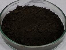
Fig. 1: 4-Amino Pyridine Fig. 2: 4-AP Copper Nanoparticles
Characterization of Prepared Nanoparticles:

Fig. 3: 4-Pyridinamine
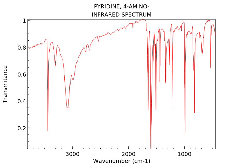
Fig. 4: 4-Aminopyridine
Zebra fish fin assay:

Control Standard


4-AP Low Dose 4-AP High dose

4-AP CuNP Low dose 4-AP CuNP high dose
Fig. 5: Effect of control, standard and 4-APCuNP on angiogenesis in regeneration of
Zebrafish fin assay.
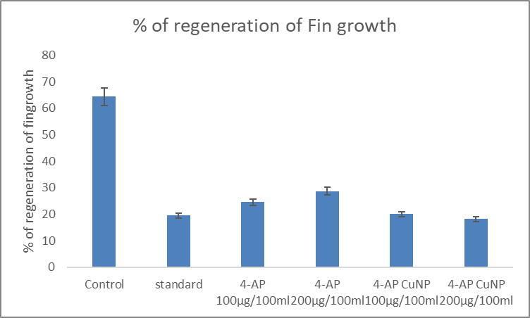
Fig. 6: Regeneration of Zebra fish Fin Assay
In zebra fish fin regeneration assay percentage fin regeneration was 19.2 in case of standard and the control was 62.3. when treated with 4-AP 100µl/100 ml fin regeneration was 22 and 4-AP 200µl/100 ml it was 28.2 which was significant when compared to control and standard. But when treated 4-AP formulated with copper capped nanoparticles 100µl/100ml and 200µl/100ml it was 18.2 and 16.4 respectively which indicates that when formulated with copper nanoparticles respectively.
Zebra fish embryo assay:


Day 1 All groups Control Day 3 4-AP 100ug


Day 3 4-AP 200ug Day 3 4-APCuNP 100ug Day 3 4-APCuNP 200ug
Fig. 7: Effect of 4-APCuNP on Zebra fish embryos.
Images of zebra fish embryos with decreased angiogenic vessel count which are treated with various drug solution.in zebra fish embryo model the percentage of angio genic vessels was taken as parameter to estimate the test drug to standard drug .

Fig. 7: Percentage of angiogenic vessels for Zebra fish embryo assay
The treated groups have shown significant, decrease in percentage of angiogenic vessels. In zebra fish model the percentage of angiogenic vessels was taken as parameter to estimate the test drug s 4-APCuNP.Sunitib is used as a standard drug.the treated group has shown significant decrease in percentage ofangiogenic vessel in the dose dependent manner. The low doses(100µg/100ml) treated group has not shown significant decrease inpercentage of angiogenic effect. Medium dose (200µg/100ml) treated groups have shown significant decrease in percentage of angiogenic vessels.
Chick chorioallantoic membrane (CAM) assay:
Points were counted under microscope. The numbers of vesseel branches points in a square region were counted and analyzed. Results have shown that significant reduction in Branching branching points at the given concentrations i.e., 100µg/100ml and 200µg/100ml of 4-AP and 4-APCuNP treated groups.

Control Sunitinib 40µg/100ml

4-AP 100µg/100ml 4-AP 200µg/100ml


4-APCuNP 100µg/100ml 4-APCuNP 100µg/100ml
Fig. 8: Effect of 4-AP and 4-APCuNP on angiogenesis in CAM assay

Fig. 9: Graph showing the effect of 4-APCuNP on the branching points in CAM assay
After treating with 4-AP 100µg/100ml and 4-AP 200µg/100ml the number of branching points per unit area were decreased to 28 and 24 and the scores was 3 which is relatively significant when compared control group.

Fig. 10: Graph representing the effect of 4-APCuNP on angiogenesis score in CAM assay
After treatment with 4-APCuNP 100µg/100ml and 4-APCuNP 200µg/100ml the branching points were reduced to 22 and 18 respectively and score was 1 which is very significant at p<0.05. This shows that formulating 4-AP with copper nanoparticles have shown to be promising effect on angiogenesis in various models of evaluation.
CONCLUSION
Based on the statistical analysis, we conclude that 4-APCuNPs is good anti-angiogenic and it is more effective in inhibiting angiogenesis. Further research on volgate gated potassium channel blockers in proliferation and migration of endothelial cells may provide better rargets for the treatment of various diseases such as cancer, rheumatid arthritis, obesity, diabetic retinopathy, psoriasis, atherosclerosis, and endomerriosis in which angiogenesis is a part of pathology. Structural modifications of the drug may result in the development of cause specific molecules with lesser side effects.
ACKNOWLEDGEMENT
The Author Suvendu Saha is grateful to the Principal, MLR Institute of Pharmacy, Dundigal, Hyderabad for providing facilities to carry out the work.
REFERENCES


Dr. Suvendu Saha, Dr. Arunabha Mallik, Evaluation Of Antiangiogenic Activity of Ion Channel Modulator Capped Copper Nano Particles, Int. J. of Pharm. Sci., 2026, Vol 4, Issue 2, 4471--4480. https://doi.org/10.5281/zenodo.18798339
 10.5281/zenodo.18798339
