View Article

Abstract

Superficial fungal infections, such as dermatophytosis and candidiasis, are prevalent and difficult to manage due to limited skin permeability, inadequate bioavailability, and the adverse effects of systemic administration of traditional antifungal agents. Herbal substances provide a safer choice, but their therapeutic effectiveness is compromised due to instability and insufficient penetration within the stratum corneum Ethosomal gel is a type of vesicular drug delivery system which has recently?been developed as an effective means to circumnavigate these hindrances. Ethosomes, consisting of phospholipids, ethanol, and water, improve the transdermal delivery, vesicle deformability, drug stability, and permit controlled and sustained?release of the encapsulated actives The aim of this study is to investigate and validate Arachis hypogaea (Peanut) leaf extract from among the herbal extracts containing flavonoids, phenolics and tannins which have?strong antifungal properties for the development of herbal ethosomal gels. The preparation consists of an optimization of gelling agents, phospholipids concentration, ethanol concentration, vesicle size and pH. The evaluation parameters include the physiochemical properties, vesicles characterization, in-vitro/ex-vivo permeation and antifungal activity. Herbal ethosomal gels exhibit better skin penetration, increased stability, paucity of systemic side effects along with the possibility of synergistic effect with multi herbal extracts, which makes them a natural and effective topical antifungal treatment with?potential .We envision further clinical evaluation and commercialization of these formulations as the new rationale in safe and effective antifungal

Keywords

Ethosomal gel, Herbal antifungal, Arachis hypogaea, Transdermal drug delivery, Skin permeation, Vesicular system.

Introduction

When pathogenic fungal species successfully infiltrate the host and proliferate within bodily tissues, fungal infections, also known as mycoses, result. These infections may just affect exterior, keratin-rich tissues like the epidermis, hair shafts, and nails, or they may spread to invasive forms that endanger internal organs, depending on the level of tissue involvement. An increase in people with compromised immune systems, such as those with HIV infection, those with long-term metabolic conditions like diabetes mellitus, and patients undergoing cytotoxic or immunosuppressive treatments, is closely linked to the rising global burden of mycotic infections [1].

Fungal infections, termed mycoses, develop when pathogenic fungal species successfully invade the host and multiply within body tissues. Based on the depth of tissue involvement, these infections may be limited to external, keratin-rich structures such as the epidermis, hair shafts, and nails, or may progress to invasive forms that compromise internal organs. The growing global burden of mycotic infections is strongly associated with an increase in individuals with impaired immune defenses, including those affected by HIV infection, chronic metabolic disorders like diabetes mellitus, and patients receiving cytotoxic or immunosuppressive treatments [1]. Fungi are heat-tolerant eukaryotic organisms that predominantly thrive in environments characterized by warmth and high humidity. Mycotic infections are categorized based on the anatomical location involved and the extent of tissue penetration, and are classified into superficial (cutaneous), subcutaneous, and systemic or deep fungal infections [2]. Mycoses are infectious conditions that arise when pathogenic fungi infiltrate human tissues and cause cellular damage through colonization and invasive growth. These infections represent a major global public-health concern, with a disproportionate impact on low- and middle-income countries and individuals with compromised immune function. Epidemiological studies indicate that superficial fungal infections affect over one billion people annually, while invasive and systemic mycoses are responsible for more than 1.5 million deaths each year [3]. Fungi comprise a diverse kingdom of eukaryotic microorganisms that include unicellular yeasts, filamentous molds, and dimorphic species capable of switching between morphological forms in response to environmental cues. In contrast to prokaryotic bacteria, fungal cells possess a membrane-bound nucleus and a structurally complex cell wall composed primarily of chitin, glucans, and mannoproteins, components that represent important targets for antifungal drug development [4]. Opportunistic mycotic infections typically develop in individuals with weakened immune defenses, resulting from conditions such as HIV/AIDS or from medical interventions including cytotoxic chemotherapy, solid organ transplantation, and prolonged exposure to antibiotics or corticosteroids [5].

Ethosomes: An Overview

Ethosomes represent a new generation of flexible vesicular carriers that have been tailored        for improved skin delivery of drugs through transdermal and dermal routes [6Ethosomes differ from conventional liposomal systems due to their elevated ethanol content, which modifies the vesicular membrane by increasing its flexibility. This enhanced deformability enables more efficient transport across the stratum corneum while maintaining the skin barrier’s structural integrity [7]. These vesicular systems are capable of delivering both water-soluble and lipid-soluble therapeutic agents, making them adaptable platforms for a wide range of pharmaceutical applications

Composition:

Ethosomes are typically composed of four main components:

Component

Example

Phospholipids

Phosphatidylcholine (soy/egg), Phosphatidylserine

Ethanol

20–45% w/w (acts as penetration enhancer)

Water (Aqueous Phase)

Purified water

Active Drug

Diclofenac, Clotrimazole, Curcumin

Optional Additives

Cholesterol, Glycerol, Propylene glycol, Stearylamine

1. Ethanol (20–45%) Co-solvents (optional) Phospholipid

Vesicular bilayers are mainly formed from phospholipids such as phosphatidylcholine derived from soy or egg sources [8]. Interactions between Ethosomes and skin lipid layers increase vesicular deformability, allowing passage through the narrow intercellular channels of the stratum corneum. In addition, ethanol at concentrations of 20–45% enhances transdermal transport by increasing membrane fluidity [9]. The amount of ethanol is carefully optimized to maintain vesicle integrity while ensuring effective skin permeation [10].

2. Water (Continuous Phase of Ethosomal Dispersions)

An aqueous medium serves as the continuous phase that disperses the phospholipid-based vesicles [11] . It promotes phospholipid molecule hydration for bilayer formation in the process of ethosome formation. The vesicles in an aqueous medium form a stable colloidal dispersion, and the medium is used to entrap hydrophilic drugs in the aqueous core of the vesicles. The size, zeta potential, and overall stability of the vesicles are also affected by the water-to-ethanol ratio. The equimolar ratio contributes to optimal vesicle self-assembly and avoids aggregation, which is important for reproducible topical or transdermal delivery of a drug [12].

Mechanism of Action

The activity of the ethosomal system for transdermal and dermal drug delivery is connected mainly with the synergistic action of ethanol, phospholipids, and the skin barrier. The process is multifactorial and includes several reinforcing events that allow the passage of hydrophilic and lipophilic molecules into the stratum corneum.[19].

  • Ethanol Softens the Stratum Corneum

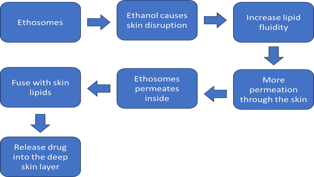
Ethanol functions as a penetration enhancer by breaking down the highly ordered stratum corneum lipid structure. It enhances lipid fluidity, decreases the transition temperature of skin lipids and their barrier function [20]. Such a softening effect facilitates the penetration of vesicular systems through the skin and enhances skin drug diffusion and partitioning. Ethanol is also responsible for the partial extraction of lipids from the stratum corneum, which increases the penetration ability of ethosomal vesicles. [21].

  • Vesicle Fusion Enhances Permeation

Phospholipid-based ethosomes are very deformable and flexible vesicles that allow them to go with the flow of skin lipids and make their way through the lipids in between cells. [22].

After reaching the skin surface, the ethosomes enriched with ethanol interact with stratum corneum lipids, and the drug is transferred into the skin through the fusion of vesicles and mixing of lipids. [23]. This fusion process improves the drug retention in deeper skin layers along with facilitating controlled and sustained release of drug in the layers of the epidermis and dermis [24].

  • Entrapment of Hydrophilic and Lipophilic Drugs

Ethosomal systems can effectively entrap both hydrophilic and lipophilic drugs, as a result of their special bilayer structure which is formed by phospholipids and ethanol. [25].

The hydrophilic core can entrap water-soluble drugs, and the lipid bilayer can be solubilized with lipophilic compounds. [26].

This bivalent encapsulation property allows ethosomes to carry various types of drugs, providing better bioavailability and improved skin deposition [27].

Advantages of Ethosome

Ethosomes are superior phospholipid-based vesicular systems that employ ethanol as a co-solvent, bestowing a number of advantages over traditional vesicular systems including liposomes and niosomes [28]. The distinctive nature of phospholipid, ethanol, and water in the TC solution endows the TC with excellent skin permeation, high drug loading, and enhanced formulation stability.

  • High Skin Permeation

Ethanol and phospholipids synergistic effect makes the per mean ability of ethosomal  content through the skin very high. Ethanol makes the lipids more fluid both in the membrane of the vesicle and in the layer of the stratum corneum and this helps the vesicular encapsulated drug to go deeper into the skin. [29],

  • Biocompatible and Non-toxic

The phospholipids (such as soy or egg phosphatidylcholine) employed in ethosomes are natural and biocompatible constituents of the cell membranes.Ethosomal systems are therefore safe in so far as their application on the skin meets the requirements of long term use in topical and transdermal drug delivery. [30].

  • Can Carry Herbal Extracts with Poor Solubility

Ethosomes are particularly efficient in carrying water-insoluble herbal extracts and phytoconstituents, which normally display less bioavailability as a consequence of poor solubility. [31].ethanol and phospholipids facilitated the solubility and permeation of lipophilic natural products including flavonoids, alkaloids and terpenloids and by forming stable ethosomal complexes. [32]

  • Enhanced Stability Compared to Liposomes

Unlike conventional liposomes, ethosomes are more physically and chemically stable in storage. [33]. Ethanol serves as a co-solvent with stabilizing properties that inhibit vesicle aggregation and the release of entrapped drugs.

Ethosomal Gel: Formulation and Composition

Ethosomal gels are a semisolid system in which ethosomal vesicles are dispersed in a suitable gel-forming agent and are appropriate for both topical and transdermal application. Ethosomal gels combine gel and ethosomal vesicles for better stability and ease of application. Compared to the liquid form of ethosomal suspensions, the ethanol and water contents in ethosomal gels are lower, and their proton NMR spectra show weaker intensity of the peaks. The gel matrix enhances patient compliance, increases the residence time of the drug on the skin, and allows for controlled release of hydrophilic and lipophilic substances [34].

Gelling Agents

Gelling agents are essential for the transformation of ethosomal dispersions into stable, convenient gels without affecting the integrity of vesicles and their spreadability. Polymers such as Carbopol, hydroxypropyl methylcellulose (HPMC), and Poloxamer are used routinely.[35]

Carbopol (Carbomer):

Carbopol polymers are synthetic high-molecular-weight cross-linked polyacrylic acid and have been widely employed for many years because of high gelation efficiency, clarity and stability [36]. They are able to closely incorporate ethosomal suspensions without vesicle rupture and offer pseudoplastic flow behaviour, which enhances the spreadability on the skin surface.

HPMC (Hydroxypropyl Methylcellulose):

HPMC is a non-ionic, biocompatible cellulose derivative that acts as a stabilizer for vesicles and helps sustain the viscosity of the gel. It is especially appropriate for ethosomal gels containing herbal or thermosensitive drugs, as it inhibits vesicle aggregation without compromising homogeneity. [37].

Poloxamer (Pluronic F127):

Poloxamer-based gels are thermosensitive, which means they are liquid at low temperatures and turn into a gel at skin temperature (~32–37 °C) [38]. This characteristic allows for simple application and prolonged drug release from ethosomal vesicles. Poloxamer also improves skin hydration and offers a mild, non-irritating feel. The type and concentration of the gelling agent depend on the required viscosity, drug compatibility, and release pattern of the formulation. [39].

Preparation Methods

Various techniques are used for the preparation of ethosomal gel based on the physicochemical properties of the drug, thermosensitive nature and the requirements of the formulation.:

a) Cold Method

The cold process is widely used and appropriate for thermolabile drugs. In this method, phospholipids and the drug are dissolved in ethanol under stirring at room temperature. Under constant stirring, water (or hydroalcoholic phase) is added dropwise to a solution of ethosomes. The resultant ethosomal suspension is then added to the gel base (Carbopol/HPMC/Poloxamer) with mild stirring until a consistent gel is formed. [40],

b) Hot Method

In the hot process, the phospholipids and drug are solubilized in ethanol at an elevated temperature (40–50 °C). The aqueous phase at the same temperature is added to the organic phase with stirring to obtain ethosomes. After cooling to room temperature, the ethosomal dispersion is mixed with the pre-hydrated gel base. [41].This technique can be applied to the drugs which are stable in gentle heating and results in homogeneous vesicles size.

Modified Ethanol Injection Method

phospholipids and drug in ethanolic solution are injected into the aqueous phase with stirring, by using a syringe or a peristaltic pump in this method. Due to solvent diffusion into the aqueous phase and the interfacial deposition of phospholipids, ethosomes are spontaneously formed [42] This modified procedure is better adapted to provide a tight size distribution of vesicles with high entrapment efficiency and no limitation for gel incorporation.

Factors Affecting Gel Performance

The ethosomal gel’s performance is affected by multiple factors of formulation and processing, collectively impacting drug release, stability, and skin permeation efficiency [43].

Phospholipid Concentration:

An increase in the concentration of phospholipids results in better vesiculation and entrapment but also leads to an increase in vesicle size and a decrease in the clarity of gel [44]. The stability and rate of drug release are both influenced by the optimum phospholipid content.

Ethanol Percentage:

Ethanol (20–45%) has a marked effect on vesicle fluidity and deformability. Higher amounts of ethanol enhance skin permeation, but could lead to reduction of vesicle stability. [45] Thus, the Control ethosomal gel dose was the most appropriate to determine the effect of the ethanol content. The effect of the ratio of ethanol was very important for the effectiveness of ethosomal gel. An ideal proportion of ethanol is critical for the effectiveness of ethosomal gel. An optimal balance between ethosome size and ethanol concentration is required for effectiveness of ethosomal gel. Consequently, the control ethosomal gel dose was considered the most suitable to evaluate the effect of the ethanol content. Maintaining a proper ratio of ethanol is essential for the efficiency of ethosomal gel. Therefore, the maintenance of the optimal ratio of ethanol is very important for effectiveness of the ethosomal gel. Therefore, an appropriate balance/compromise between ethosome size and concentration of ethanol is necessary for effectiveness of the ethosomal gel. Hence, the control ethosomal gel dose was the best candidate to evaluate the effect of the ethanol content. Keeping a proper ethanol proportion is crucial for ethanol gel application.

Vesicle Size:

The smaller vesicles (100–200 nm) allow for better skin penetration and homogeneous distribution in the gel matrix while larger vesicles could increase drug retention on skin surface [46].

pH:

The pH of the ethosomal gel (usually 5.5–7.4) should be skin-friendly to prevent skin irritation and to maintain the stability of the phospholipids [47]. Departure from this range may influence the charge and the vesicle aggregation

Viscosity:

The viscosity of gels affects both drug release rate and skin adhesion. Drug diffusion is reduced with increasing viscosity, but residence time on the skin is increased. Thus, it must be optimized to achieve an appropriate balance between spreadability and sustained release [ 48].

Incorporation of Herbal Extracts

Neem (Azadirachta indica)

The neem extract contains high concentration of bioactive compounds like azadirachtin, nimbin and nimbidin which have antimicrobial (antibacterial and antifungal) and anti-inflammatory properties. But these substances are poorly water soluble and have limited skin absorption. Loading into ethosomal gels significantly improves skin delivery and antimicrobial activity, in particular against Staphylococcus aureus and Candida albicans. [49]. Ethosomes enhance drug retention in the epidermal layers and thereby increase the duration of action of formulations based on neem extract [50]

Potential for Arachis hypogaea (Peanut) Leaf Extract

Resveratrol, luteolin, and kaempferol, which have antioxidant, anti-inflammatory, and antimicrobial activities, are among the bioactive polyphenols and flavonoids present in the leaves of Arachis hypogaea (peanut plant)[51]. Although the pharmacological potential of these compounds is high, it has a poor solubility in water and low permeability through skin which making it unable to be directly applied on skin.

Incorporation of peanut leaf extract into ethosomal gel systems could offer the following advantages:

Enhanced Solubility: Ethanol is a co-solvent in ethosome that enhanced the solubility of polyphenolic compounds like resveratrol and luteolin [52].

Improved Skin Penetration: The synergy of ethanol and phospholipids enhances fluidization of skin lipids, which, in turn, facilitates the deeper dermal penetration of phytoconstituents. [53].

Stabilization of Antioxidants: Phospholipids shield vulnerable flavonoids against oxidation and degradation in the process of formulation and storage. [54].

Sustained Release: The gel matrix allows the drug to reside for an extended period of time at the site of application, producing a sustained antioxidant and anti-inflammatory effect. [55].

Consequently the present work was to formulate ethosomal gel of Arachis hypogaea leaf extract as a new safe biocompatible approach for anti-inflammatory, antioxidant and skin protective application. Further studies investigating stability of phytochemicals and skin permeation as well as in vivo efficacy would be desirable to confirm its therapeutic potential.

Herbal Antifungal Agents Used in Ethosomal Gels

Herbal antifungals possess high anti-fungal activity, are biocompatible and have fewer side effect and thus became the focus of attention in the last years. Loading such compounds into ethosomal gels leads to an improvement of their cutaneous delivery, stability and therapeutic potential, especially in the treatment of superficial and topical mycotic infections. [56]. Ethosomes, which are made up of phospholipids and a high concentration of ethanol (20–45%), act as good carriers for delivering the substances contained in them deep in to the skin and also protect phytochemicals from degradation. [57].

Mechanism of Action of Herbal Antifungal Agents

Anti-fungal action of herbal substances is mainly through various synergistic actions which interfere the life cycle and the growth of fungus:

Cell Membrane Disruption

Inhibition of Fungal Enzymes

Herbal antifungals may disrupt fungal metabolism by acting on crucial enzymes, such as chitin synthase, β-glucan synthas e, and cytochrome P450-dependent enzymes involved in the biosynthesis of ergostrol.Inhibition of these enzymes leads to defective synthesis of the cell wall and membrane formation, which in turn halts the growth and reproduction of the fungus [60]. Certain compounds such as curcumin and neem extract components are said to target various enzyme systems in the fungi [61]

Reactive Oxygen Species (ROS) Generation

Several herbal bioactives induce oxidative stress in fungal cells by elevating the levels of intracellular ROS, resulting in oxidative damage of proteins, lipids and DNA. Excessive ROS buildup leads to apoptosis-like cell death in fungal cells [62]. For example, allicin1 from garlic and resveratrol from peanut leaves are reported to elicit ROS2-induced antifungal activity [63]

Advantages of Herbal Antifungal Agents in Ethosomal Gels

Improved Skin Penetration: Ethosomes facilitate the transport of hydrophilic and lipophilic herbal antifungal drugs through the stratum corneum by disrupting the barrier function of skin lipids. [64].

Phytochemical stability: Phospholipid vesicle encapsulation shields phenolic and other sensitive compounds (curcumin, eugenol, allicin, etc.) from oxidation and hydrolysis[65].

 Sustained Release: The gel medium permits a sustained release of the antifungal with concomitant prolongation of the therapeutic effect and a decrease in dosing frequency. [66].

Reduced Irritation: Ethosomal gel-based delivery of herbal substances [ETHOSOMAL GEL] shows minimal skin irritation as opposed to alcohol-based tinctures or traditional creams. [67].

Evaluation of Herbal Ethosomal Gels

The assessment of herbal ethosomal gels is necessary to maintain quality and to confirm the safety and efficacy of the formulation. Ethosomal gels are sophisticated delivery systems in which phospholipid vesicles entrap herbal actives and enable enhanced dermal delivery.Proper evaluation results in these gels being physically stable, pharmaceutically acceptable, and therapeutically efficient. [68].

Physicochemical Evaluation

Both patient acceptance and drug performance are influenced by physicochemical properties. Typical parameters are:

Appearance and Homogeneity

The gel should be smoothly textured without lumps, phase separation or air bubbles. Uniform gels represent uniform dispersion of ethosomal vesicles and are essential for uniform drug delivery. [69].

pH Measurement

The pH value of the topical gels should be near to the skin pH (5.5–6.5) to prevent irritation and to maintain its bio compatibility. pH can also affect the stability of vesicles and the release of drugs. [70].

Viscosity

Viscosity affects the spreadability, retention, and release profile of the formulation and can be determined by rotational viscometer or Brookfield viscometer. The optimal viscosity allows an easy application and provides a long skin contact time to achieve an adequate bioavailability of the drug. [71].

Spreadability

While applying gel to skin, tests the Slip and Drag or parallel plate methodologies. Patient compliance: How evenly the gel can be spread over the affected area is an important factor for uniform coverage. [72].

Vesicle Characterization

Ethosomes are vesicular carriers for both hydrophilic and lipophilic herbal actives.The key parameters are:

Vesicle Size

Measured by dynamic light scattering (DLS) or electron microscopy. Smaller vesicles (usually 100–300 nm) have a better penetration ability for stratum corneum. [73].

Zeta Potential

Determine the surface charge of the vesicles, Which is related to the physical stability of the vesicles. Values of ±30 mV are indicative of stability for colloidal system as particles will repel each other due to the electrostatic forces and hence will not aggregate. [74].

Polydispersity Index (PDI)

Reflects the size distribution of vesicles; PDI < 0.3 indicates uniform vesicle population, essential for reproducible skin penetration [75].

In-vitro and Ex-vivo Studies

This investigations offer insights into drug release, permeation and stability

In-vitro Drug Release

Performed in Franz diffusion cells using synthetic membranes. Evaluates the release rate and release extent of herbal actives from ethosomal gel with time.Aids in determination of best formulation composition and release kinetic profiles [76].

Ex-vivo Skin Permeation

Conducted on rat, porcine, or human cadaver skin to evaluate dermal penetration, flux, and retention. Crucial to verify that herbal actives penetrate deeper skin layers. [77].

Stability Studies

Assess physical, chemical and microbial stability under various conditions (e.g., temperature, humidity, light). o Stability studies confirm that the herbal actives are still potent in storage. [78].

Antifungal Activity Testing

Evaluation of antifungal activity is to make sure that the herbal ethosomal gel is effective in treatment.:

Agar Diffusion Method

The results of the study on the inhibition of Theòrnelysin resistance development were examined. The zone of inhibition of fungal growth was measured with strains such as C. albicans, A. niger, and dermatophytes. The ethosomal gel demonstrated superior antifungal action compared to the free extract. [79].

Broth Dilution Method

The broth dilution method is a widely accepted quantitative assay used to determine the Minimum Inhibitory Concentration (MIC) of antimicrobial agents. In this procedure, serial dilutions of the test substance are prepared in a nutrient broth, followed by inoculation with a standardized microbial suspension. The MIC is defined as the lowest concentration of the compound that prevents visible microbial growth after incubation. This method is highly reliable for evaluating antimicrobial potency, performing dose–response studies, and optimizing therapeutic doses for experimental or pharmaceutical applications [80]. Both microdilution and macrodilution formats are used depending on sample volume requirements, with microdilution preferred for higher throughput and precision  [81]

Enhanced Skin Penetration

The high content of ethanol in ethosomal gels renders both the vesicular bilayer and the skin lipid matrix fluidized and subsequently, vesicles are allowed to penetrate the stratum corneum more deeply and in an easy manner.  Herbal actives, which generally possess poor water solubility and skin permeability, are more effectively delivered into the deep epidermal layers and dermis, leading to enhanced therapeutic outcomes. Studies have demonstrated that ethosomal systems enhance transdermal delivery of herbal extracts such as curcumin, neem, Aloe vera with rapid onset of action [81].

Controlled and Sustained Release

The vesicular nature of ethosomes allows for the controlled release of herbal actives over a longer time span. This release profile is responsible for maintaining therapeutic drug concentration in the skin and leads to less frequent applications, which subsequently improves patient compliance. Controlled release also prevents drug degradation and prolongs antifungal activity.[82].

Reduced Systemic Side Effects

The systemic exposure will be reduced by targeting the delivery of herbal actives to the target site (skin or superficial fungal infections). This lowers the chance of undesirable systemic effects, which is important for strong antifungal substances. Localized action is also advantageous for sensitive groups of patients, such as children or those with liver or kidney problems [83].

Improved Stability and Bioavailability of Herbal Actives

Several herbal constituents (such as curcumin, flavonoids, terpenoids) are chemically unstable in traditional topical formulations. Ethosomal encapsulation shields these actives against oxidation, hydrolysis and photodegradation, leading to enhanced shelf-life and therapeutic efficacy. The improved skin penetration and controlled delivery leads to a better bioavailability and consequently to an enhanced pharmacological effect of the herbal extract, thus the herbal extract pharmacological effect is maximized. [84].

CONCLUSION

Herbal ethosomal gels could be a better and innovative platform for topical antifungal treatment having therapeutic efficacy of natural herbal phytoconstituents and peculiarity of modern transdermal delivery system. These gels improve the penetration, stability and bioavailability of herbal actives and provide site specific and sustained drug delivery with reduction in systemic side effects. The use of peanut (Arachis hypogaea) leaf extract as an antifungal agent is a natural and new approach and is based on its bioactive components that include flavonoids, phenolics, and tannins which said to have the strongest antifungal activities.. In addition, the ethosomal gel system could be also combined with other herbal extracts for the potential synergistic antifungal and clinical effects. Thus, the peanut leaf based herbal ethosomal gel can be a non-toxic, efficacious, and a marketable solution to combating superficial fungal infections, with prospects of translating the work into multi-herbal formulations, clinical trial and large scale production for wider therapeutic application in the near future.

REFERENCES

  1. J. R. Perfect, “The impact of the host on fungal infections,” The American Journal of Medicine, vol. 125, no. 1, pp. S39–S51, 2012.
  2. D. W. Denning and W. W. Hope, “Therapy for fungal diseases: Opportunities and priorities,” Trends in Microbiology, vol. 18, no. 5, pp. 195–204, 2010
  3.  S. Shwetha and R. P. Jadhav, “Classification and treatment of fungal infections: A review,” Journal of Pharmaceutical Sciences and Research, vol. 12, G no. 7, pp. 881–888, 2020.
  4. K. M. Thambugala, D. A. Daranagama, D. S. Tennakoon, D. P. W. Jayatunga, S. Hongsanan and N. Xie, “Humans vs. Fungi: An Overview of Fungal Pathogens against Humans,” Pathogens, vol. 13, no. 5, article 426, May 2024. doi:10.3390/pathogens13050426.
  5. M. G. Netea et al., “Immune defense against Candida fungal infections,” Nat. Rev. Immunol., vol. 15, no. 10, pp. 630–642, 2015.
  6. D. W. Denning and W. W. Hope, “Therapy for fungal diseases: Opportunities and priorities,” Trends Microbiol., vol. 18, no. 5, pp. 195–204, 2010.
  7. E. Touitou, N. Dayan, L. Bergelson, B. Godin, and M. Eliaz, “Ethosomes—novel vesicular carriers for enhanced delivery: characterization and skin penetration properties,” Journal of Controlled Release, vol. 65, no. 3, pp. 403–418, Dec. 2000. DOI: 10.1016/s0168-3659(99)00222-9 
  8. K. B. Ita, “Current Status of Ethosomes and Elastic Liposomes in Dermal and Transdermal Drug Delivery,” Current Pharmaceutical Design, vol. 22, no. 999, pp. 1–14, Apr. 2016. DOI: 10.2174/1381612822666160511150228.
  9. A review on phospholipids and their main applications in drug delivery systems by Jing Li, Xulin Wang, Ting Zhang, Chunling Wang, Zhenjun Huang, Xiang Luo & Yihui Deng. It was published in Asian Journal of Pharmaceutical Sciences, Vol.?10(2), pp.?81–98, DOI:10.1016/j.ajps.2014.09.004
  10. P. Verma and K. Pathak, “Therapeutic and cosmeceutical potential of ethosomes: An overview,” J. Adv. Pharm. Technol. Res., vol. 1, no. 3, pp. 274–282, Jul.-Sep. 2010, doi: 10.4103/0110-5558.72415.
  11. Paiva Santos, A.?C., Silva, A.?L., Guerra, C., Peixoto, D., Pereira Silva, M., Zeinali, M., Mascarenhas Melo, F., Castro, R., &?Veiga, F. (2021). Ethosomes as nanocarriers for the development of skin delivery formulations.?Pharmaceutical Research,?38(6), 947–970
  12. L. Kumar, S.?Verma, K. Singh, D.?N. Prasad and A.?K. Jain, “Ethanol Based Vesicular Carriers in Transdermal Drug Delivery: Nanoethosomes and Transethosomes in Focus,” NanoWorld J., vol.?2, no.?3, pp. 41–51, Oct. 26, 2016.
  13. M.?H.?Abdallah, M.?M.?Shahien, H.?E.-S.?El Horany and E.?H.?Ahmed, “Modified Phospholipid Vesicular Gel for Transdermal Drug Delivery: The Influence of Glycerin and/or Ethanol on Their Lipid Bilayer Fluidity and Penetration Characteristics,” Gels, vol.?11, no.?5, Art.?no.?358, May?2025. doi:10.3390/gels11050358
  14. R. M. Almuqbil and B. Aldhubiab, “Ethosome-based transdermal drug delivery: its structural components, preparation techniques, and therapeutic applications across metabolic, chronic, and oncological conditions,” Pharmaceutics, vol. 17, no. 5, p. 583, Apr. 2025, doi: 10.3390/pharmaceutics17050583.
  15. M. Manpreet, S. Sachdeva, H. Kaur, R. Garg, and J. Singh, “Ethosomes: A revolutionary approach in advanced drug delivery systems,” Department of Pharmaceutics, Lord Shiva College of Pharmacy, Sirsa, Haryana, Feb. 15, 2025.
  16. A. A. El-Shenawy, W. A. Abdelhafez, A. Ismail, and A. Kassem, “Formulation and characterization of nanosized ethosomal formulations of antigout model drug (Febuxostat) prepared by cold method: in vitro/ex vivo and in vivo assessment,” AAPS PharmSciTech, vol. 21, no. 1, Jan. 2020, doi: 10.1208/s12249-019-1556-z.
  17. E. Magnusson, L. Nilsson, and B. Bergenståhl, “Effect of the dispersed state of phospholipids on emulsification—Part 1. Phosphatidylcholine,” Colloids and Surfaces A: Physicochemical and Engineering Aspects, vol. 506, pp. 603–611, July 2016, doi: 10.1016/j.colsurfa.2016.07.055.
  18. I. M. Abdulbaqi, Y. Darwis, N. A. K. Khan, R. A. Assi, and A. A. Khan, “Ethosomal nanocarriers: The impact of constituents and formulation techniques on ethosomal properties, in vivo studies, and clinical trials,” International Journal of Nanomedicine, vol. 11, pp. 2279–2304, May 2016, doi: 10.2147/IJN.S105016.
  19. K. T. Savjani, A. K. Gajjar, and J. K. Savjani, “Drug solubility: Importance and enhancement techniques,” ISRN Pharmaceutics, vol. 2012, p. 195727, July 2012, doi: 10.5402/2012/195727.
  20. H. Natsheh and E. Touitou, “Phospholipid vesicles for dermal/transdermal and nasal administration of active molecules: The effect of surfactants and alcohols on the fluidity of their lipid bilayers and penetration enhancement properties,” Molecules, vol. 25, no. 13, p. 2959, June 2020, doi: 10.3390/molecules25132959.
  21. V. Hmingthansanga, N. Singh, S. Banerjee, S. Manickam, R. Velayutham, and S. Natesan, “Improved topical drug delivery: Role of permeation enhancers and advanced approaches,” Pharmaceutics, vol. 14, no. 12, p. 2818, Dec. 2022, doi: 10.3390/pharmaceutics14122818.
  22. Zhan?B., Wang?J., Li?H., Xiao?K., Fang?X., Shi?Y., &?Jia?Y. (2024). Ethosomes: A Promising Drug Delivery Platform for Transdermal Application. Chemistry, 6(5), 993 1019. https://doi.org/10.3390/chemistry6050058
  23. Jafari?A., Daneshamouz?S., Ghasemiyeh?P., &?Mohammadi Samani?S. (2023). Ethosomes as dermal/transdermal drug delivery systems: applications, preparation and characterization. Journal of Liposome Research, 33(1), 34 52. doi: 10.1080/08982104.2022.2085742.
  24. P.?Patra, “Cutting edge advances in transethosomes and nanoethosomes for transdermal drug delivery,” Discover Chemistry, vol.?2, art.?no.?148, Jun.?19?2025, doi:?10.1007/s44371 025 00232 w.
  25. D. Bommannan, R. O. Potts, and R. H. Guy, “Examination of the effect of ethanol on human stratum corneum in vivo using infrared spectroscopy,” Journal of Controlled Release, vol. 16, no. 3, pp. 299–304, Aug. 1991, doi: 10.1016/0168-3659(91)90060-B.
  26. D. D. Verma & A. Fahr (2004). Synergistic penetration enhancement effect of ethanol and phospholipids on the topical delivery of Cyclosporin?A. Journal of Controlled Release, Vol.?97, No.?1, pp.?55 66. doi:10.1016/j.jconrel.2004.02.028.
  27. Natsheh, H., & Touitou, E. (2020). Phospholipid vesicles for dermal/transdermal and nasal administration of active molecules: The effect of surfactants and alcohols on the fluidity of their lipid bilayers and penetration enhancement properties. Molecules, 25(13), 2959.
  28. Chauhan, M., Chandra, J., Gupta, G., Ramaiah, R., Hani, U., & Kesharwani, P. (2025). Harnessing phytoconstituents in ethosomes: A new frontier in skin disorder management. International Journal of Pharmaceutics, 671, 125273.
  29. Lee, D. H., Lim, S., Kwak, S. S., & Kim, J. (2023). Advancements in Skin?Mediated Drug Delivery: Mechanisms, Techniques, and Applications. Advanced Healthcare Materials, 13(7), 2302375.
  30. S. Narasapuram, D. R. Peram, and S. Dugasani, "Ethosomes—An innovative method for drug delivery systems," Future Journal of Pharmaceuticals and Health Sciences, vol. 5, no. 2, pp. 138-146, 2025. https://doi.org/10.26452/fjphs.v5i2.747.
  31. R. A. Schwendener and H. Schott, "Liposome formulations of hydrophobic drugs," in Methods in Molecular Biology, vol. 605, pp. 129-138, 2010. https://doi.org/10.1007/978-1-60327-360-2_8.
  32. A. C. Paiva-Santos, A. L. Silva, C. Guerra, D. Peixoto, M. Pereira-Silva, M. Zeinali, F. Mascarenhas-Melo, R. Castro, and F. Veiga, "Ethosomes as nanocarriers for the development of skin delivery formulations," Pharmaceutical Research, vol. 38, pp. 947–970, 2021. https://doi.org/10.1007/s11095-021-03169-5.
  33. L. Antonara, E. Triantafyllopoulou, M. Chountoulesi, N. Pippa, N. Lagopati, P. P. Dallas, D. M. Rekkas, and M. Gazouli, "Recent advances in niosome-based transdermal drug delivery systems," Current Opinion in Biomedical Engineering, vol. 35, Article 100603, 2025.
  34. N. Chauhan, P. Vasava, S. L. Khan, F. A. Siddiqui, F. Islam, H. Chopra, and T. Bin Emran, "Ethosomes: A novel drug carrier," Annals of Medicine and Surgery, vol. 82, Article 104595, Sep. 2022..
  35. M. Gupta, D. N. Chauhan, V. Sharma, and N. S. Chauhan, Eds., "Development of ethosome formulation for topical therapeutic applications," in Novel Drug Delivery Systems for Phytoconstituents, 2020.
  36. A. Anita, T. Jain, C. Saini, N. Devi, and others, "Enhanced delivery of herbal medications by using ethosomes," Archives of Biochemistry and Biophysics, vol. 24, no. 1, pp. 541–552, Apr. 2024. https://doi.org/10.51470/bca.2024.24.1.541.
  37. E. Esposito, A. Pecorelli, F. Ferrara, and M. A. Lila, "Feeding the body through the skin: Ethosomes and transethosomes as a new topical delivery system for bioactive compounds," Annual Review of Food Science and Technology, vol. 15, no. 1, pp. 53–78, Jun. 2024. https://doi.org/10.1146/annurev-food-072023-034528.
  38. E. Musielak and V. Krajka-Ku?niak, "Liposomes and ethosomes: Comparative potential in enhancing skin permeability for therapeutic and cosmetic applications," Cosmetics, vol. 11, no. 6, p. 191, Nov. 2024..
  39. S. Gharat, M. Momin, U. Panchal, and A. Omri, "Novel ethosomal gel formulation for enhanced transdermal delivery of curcumin and cyclosporine: a preclinical approach to rheumatoid arthritis management," Drug Delivery, vol. 32, no. 1, pp. 251–2620, Jun. 2025. https://doi.org/10.1080/10717544.2025.2512620.
  40. T. K. Sastri, V. N. Gupta, S. Chakraborty, S. Madhusudhan, H. Kumar, P. Chand, V. Jain, B. Veeranna, and D. V. Gowda, "Novel gels: An emerging approach for delivering of therapeutic molecules and recent trends," Gels, vol. 8, no. 5, p. 316, May 2022.
  41. A. George, V. Limbachiya, and P. S. Shrivastav, "Current status and role of carbopols in oral, nasal, transdermal, topical and ophthalmic drug delivery systems," Next Materials, vol. 9, Oct. 2025, Art. no. 100848.
  42. E. A. Ozon, A. M. Burloiu, A. M. Musuc, G. Manda, V. Anuta, C. E. Dinu-Pîrvu, D. Lupuliasa, I. V. Neagoe, M. Anastasescu, R. P. Socoteanu, I. Atkinson, R.-A. Mitran, D. C. Culita, and R. Boscencu, "Cellulose-derived gels for topical delivery: HPMC as a functional matrix for porphyrinic photosensitizers," Gels, vol. 11, no. 10, p. 824, Oct. 2025, doi: 10.3390/gels11100824.
  43. N. Hirun, P. Kraisit, and V. Tantishaiyakul, "Thermosensitive polymer blend composed of Poloxamer 407, Poloxamer 188 and Polycarbophil for the use as mucoadhesive in situ gel," Polymers (Basel), vol. 14, no. 9, p. 1836, Apr. 2022, doi: 10.3390/polym14091836.
  44. N. M. Daood, Z. E. Jassim, M. Ghareeb, and H. Zaki, "Studying the effect of different gelling agent on the preparation and characterization of metronidazole as topical emulgel," Asian J. Pharm. Clin. Res., vol. 12, no. 3, Feb. 2019, doi: 10.22159/ajpcr.2019.v12i3.31504.
  45. N. Akhtar, N. Akhtar, F. Menaa, W. Alharbi, F. S. S. Alaryani, A. M. Alqahtani, and F. Ahmad, "Fabrication of ethosomes containing tocopherol acetate to enhance transdermal permeation: In vitro and ex vivo characterizations," Gels, vol. 8, no. 6, p. 335, 2022, doi: 10.3390/gels8060335.
  46. S. Jacob, F. S. Kather, S. H. S. Boddu, R. Rao, and A. B. Nair, "Vesicular carriers for phytochemical delivery: A comprehensive review of techniques and applications," Pharmaceutics, vol. 17, no. 4, p. 464, Apr. 2025, doi: 10.3390/pharmaceutics17040464.
  47. G. Du and X. Sun, "Ethanol injection method for liposome preparation," Methods Mol. Biol., vol. 2622, pp. 65-70, 2023, doi: 10.1007/978-1-0716-2954-3_5.
  48. A. A. Aljohani, M. A. Alanazi, L. A. Munahhi, J. D. Hamroon, Y. Mortagi, M. Qushawy, and G. M. Soliman, "Binary ethosomes for the enhanced topical delivery and antifungal efficacy of ketoconazole," OpenNano, vol. 11, p. 100145, May 2023, doi: 10.1016/j.onano.2023.100145.
  49. N. Akombaetwa, A. B. Ilangala, L. Thom, P. B. Memvanga, B. A. Witika, A. B. Buya, "Current advances in lipid nanosystems intended for topical and transdermal drug delivery applications," Pharmaceutics, vol. 15, no. 2, p. 656, Feb. 2023, doi: 10.3390/pharmaceutics15020656.
  50. R. Seenivasan, P. Halagali, D. Nayak, and V. K. Tippavajhala, "Transethosomes: A comprehensive review of ultra-deformable vesicular systems for enhanced transdermal drug delivery," AAPS PharmSciTech, vol. 26, art. no. 41, Jan. 2025, doi: 10.1007/s43440-025-00041-7.
  51. A. Choudhury, A. Kirti, S. S. Lenka, S. S. Naser, A. Sinha, S. Kumari, N. K. Kaushik, A. Ghosh, and S. K. Verma, "Strategic advances in liposomes technology: translational paradigm in transdermal delivery for skin dermatosis," J. Nanobiotechnol., vol. 23, p. 576, Aug. 2025, doi: 10.1186/s12951-025-03660-z.
  52. Y. Mehmood, H. Shahid, S. Ahmed, A. Khursheed, T. Jamshaid, M. Jamshaid, A. A. Mengistie, T. M. Dawoud, and F. Siddique, “Synthesis of vitamin D3 loaded ethosomes  gel to cure chronic immune-mediated inflammatory skin disease: physical characterization, in vitro and ex vivo studies,” Scientific Reports, vol. 14, Art. no. 23866, Oct. 2024. [Online]. Available:
  53. L. Binder, J. Mazál, R. Petz, V. Klang, and C. Valenta, “The role of viscosity on skin penetration from cellulose ether?based hydrogels,” Skin Research and Technology, vol. 25, no. 5, pp. 725–734, May 2019, doi: 10.1111/srt.12709.
  54.  M. A. Alzohairy, “Therapeutics role of Azadirachta indica (Neem) and their active constituents in diseases prevention and treatment,” Evidence-Based Complementary and Alternative Medicine, vol. 2016, Art. no. 7382506, Mar. 2016, doi: 10.1155/2016/7382506.
  55. S. Ullah, S. A. Hussain, F. Shaukat, A. Hameed, W. Yang, and Y. Song, “Antioxidant potential and the characterization of Arachis hypogaea roots,” BioMed Research International, vol. 2019, Art. no. 7073456, Dec. 2019, doi: 10.1155/2019/7073456.
  56. M. Martínez-Ávila, D. Guajardo-Flores, and M. Antunes-Ricardo, “Phytochemical nanodelivery systems as potential biopharmaceuticals,” in Nanotechnology in Biomedicine, Chapter 8 – Nanoformulations Applied to the Delivery of Phenolic Compounds, pp. 283–325, 2023, doi: 10.1016/B978-0-323-90390-5.00009-8.
  57. M. Emanet, M. Battaglini, A. Carmignani, F. Catalano, M. Bartolucci, A. Petretto, and G. Ciofani, “Evaluation of the effects of sumac (Rhus coriaria) extract-loaded ethosomes on an in vitro wound healing model,” ACS Omega, vol. 10, no. 25, pp. 26669–26683, Jun. 2025, doi: 10.1021/acsomega.5c00910.
  58. E. Durand, M. Laguerre, C. Bourlieu-Lacanal, J. Lecomte, and P. Villeneuve, “Navigating the complexity of lipid oxidation and antioxidation: A review of evaluation methods and emerging approaches,” Progress in Lipid Research, vol. 97, Art. no. 101317, Jan. 2025, doi: 10.1016/j.plipres.2024.101317.
  59. H. Omidian and R. L. Wilson, “Long-acting gel formulations: Advancing drug delivery across diverse therapeutic areas,” Pharmaceuticals (Basel), vol. 17, no. 4, Art. no. 493, Apr. 2024, doi: 10.3390/ph17040493
  60. N. Kaur, A. Bains, R. Kaushik, S. B. Dhull, F. Melinda, and P. Chawla, “A review on antifungal efficiency of plant extracts entrenched polysaccharide-based nanohydrogels,” Nutrients, vol. 13, no. 6, Art. no. 2055, Jun. 2021, doi: 10.3390/nu13062055.
  61.  S. T. Galatage, A. S. Manjappa, K. R. Sankula, S. J. Nadaf, N. S. Rao, S. N., S. Gunnam, P. Shyamsundar, R. J. Kadam, K. Gourisankar, P. Lakshmanarao, and M. R. Kaipu, “Development and characterization of ethosomes of Acacia senegal for improved topical treatment of breast cancer,” Next Materials, vol. 8, Art. no. 100556, Jul. 2025, doi: 10.1016/j.nxmate.2025.100556.
  62. P. Shinde, A. Page, and S. Bhattacharya, “Ethosomes and their monotonous effects on skin cancer disruption,” Frontiers in Nanotechnology, vol. 5, Jan. 2023, Art. no. 1087413, doi: 10.3389/fnano.2023.1087413.
  63. H. B. Konuk and B. Ergüden, “Phenolic –OH group is crucial for the antifungal activity of terpenoids via disruption of cell membrane integrity,” Folia Microbiologica, vol. 65, no. 4, pp. 619–631, Mar. 2020, doi: 10.1007/s12223-020-00787-4.
  64.  H. Yehia, M. A. Ibrahim, S. A. Donia, N. M. Hassanein, and E. Ibrahim, “Antimicrobial activity, ergosterol content and phytochemical screening of Rorippa islandica (Oeder ex Murr.) and Carrichtera annua (L.),” Scientific Reports, vol. 15, Art. no. 34890, Oct. 2025, doi: 10.1038/s41598-025-34890-x.
  65. T. K. Mazu, B. A. Bricker, H. Flores-Rozas, and S. Y. Ablordeppey, “The mechanistic targets of antifungal agents: An overview,” Mini Reviews in Medicinal Chemistry, vol. 16, no. 7, pp. 555–578, 2016, doi: 10.2174/1389557516666160118112103
  66. S. Sarkar, R. P. Singh, and G. Bhattacharya, “Exploring the role of Azadirachta indica (neem) and its active compounds in the regulation of biological pathways: An update on molecular approach,” 3 Biotech, vol. 11, no. 4, Art. no. 178, Mar. 2021, doi: 10.1007/s13205-021-02745-4.
  67. M. Muchtaridi, F. Az-Zahra, H. Wongso, L. U. Setyawati, D. Novitasari, and E. H. K. Ikram, “Molecular mechanism of natural food antioxidants to regulate ROS in treating cancer: A review,” Antioxidants (Basel), vol. 13, no. 2, Art. no. 207, Feb. 2024, doi: 10.3390/antiox13020207
  68. G. E.-S. Batiha, A. M. Beshbishy, L. G. Wasef, Y. H. A. Elewa, A. A. Al-Sagan, M. E. Abd El-Hack, A. E. Taha, Y. M. Abd-Elhakim, and H. P. Devkota, “Chemical constituents and pharmacological activities of garlic (Allium sativum L.): A review,” Nutrients, vol. 12, no. 3, Art. no. 872, Mar. 2020, doi: 10.3390/nu12030872.
  69. E. Musielak and V. Krajka-Ku?niak, “Liposomes and ethosomes: Comparative potential in enhancing skin permeability for therapeutic and cosmetic applications,” Cosmetics, vol. 11, no. 6, Art. no. 191, 2024, doi: 10.3390/cosmetics11060191.
  70. S. Jacob, F. S. Kather, S. H. S. Boddu, and R. Rao, “Vesicular carriers for phytochemical delivery: A comprehensive review of techniques and applications,” Pharmaceutics, vol. 17, no. 4, Art. no. 464, Apr. 2025, doi: 10.3390/pharmaceutics17040464.
  71. A. Nokhodchi, S. Raja, P. Patel, and K. Asare-Addo, “The role of oral controlled release matrix tablets in drug delivery systems,” Bioimpacts, vol. 2, no. 4, pp. 175–187, Nov. 2012, doi: 10.5681/bi.2012.027.
  72. D. Dwivedi, “Vesicular drug delivery system for the herbal treatment of skin disease,” J. Drug Deliv. Sci. Technol., vol. 11, no. 4, 2022.
  73. K. V. Shukla, A. Sharma, and M. Yadav, “Formulation development and evaluation of ethosomal gel of acyclovir for the treatment of herpes zoster,” J. Drug Deliv. Ther., vol. 9, no. 4-A, Jul.–Aug. 2019, Supplement Issue-2.
  74. A. Tiwari, P. Bag, M. Sarkar, V. Chawla, and P. A. Chawla, “Formulation, validation and evaluation studies on metaxalone and diclofenac potassium topical gel,” Environ. Anal. Health Toxicol., vol. 36, no. 1, e2021001, Jan. 2021, doi: 10.5620/eaht.2021001.
  75. S. M., H. P. P., R. L., P. N. Murthy, P. S. G., A. V. Matadh, S. N. Murthy, S. R., H. N. Shivakumar, H. Maibach, and S. N. Murthy, “Skin can modulate the pH of topical creams and gels,” AAPS PharmSciTech, vol. 26, no. 5, p. 166, Jun. 2025, doi: 10.1208/s12249-025-03161-0.
  76. R. Pal and P. Pandey, “Physical pharmaceutics experiments: Determination of viscosity of semisolid by using Brookfield viscometer,” ResearchGate, May 2023, doi: 10.13140/RG.2.2.28874.54729.
  77. E. Bakhrushina, M. N. Anurova, M. S. Zavalniy, and N. B. Demina, “Dermatologic gels spreadability measuring methods comparative study,” International Journal of Applied Pharmaceutics, vol. 14, no. 1, Jan. 2022, doi: 10.22159/ijap.2022v14i1.41267.
  78. S. A. T. Opatha, V. Titapiwatanakun, and R. Chutoprapat, “Transfersomes: A promising nanoencapsulation technique for transdermal drug delivery,” Pharmaceutics, vol. 12, no. 9, p. 855, Sep. 2020, doi: 10.3390/pharmaceutics12090855.
  79. Z. Németh, I. Csóka, R. S. Jazani, B. Sipos, H. Haspel, G. Kozma, Z. Kónya, and D. G. Dobó, “Quality by design-driven zeta potential optimisation study of liposomes with charge imparting membrane additives,” Pharmaceutics, vol. 14, no. 9, p. 1798, Aug. 2022, doi: 10.3390/pharmaceutics14091798.
  80. M. Danaei, M. Dehghankhold, S. Ataei, F. H. Davarani, R. Javanmard, A. Dokhani, S. Khorasani, and M. R. Mozafari, “Impact of particle size and polydispersity index on the clinical applications of lipidic nanocarrier systems,” Pharmaceutics, vol. 10, no. 2, p. 57, May 2018, doi: 10.3390/pharmaceutics10020057.
  81.  R. Neupane, S. H. S. Boddu, J. Renukuntla, R. J. Babu, and A. K. Tiwari, “Alternatives to biological skin in permeation studies: Current trends and possibilities,” Pharmaceutics, vol. 12, no. 2, p. 152, Feb. 2020, doi: 10.3390/pharmaceutics12020152.
  82.  S. Aashigari, R. G. Goud, S. Sneha, and V. Uppala, “Stability studies of pharmaceutical products,” World Journal of Pharmaceutical Research, vol. 8, no. 8, pp. 479–492, Jan. 2019, doi: 10.20959/wjpr20191-13872.
  83. C. Kaur and P. Maurya, “Antifungal activity of amphotericin-B ethosomal gel against Candida albicans: A comparative study,” International Journal of Pharmaceutical Sciences and Research (IJPSR), vol. 11, no. 1, pp. —, Jan. 2020.
  84. Wiegand, I., Hilpert, K., & Hancock, R. E. W. (2008). Agar and broth dilution methods to determine the minimal inhibitory concentration (MIC) of antimicrobial substances. Nature Protocols, 3(2), 163–175.

Reference

  1. J. R. Perfect, “The impact of the host on fungal infections,” The American Journal of Medicine, vol. 125, no. 1, pp. S39–S51, 2012.
  2. D. W. Denning and W. W. Hope, “Therapy for fungal diseases: Opportunities and priorities,” Trends in Microbiology, vol. 18, no. 5, pp. 195–204, 2010
  3.  S. Shwetha and R. P. Jadhav, “Classification and treatment of fungal infections: A review,” Journal of Pharmaceutical Sciences and Research, vol. 12, G no. 7, pp. 881–888, 2020.
  4. K. M. Thambugala, D. A. Daranagama, D. S. Tennakoon, D. P. W. Jayatunga, S. Hongsanan and N. Xie, “Humans vs. Fungi: An Overview of Fungal Pathogens against Humans,” Pathogens, vol. 13, no. 5, article 426, May 2024. doi:10.3390/pathogens13050426.
  5. M. G. Netea et al., “Immune defense against Candida fungal infections,” Nat. Rev. Immunol., vol. 15, no. 10, pp. 630–642, 2015.
  6. D. W. Denning and W. W. Hope, “Therapy for fungal diseases: Opportunities and priorities,” Trends Microbiol., vol. 18, no. 5, pp. 195–204, 2010.
  7. E. Touitou, N. Dayan, L. Bergelson, B. Godin, and M. Eliaz, “Ethosomes—novel vesicular carriers for enhanced delivery: characterization and skin penetration properties,” Journal of Controlled Release, vol. 65, no. 3, pp. 403–418, Dec. 2000. DOI: 10.1016/s0168-3659(99)00222-9 
  8. K. B. Ita, “Current Status of Ethosomes and Elastic Liposomes in Dermal and Transdermal Drug Delivery,” Current Pharmaceutical Design, vol. 22, no. 999, pp. 1–14, Apr. 2016. DOI: 10.2174/1381612822666160511150228.
  9. A review on phospholipids and their main applications in drug delivery systems by Jing Li, Xulin Wang, Ting Zhang, Chunling Wang, Zhenjun Huang, Xiang Luo & Yihui Deng. It was published in Asian Journal of Pharmaceutical Sciences, Vol.?10(2), pp.?81–98, DOI:10.1016/j.ajps.2014.09.004
  10. P. Verma and K. Pathak, “Therapeutic and cosmeceutical potential of ethosomes: An overview,” J. Adv. Pharm. Technol. Res., vol. 1, no. 3, pp. 274–282, Jul.-Sep. 2010, doi: 10.4103/0110-5558.72415.
  11. Paiva Santos, A.?C., Silva, A.?L., Guerra, C., Peixoto, D., Pereira Silva, M., Zeinali, M., Mascarenhas Melo, F., Castro, R., &?Veiga, F. (2021). Ethosomes as nanocarriers for the development of skin delivery formulations.?Pharmaceutical Research,?38(6), 947–970
  12. L. Kumar, S.?Verma, K. Singh, D.?N. Prasad and A.?K. Jain, “Ethanol Based Vesicular Carriers in Transdermal Drug Delivery: Nanoethosomes and Transethosomes in Focus,” NanoWorld J., vol.?2, no.?3, pp. 41–51, Oct. 26, 2016.
  13. M.?H.?Abdallah, M.?M.?Shahien, H.?E.-S.?El Horany and E.?H.?Ahmed, “Modified Phospholipid Vesicular Gel for Transdermal Drug Delivery: The Influence of Glycerin and/or Ethanol on Their Lipid Bilayer Fluidity and Penetration Characteristics,” Gels, vol.?11, no.?5, Art.?no.?358, May?2025. doi:10.3390/gels11050358
  14. R. M. Almuqbil and B. Aldhubiab, “Ethosome-based transdermal drug delivery: its structural components, preparation techniques, and therapeutic applications across metabolic, chronic, and oncological conditions,” Pharmaceutics, vol. 17, no. 5, p. 583, Apr. 2025, doi: 10.3390/pharmaceutics17050583.
  15. M. Manpreet, S. Sachdeva, H. Kaur, R. Garg, and J. Singh, “Ethosomes: A revolutionary approach in advanced drug delivery systems,” Department of Pharmaceutics, Lord Shiva College of Pharmacy, Sirsa, Haryana, Feb. 15, 2025.
  16. A. A. El-Shenawy, W. A. Abdelhafez, A. Ismail, and A. Kassem, “Formulation and characterization of nanosized ethosomal formulations of antigout model drug (Febuxostat) prepared by cold method: in vitro/ex vivo and in vivo assessment,” AAPS PharmSciTech, vol. 21, no. 1, Jan. 2020, doi: 10.1208/s12249-019-1556-z.
  17. E. Magnusson, L. Nilsson, and B. Bergenståhl, “Effect of the dispersed state of phospholipids on emulsification—Part 1. Phosphatidylcholine,” Colloids and Surfaces A: Physicochemical and Engineering Aspects, vol. 506, pp. 603–611, July 2016, doi: 10.1016/j.colsurfa.2016.07.055.
  18. I. M. Abdulbaqi, Y. Darwis, N. A. K. Khan, R. A. Assi, and A. A. Khan, “Ethosomal nanocarriers: The impact of constituents and formulation techniques on ethosomal properties, in vivo studies, and clinical trials,” International Journal of Nanomedicine, vol. 11, pp. 2279–2304, May 2016, doi: 10.2147/IJN.S105016.
  19. K. T. Savjani, A. K. Gajjar, and J. K. Savjani, “Drug solubility: Importance and enhancement techniques,” ISRN Pharmaceutics, vol. 2012, p. 195727, July 2012, doi: 10.5402/2012/195727.
  20. H. Natsheh and E. Touitou, “Phospholipid vesicles for dermal/transdermal and nasal administration of active molecules: The effect of surfactants and alcohols on the fluidity of their lipid bilayers and penetration enhancement properties,” Molecules, vol. 25, no. 13, p. 2959, June 2020, doi: 10.3390/molecules25132959.
  21. V. Hmingthansanga, N. Singh, S. Banerjee, S. Manickam, R. Velayutham, and S. Natesan, “Improved topical drug delivery: Role of permeation enhancers and advanced approaches,” Pharmaceutics, vol. 14, no. 12, p. 2818, Dec. 2022, doi: 10.3390/pharmaceutics14122818.
  22. Zhan?B., Wang?J., Li?H., Xiao?K., Fang?X., Shi?Y., &?Jia?Y. (2024). Ethosomes: A Promising Drug Delivery Platform for Transdermal Application. Chemistry, 6(5), 993 1019. https://doi.org/10.3390/chemistry6050058
  23. Jafari?A., Daneshamouz?S., Ghasemiyeh?P., &?Mohammadi Samani?S. (2023). Ethosomes as dermal/transdermal drug delivery systems: applications, preparation and characterization. Journal of Liposome Research, 33(1), 34 52. doi: 10.1080/08982104.2022.2085742.
  24. P.?Patra, “Cutting edge advances in transethosomes and nanoethosomes for transdermal drug delivery,” Discover Chemistry, vol.?2, art.?no.?148, Jun.?19?2025, doi:?10.1007/s44371 025 00232 w.
  25. D. Bommannan, R. O. Potts, and R. H. Guy, “Examination of the effect of ethanol on human stratum corneum in vivo using infrared spectroscopy,” Journal of Controlled Release, vol. 16, no. 3, pp. 299–304, Aug. 1991, doi: 10.1016/0168-3659(91)90060-B.
  26. D. D. Verma & A. Fahr (2004). Synergistic penetration enhancement effect of ethanol and phospholipids on the topical delivery of Cyclosporin?A. Journal of Controlled Release, Vol.?97, No.?1, pp.?55 66. doi:10.1016/j.jconrel.2004.02.028.
  27. Natsheh, H., & Touitou, E. (2020). Phospholipid vesicles for dermal/transdermal and nasal administration of active molecules: The effect of surfactants and alcohols on the fluidity of their lipid bilayers and penetration enhancement properties. Molecules, 25(13), 2959.
  28. Chauhan, M., Chandra, J., Gupta, G., Ramaiah, R., Hani, U., & Kesharwani, P. (2025). Harnessing phytoconstituents in ethosomes: A new frontier in skin disorder management. International Journal of Pharmaceutics, 671, 125273.
  29. Lee, D. H., Lim, S., Kwak, S. S., & Kim, J. (2023). Advancements in Skin?Mediated Drug Delivery: Mechanisms, Techniques, and Applications. Advanced Healthcare Materials, 13(7), 2302375.
  30. S. Narasapuram, D. R. Peram, and S. Dugasani, "Ethosomes—An innovative method for drug delivery systems," Future Journal of Pharmaceuticals and Health Sciences, vol. 5, no. 2, pp. 138-146, 2025. https://doi.org/10.26452/fjphs.v5i2.747.
  31. R. A. Schwendener and H. Schott, "Liposome formulations of hydrophobic drugs," in Methods in Molecular Biology, vol. 605, pp. 129-138, 2010. https://doi.org/10.1007/978-1-60327-360-2_8.
  32. A. C. Paiva-Santos, A. L. Silva, C. Guerra, D. Peixoto, M. Pereira-Silva, M. Zeinali, F. Mascarenhas-Melo, R. Castro, and F. Veiga, "Ethosomes as nanocarriers for the development of skin delivery formulations," Pharmaceutical Research, vol. 38, pp. 947–970, 2021. https://doi.org/10.1007/s11095-021-03169-5.
  33. L. Antonara, E. Triantafyllopoulou, M. Chountoulesi, N. Pippa, N. Lagopati, P. P. Dallas, D. M. Rekkas, and M. Gazouli, "Recent advances in niosome-based transdermal drug delivery systems," Current Opinion in Biomedical Engineering, vol. 35, Article 100603, 2025.
  34. N. Chauhan, P. Vasava, S. L. Khan, F. A. Siddiqui, F. Islam, H. Chopra, and T. Bin Emran, "Ethosomes: A novel drug carrier," Annals of Medicine and Surgery, vol. 82, Article 104595, Sep. 2022..
  35. M. Gupta, D. N. Chauhan, V. Sharma, and N. S. Chauhan, Eds., "Development of ethosome formulation for topical therapeutic applications," in Novel Drug Delivery Systems for Phytoconstituents, 2020.
  36. A. Anita, T. Jain, C. Saini, N. Devi, and others, "Enhanced delivery of herbal medications by using ethosomes," Archives of Biochemistry and Biophysics, vol. 24, no. 1, pp. 541–552, Apr. 2024. https://doi.org/10.51470/bca.2024.24.1.541.
  37. E. Esposito, A. Pecorelli, F. Ferrara, and M. A. Lila, "Feeding the body through the skin: Ethosomes and transethosomes as a new topical delivery system for bioactive compounds," Annual Review of Food Science and Technology, vol. 15, no. 1, pp. 53–78, Jun. 2024. https://doi.org/10.1146/annurev-food-072023-034528.
  38. E. Musielak and V. Krajka-Ku?niak, "Liposomes and ethosomes: Comparative potential in enhancing skin permeability for therapeutic and cosmetic applications," Cosmetics, vol. 11, no. 6, p. 191, Nov. 2024..
  39. S. Gharat, M. Momin, U. Panchal, and A. Omri, "Novel ethosomal gel formulation for enhanced transdermal delivery of curcumin and cyclosporine: a preclinical approach to rheumatoid arthritis management," Drug Delivery, vol. 32, no. 1, pp. 251–2620, Jun. 2025. https://doi.org/10.1080/10717544.2025.2512620.
  40. T. K. Sastri, V. N. Gupta, S. Chakraborty, S. Madhusudhan, H. Kumar, P. Chand, V. Jain, B. Veeranna, and D. V. Gowda, "Novel gels: An emerging approach for delivering of therapeutic molecules and recent trends," Gels, vol. 8, no. 5, p. 316, May 2022.
  41. A. George, V. Limbachiya, and P. S. Shrivastav, "Current status and role of carbopols in oral, nasal, transdermal, topical and ophthalmic drug delivery systems," Next Materials, vol. 9, Oct. 2025, Art. no. 100848.
  42. E. A. Ozon, A. M. Burloiu, A. M. Musuc, G. Manda, V. Anuta, C. E. Dinu-Pîrvu, D. Lupuliasa, I. V. Neagoe, M. Anastasescu, R. P. Socoteanu, I. Atkinson, R.-A. Mitran, D. C. Culita, and R. Boscencu, "Cellulose-derived gels for topical delivery: HPMC as a functional matrix for porphyrinic photosensitizers," Gels, vol. 11, no. 10, p. 824, Oct. 2025, doi: 10.3390/gels11100824.
  43. N. Hirun, P. Kraisit, and V. Tantishaiyakul, "Thermosensitive polymer blend composed of Poloxamer 407, Poloxamer 188 and Polycarbophil for the use as mucoadhesive in situ gel," Polymers (Basel), vol. 14, no. 9, p. 1836, Apr. 2022, doi: 10.3390/polym14091836.
  44. N. M. Daood, Z. E. Jassim, M. Ghareeb, and H. Zaki, "Studying the effect of different gelling agent on the preparation and characterization of metronidazole as topical emulgel," Asian J. Pharm. Clin. Res., vol. 12, no. 3, Feb. 2019, doi: 10.22159/ajpcr.2019.v12i3.31504.
  45. N. Akhtar, N. Akhtar, F. Menaa, W. Alharbi, F. S. S. Alaryani, A. M. Alqahtani, and F. Ahmad, "Fabrication of ethosomes containing tocopherol acetate to enhance transdermal permeation: In vitro and ex vivo characterizations," Gels, vol. 8, no. 6, p. 335, 2022, doi: 10.3390/gels8060335.
  46. S. Jacob, F. S. Kather, S. H. S. Boddu, R. Rao, and A. B. Nair, "Vesicular carriers for phytochemical delivery: A comprehensive review of techniques and applications," Pharmaceutics, vol. 17, no. 4, p. 464, Apr. 2025, doi: 10.3390/pharmaceutics17040464.
  47. G. Du and X. Sun, "Ethanol injection method for liposome preparation," Methods Mol. Biol., vol. 2622, pp. 65-70, 2023, doi: 10.1007/978-1-0716-2954-3_5.
  48. A. A. Aljohani, M. A. Alanazi, L. A. Munahhi, J. D. Hamroon, Y. Mortagi, M. Qushawy, and G. M. Soliman, "Binary ethosomes for the enhanced topical delivery and antifungal efficacy of ketoconazole," OpenNano, vol. 11, p. 100145, May 2023, doi: 10.1016/j.onano.2023.100145.
  49. N. Akombaetwa, A. B. Ilangala, L. Thom, P. B. Memvanga, B. A. Witika, A. B. Buya, "Current advances in lipid nanosystems intended for topical and transdermal drug delivery applications," Pharmaceutics, vol. 15, no. 2, p. 656, Feb. 2023, doi: 10.3390/pharmaceutics15020656.
  50. R. Seenivasan, P. Halagali, D. Nayak, and V. K. Tippavajhala, "Transethosomes: A comprehensive review of ultra-deformable vesicular systems for enhanced transdermal drug delivery," AAPS PharmSciTech, vol. 26, art. no. 41, Jan. 2025, doi: 10.1007/s43440-025-00041-7.
  51. A. Choudhury, A. Kirti, S. S. Lenka, S. S. Naser, A. Sinha, S. Kumari, N. K. Kaushik, A. Ghosh, and S. K. Verma, "Strategic advances in liposomes technology: translational paradigm in transdermal delivery for skin dermatosis," J. Nanobiotechnol., vol. 23, p. 576, Aug. 2025, doi: 10.1186/s12951-025-03660-z.
  52. Y. Mehmood, H. Shahid, S. Ahmed, A. Khursheed, T. Jamshaid, M. Jamshaid, A. A. Mengistie, T. M. Dawoud, and F. Siddique, “Synthesis of vitamin D3 loaded ethosomes  gel to cure chronic immune-mediated inflammatory skin disease: physical characterization, in vitro and ex vivo studies,” Scientific Reports, vol. 14, Art. no. 23866, Oct. 2024. [Online]. Available:
  53. L. Binder, J. Mazál, R. Petz, V. Klang, and C. Valenta, “The role of viscosity on skin penetration from cellulose ether?based hydrogels,” Skin Research and Technology, vol. 25, no. 5, pp. 725–734, May 2019, doi: 10.1111/srt.12709.
  54.  M. A. Alzohairy, “Therapeutics role of Azadirachta indica (Neem) and their active constituents in diseases prevention and treatment,” Evidence-Based Complementary and Alternative Medicine, vol. 2016, Art. no. 7382506, Mar. 2016, doi: 10.1155/2016/7382506.
  55. S. Ullah, S. A. Hussain, F. Shaukat, A. Hameed, W. Yang, and Y. Song, “Antioxidant potential and the characterization of Arachis hypogaea roots,” BioMed Research International, vol. 2019, Art. no. 7073456, Dec. 2019, doi: 10.1155/2019/7073456.
  56. M. Martínez-Ávila, D. Guajardo-Flores, and M. Antunes-Ricardo, “Phytochemical nanodelivery systems as potential biopharmaceuticals,” in Nanotechnology in Biomedicine, Chapter 8 – Nanoformulations Applied to the Delivery of Phenolic Compounds, pp. 283–325, 2023, doi: 10.1016/B978-0-323-90390-5.00009-8.
  57. M. Emanet, M. Battaglini, A. Carmignani, F. Catalano, M. Bartolucci, A. Petretto, and G. Ciofani, “Evaluation of the effects of sumac (Rhus coriaria) extract-loaded ethosomes on an in vitro wound healing model,” ACS Omega, vol. 10, no. 25, pp. 26669–26683, Jun. 2025, doi: 10.1021/acsomega.5c00910.
  58. E. Durand, M. Laguerre, C. Bourlieu-Lacanal, J. Lecomte, and P. Villeneuve, “Navigating the complexity of lipid oxidation and antioxidation: A review of evaluation methods and emerging approaches,” Progress in Lipid Research, vol. 97, Art. no. 101317, Jan. 2025, doi: 10.1016/j.plipres.2024.101317.
  59. H. Omidian and R. L. Wilson, “Long-acting gel formulations: Advancing drug delivery across diverse therapeutic areas,” Pharmaceuticals (Basel), vol. 17, no. 4, Art. no. 493, Apr. 2024, doi: 10.3390/ph17040493
  60. N. Kaur, A. Bains, R. Kaushik, S. B. Dhull, F. Melinda, and P. Chawla, “A review on antifungal efficiency of plant extracts entrenched polysaccharide-based nanohydrogels,” Nutrients, vol. 13, no. 6, Art. no. 2055, Jun. 2021, doi: 10.3390/nu13062055.
  61.  S. T. Galatage, A. S. Manjappa, K. R. Sankula, S. J. Nadaf, N. S. Rao, S. N., S. Gunnam, P. Shyamsundar, R. J. Kadam, K. Gourisankar, P. Lakshmanarao, and M. R. Kaipu, “Development and characterization of ethosomes of Acacia senegal for improved topical treatment of breast cancer,” Next Materials, vol. 8, Art. no. 100556, Jul. 2025, doi: 10.1016/j.nxmate.2025.100556.
  62. P. Shinde, A. Page, and S. Bhattacharya, “Ethosomes and their monotonous effects on skin cancer disruption,” Frontiers in Nanotechnology, vol. 5, Jan. 2023, Art. no. 1087413, doi: 10.3389/fnano.2023.1087413.
  63. H. B. Konuk and B. Ergüden, “Phenolic –OH group is crucial for the antifungal activity of terpenoids via disruption of cell membrane integrity,” Folia Microbiologica, vol. 65, no. 4, pp. 619–631, Mar. 2020, doi: 10.1007/s12223-020-00787-4.
  64.  H. Yehia, M. A. Ibrahim, S. A. Donia, N. M. Hassanein, and E. Ibrahim, “Antimicrobial activity, ergosterol content and phytochemical screening of Rorippa islandica (Oeder ex Murr.) and Carrichtera annua (L.),” Scientific Reports, vol. 15, Art. no. 34890, Oct. 2025, doi: 10.1038/s41598-025-34890-x.
  65. T. K. Mazu, B. A. Bricker, H. Flores-Rozas, and S. Y. Ablordeppey, “The mechanistic targets of antifungal agents: An overview,” Mini Reviews in Medicinal Chemistry, vol. 16, no. 7, pp. 555–578, 2016, doi: 10.2174/1389557516666160118112103
  66. S. Sarkar, R. P. Singh, and G. Bhattacharya, “Exploring the role of Azadirachta indica (neem) and its active compounds in the regulation of biological pathways: An update on molecular approach,” 3 Biotech, vol. 11, no. 4, Art. no. 178, Mar. 2021, doi: 10.1007/s13205-021-02745-4.
  67. M. Muchtaridi, F. Az-Zahra, H. Wongso, L. U. Setyawati, D. Novitasari, and E. H. K. Ikram, “Molecular mechanism of natural food antioxidants to regulate ROS in treating cancer: A review,” Antioxidants (Basel), vol. 13, no. 2, Art. no. 207, Feb. 2024, doi: 10.3390/antiox13020207
  68. G. E.-S. Batiha, A. M. Beshbishy, L. G. Wasef, Y. H. A. Elewa, A. A. Al-Sagan, M. E. Abd El-Hack, A. E. Taha, Y. M. Abd-Elhakim, and H. P. Devkota, “Chemical constituents and pharmacological activities of garlic (Allium sativum L.): A review,” Nutrients, vol. 12, no. 3, Art. no. 872, Mar. 2020, doi: 10.3390/nu12030872.
  69. E. Musielak and V. Krajka-Ku?niak, “Liposomes and ethosomes: Comparative potential in enhancing skin permeability for therapeutic and cosmetic applications,” Cosmetics, vol. 11, no. 6, Art. no. 191, 2024, doi: 10.3390/cosmetics11060191.
  70. S. Jacob, F. S. Kather, S. H. S. Boddu, and R. Rao, “Vesicular carriers for phytochemical delivery: A comprehensive review of techniques and applications,” Pharmaceutics, vol. 17, no. 4, Art. no. 464, Apr. 2025, doi: 10.3390/pharmaceutics17040464.
  71. A. Nokhodchi, S. Raja, P. Patel, and K. Asare-Addo, “The role of oral controlled release matrix tablets in drug delivery systems,” Bioimpacts, vol. 2, no. 4, pp. 175–187, Nov. 2012, doi: 10.5681/bi.2012.027.
  72. D. Dwivedi, “Vesicular drug delivery system for the herbal treatment of skin disease,” J. Drug Deliv. Sci. Technol., vol. 11, no. 4, 2022.
  73. K. V. Shukla, A. Sharma, and M. Yadav, “Formulation development and evaluation of ethosomal gel of acyclovir for the treatment of herpes zoster,” J. Drug Deliv. Ther., vol. 9, no. 4-A, Jul.–Aug. 2019, Supplement Issue-2.
  74. A. Tiwari, P. Bag, M. Sarkar, V. Chawla, and P. A. Chawla, “Formulation, validation and evaluation studies on metaxalone and diclofenac potassium topical gel,” Environ. Anal. Health Toxicol., vol. 36, no. 1, e2021001, Jan. 2021, doi: 10.5620/eaht.2021001.
  75. S. M., H. P. P., R. L., P. N. Murthy, P. S. G., A. V. Matadh, S. N. Murthy, S. R., H. N. Shivakumar, H. Maibach, and S. N. Murthy, “Skin can modulate the pH of topical creams and gels,” AAPS PharmSciTech, vol. 26, no. 5, p. 166, Jun. 2025, doi: 10.1208/s12249-025-03161-0.
  76. R. Pal and P. Pandey, “Physical pharmaceutics experiments: Determination of viscosity of semisolid by using Brookfield viscometer,” ResearchGate, May 2023, doi: 10.13140/RG.2.2.28874.54729.
  77. E. Bakhrushina, M. N. Anurova, M. S. Zavalniy, and N. B. Demina, “Dermatologic gels spreadability measuring methods comparative study,” International Journal of Applied Pharmaceutics, vol. 14, no. 1, Jan. 2022, doi: 10.22159/ijap.2022v14i1.41267.
  78. S. A. T. Opatha, V. Titapiwatanakun, and R. Chutoprapat, “Transfersomes: A promising nanoencapsulation technique for transdermal drug delivery,” Pharmaceutics, vol. 12, no. 9, p. 855, Sep. 2020, doi: 10.3390/pharmaceutics12090855.
  79. Z. Németh, I. Csóka, R. S. Jazani, B. Sipos, H. Haspel, G. Kozma, Z. Kónya, and D. G. Dobó, “Quality by design-driven zeta potential optimisation study of liposomes with charge imparting membrane additives,” Pharmaceutics, vol. 14, no. 9, p. 1798, Aug. 2022, doi: 10.3390/pharmaceutics14091798.
  80. M. Danaei, M. Dehghankhold, S. Ataei, F. H. Davarani, R. Javanmard, A. Dokhani, S. Khorasani, and M. R. Mozafari, “Impact of particle size and polydispersity index on the clinical applications of lipidic nanocarrier systems,” Pharmaceutics, vol. 10, no. 2, p. 57, May 2018, doi: 10.3390/pharmaceutics10020057.
  81.  R. Neupane, S. H. S. Boddu, J. Renukuntla, R. J. Babu, and A. K. Tiwari, “Alternatives to biological skin in permeation studies: Current trends and possibilities,” Pharmaceutics, vol. 12, no. 2, p. 152, Feb. 2020, doi: 10.3390/pharmaceutics12020152.
  82.  S. Aashigari, R. G. Goud, S. Sneha, and V. Uppala, “Stability studies of pharmaceutical products,” World Journal of Pharmaceutical Research, vol. 8, no. 8, pp. 479–492, Jan. 2019, doi: 10.20959/wjpr20191-13872.
  83. C. Kaur and P. Maurya, “Antifungal activity of amphotericin-B ethosomal gel against Candida albicans: A comparative study,” International Journal of Pharmaceutical Sciences and Research (IJPSR), vol. 11, no. 1, pp. —, Jan. 2020.
  84. Wiegand, I., Hilpert, K., & Hancock, R. E. W. (2008). Agar and broth dilution methods to determine the minimal inhibitory concentration (MIC) of antimicrobial substances. Nature Protocols, 3(2), 163–175.

Photo
Sadhavi Tripathi
Corresponding author

Institute of Technology & Management, GIDA, Gorakhpur, Uttar Pradesh, India 273209

Photo
Dr. Prabhudatta Panda
Co-author

Institute of Technology & Management, GIDA, Gorakhpur, Uttar Pradesh, India 273209

Sadhavi Tripathi, Dr. Prabhudatta Panda, Ethosomal Herbal Gels: A Novel Approach for Topical Delivery of Antifungal Agents, Int. J. of Pharm. Sci., 2026, Vol 4, Issue 3, 2755-2771. https://doi.org/10.5281/zenodo.19190379

More related articles
An Elaborative Review On Medicinal Values Of Aegle...
Chitra janardan chavan , Chaitrali G.Markad , Deepti R.Indalkar ,...
The Ash Gourd: Unlocking It’s Health Benefits...
Vaishnavi Shinde , Anil Jadhav , ...
Related Articles
A Review On Polypropylene Microplastics and Respiratory Toxicity...
Nikita Wankhade , Dr. Vivek Paithankar, Dr. Anjali Wankhade, J. V. Vyas, ...
The Prescribing Pattern of Antibiotics Agents in Adult Population Using WHO AWaR...
Renushri B.V., Umea Hani B., Dinesh Kumar V., Mohammed Mateen Maaz, ...
Recent Advances And Emergency Trends In Anticoagulant Therapy...
Bhavani Suba Niranjana, Julliyan Dilleban , Deephiha Elango, Balakrishnan karuppiah, ...
An Elaborative Review On Medicinal Values Of Aegle Marmelos (BAEL)...
Chitra janardan chavan , Chaitrali G.Markad , Deepti R.Indalkar , Kishor V. Otari, Trushali A. Mand...
More related articles
An Elaborative Review On Medicinal Values Of Aegle Marmelos (BAEL)...
Chitra janardan chavan , Chaitrali G.Markad , Deepti R.Indalkar , Kishor V. Otari, Trushali A. Mand...
The Ash Gourd: Unlocking It’s Health Benefits...
Vaishnavi Shinde , Anil Jadhav , ...
An Elaborative Review On Medicinal Values Of Aegle Marmelos (BAEL)...
Chitra janardan chavan , Chaitrali G.Markad , Deepti R.Indalkar , Kishor V. Otari, Trushali A. Mand...
The Ash Gourd: Unlocking It’s Health Benefits...
Vaishnavi Shinde , Anil Jadhav , ...