Post Graduate Teaching Department of Zoology, RTM Nagpur University, Nagpur (M.S.)-440033
Tuberculosis is infectious disease treated by the first line antituberculosis medications, rifampicin and isoniazid. Despite their high efficacy, prolonged use of these drugs either individually or in combination is frequently associated with hepatotoxicity, which represents a major limitation in tuberculosis treatment. One of the vital organs in the human body is liver, that controls various metabolic and biochemical methods, such as breakdown of lipid, proteins, carbohydrates and amino acids. Isoniazid, an effective mycobactericidal drug and rifampicin, a semisynthetic rifamycin antibacterial, are known for causing liver injury by producing reactive metabolites damaging hepatocellular integrity. Rifampicin and isoniazid produce major biochemical, histology and molecular changes in the liver, as revealed the reviewed studies consistently. These include decreased glutathione, superoxide dismutase and catalase whereas increased blood liver enzymes, bilirubin, lipid peroxidation, and inflammatory cytokines. Histopathological studies revealed hepatocellular necrosis, steatosis, cholestasis, inflammation, portal triaditis and in cases of prolonged exposure, fibrosis are frequently seen. Previous literature showed hepatotoxicity mediated by oxidative stress, endoplasmic reticulum stress, cytochrome P450 enzyme (CYP2E1) activation, lipid metabolism dysregulation, bile acid transport imbalance, mitochondrial malfunction and immune inflammatory reactions. Rifampicin is shown to enhance isoniazid induced hepatotoxicity via increasing bioactivation pathways, whereas combination therapy often induces severe liver damage than either medication alone. There is strong evidence that the hepatotoxicity resulting from isoniazid and rifampicin is complex and dose and duration dependent. Antitubercular therapy all need an understanding of TB therapy creating successful hepatoprotective precautions.
Liver is one of the largest and most vital organs in the human body located between the absorptive surface of the gastrointestinal tract responsible for the regulation of several crucial biochemical activities such as carbohydrates, proteins, amino acid and lipids metabolism. Being frequently exposed to toxic substances and therefore vulnerable to tissue damage. Any damage to liver is associated with various metabolic dysfunctions [1, 2, 3]. Tuberculosis (TB) is one of the leading global health threats caused by Mycobacterium tuberculosis infecting nearly about one third of the world’s population and is relatively resistant to many antibiotics [4, 5].
Rifampicin (RIF) and Isoniazid (ISO) drugs are the primary medications used for the management of TB, where liver injury is the major adverse effect [6]. RIF is a bacteriostatic semisynthetic antibiotic drug derived from Streptomyces mediterranei. RIF was introduced in 1967 as a main drug in addition to isoniazid, ethambutol, pyrazinamide and streptomycin for the treatment of tuberculosis and inactive meningitis [1]. RIF a brownish red crystalline powder, was first introduced as an effective agent for the treatment of TB in 1968 [2] and found to lower the growth of most positive and negative Gram bacteria [5]. RIF compounds include rifabutin, rifapentine, rifalazil and rifaximin [6]. RIF has a chemical formula C43H58 N4O12 and molecular weight of 822.9 g/mol [8] (Fig. 1). Isoniazid is the most reliable and commonly used medication for tuberculosis. It is mycobactericidal in nature [8]. It remains inactive when administered in the body, later metabolized into a biologically active compound by a bacterial catalase peroxidase enzyme which pairs the isonicotinic acid with nicotinamide adenine dinucleotide (NADH) to form isonicotinic acyl-NADH complex [9]. ISO is a carbohydrazide obtained by formal condensation between pyridine-4-carboxylic acid and hydrazine. It derives from an isonicotinic acid. Isoniazid chemically known pyridine-4-carbohydrazide having chemical formula of C6H7N3O with a molecular weight of 137.139g/mol. [8] (Fig. 2).
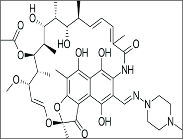
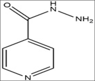
Fig .1 and 2 The Chemical structure of Rifampicin and Isoniazid [8].
METHODOLOGY
The present review is a thorough assessment of RIF and ISO toxicity and associated risks, with a particular emphasis on its effect on the liver. The literature includes both experimental and non-experimental data extracted by using databases and journals indexed in Google Scholar, Science Direct, PubMed, Web of science, covering studies published between 1994 and 2025. The extracted data was then analysed and compiled to assess rifampicin and isoniazid induced hepatotoxicity.
EFFECT OF RIF AND ISO
Liver damage in various gradations could be the cause of administration of both RIF and ISO [5]. ISO treatment causes skin eruption, jaundice, back pain, infection and optic nerve degeneration and destructive influence on the peripheral and the central nervous system. Hepatotoxins transform into chemically reactive metabolites in the liver, that have the potential to interfere with other cellular components such as protein, lipids and nucleic acids leading to dysfunction [1]. RIF exposure may lead to a number of symptoms, such as thrombocytopenia, which is defined by skin and mucosal bleeding and metabolic acidosis. Oligouric disturbances in the kidney, spasms, jaundice like symptoms such as yellowing of skin and eyes due to bile component accumulation, edema around the eyes, skin rashes and facial inflammation are examples of RIF and ISO induced toxicity [10]. Hepatitis is one of the documented side effects of rifampicin, which also includes nausea, vomiting, lack of appetite, stomach cramps and diarrhoea. Other adverse effects, are vertigo, headache, exhaustion, disorientation and menstruation disruption [11].
Body and organ weight
The body weight of rats co-administered with isoniazid and rifampicin significantly (p < 0.001) decreased to 171.8 ± 3.12g compared to that of control to 218 ± 7.18 g. whereas liver weight significantly (p < 0.001) increased to 8.77 ± 0.11 g compared to that of control with 6.61 ± 0.21 g [12,13,2]. Increase of mean body weight and liver weight was very slow in RIF and ISO treated group as compared to control [14]. Decrease in mean final body weight, liver weight, length, width and thickness noted in treated group compared to control group [15]. Significant increase in liver/body weight index after RIF and ISO treatment was noted as compared to control [16].
Morphological and behavioural observation
Animals treated with ISO alone and in combination with RIF exhibited decreased food intake, quietness, less motor activity, rough hair coat, inactiveness and slow response to external stimuli [17] [18].
Biochemical Analysis
Previous experimental studies on animals suggest that ISO administration in toxic doses distress hepatocellular membrane integrity. The effect of rifampicin at high dose of 100 mg/kg body weight for 6 days showed noteworthy rise in the level of γ-glutamyl transpeptidase, acid ribonuclease, acid phosphatase whereas the level of succinate dehydrogenase diminished. The serum level of RNA, total proteins and lipids, bilirubin, phospholipids, cholesterol, lipid peroxides increased while hepatic glycogen decreased and the lower dose of 50 mg/kg body weight showed less significant changes as compared to higher dose [19]. [20, 9] evaluated liver injury in rats induced by ISO and RIF and detected three times increase in serum aspartate aminotransaminase, alanine transaminase, alkaline phosphatase and bilirubin and altered the lipid levels in serum and liver compared to control group, while triglycerides were significantly elevated. In contrast, phospholipids were significantly decreased in liver. Serum bilirubin, peroxidation of lipids (P < 0.01) and low levels of hepatic non protein thiols (P < 0.01) in all the animals of RIF and ISO treated group significantly increased as compared to control [21,22]. According to [23] administration of RIF and ISO at a high and low dose of 250 and 50 mg/kg body weight respectively for 90 days daily resulted in abnormal rises or falls in the liver marker enzymes with mild hyperlipidemia, hypercholesterolemia and hyperuricemia, leading to membrane damage causing outflow of alanine transaminase, alkaline phosphatase and bilirubin, increased lipid peroxidation and glutathione homeostasis, and enhanced the CYP 2E1 mediated bioactivation mechanism [24]. [25] reported significant elevation in the level of oxidative stress and hepatocellular injury followed by RIF and ISO treatment. [26, 27] demonstrated significant increase in the thiobarbituric acid reactive substances, the oxidative stress markers along with liver marker enzymes in experimental rats. The RIF and ISO administered animals exhibited significantly (P<0.01) reduced hepatic glutathione levels at100 mg/kg body weight intra peritoneally.
[12] reported increased total bilirubin level, blood urea nitrogen, serum creatinine and serum uric acid along with glutathione pyruvate transaminase, oxaloacetate transaminase and alkaline phosphatase. [28] stated that RIF and ISO elevated the level of alanine aminotransferase activity, steatosis, apoptosis and oxidative stress markers in experimental animals, whereas decreased glutathione content provoked liver damage. Significant increase in the total cholesterol, triglycerides and low density lipoprotein cholesterol levels, whereas high density lipoprotein cholesterol level showed a significant decrease. The level of total proteins, albumin and alpha 1-globulin decreased significantly. But no noticeable changes were documented in the other elements of globulins such as alpha, beta and gamma globulin, as well as albumin to globulin ratio and creatinine level [29, 30]. [31] revealed significantly increased serum levels of liver marker enzymes. Whereas activity of superoxide dismutase and glutathione was reduced followed by ISO and RIF treatment that indicate the oxidative stress induced liver damage. The level of serum bilirubin, serum enzyme markers, total protein, albumin and total globulin increased as compared to control [1, 3, 16, 32, 18, 33, 15, 34, 35, 3]. Furthermore, mRNA levels of Fas, Acc and Scd-1genes, some significant genes responsible for fatty acid synthesis, class B scavenger receptor CD36 rise in RIF treated group. Inactivation of transcription factors SREBP-1c and LXR-α that regulate the genes for synthesis of hepatic fatty acid was reported in RIF treated group. Whereas hepatic PXR was activated quickly in RIF treated mice. Hepatic PPARγ, a downstream target of PXR, was transcriptionally upregulated. Thus, the augmented level of lipid synthesis in liver and absorption of fatty acids into liver from circulation collectively attributed to RIF induced hepatic lipid accumulation. This partially attributed to RIF induced up regulation of PPARγ and its target genes [36]. [37] analysed and enumerated various proteins and reported that a total of 29 and 40 proteins expressed significantly whereas proteins 27 and 118 were not expressed in response to 177 and 442.5 mg/kg RIF, respectively. Analysis by Gene Ontology and Kyoto Encyclopedia of Genes and Genomes pathway enhancement showed up regulation of glutathione transferase activity and down regulation of proteins associated with arachidonic acid metabolism whereas signalling pathway of transcription factor activated by a ligand and regulator of adipogenesis and lipid metabolism, CYP enzymes, tripeptide glutathione, chemical carcinogenesis and relative proteins amplified in RIF treated liver.
[2,18,14] reported that the level of antioxidants like superoxide dismutase and catalase in RIF and ISO treated groups were significantly decreased whereas liver marker enzymes increased significantly (p < 0.001). The serum level of protein and albumin decreased significantly (p < 0.001). 50% decline in the ratio of albumin to globulin to that of its original value observed in RIF treated group as compared to control group. The serum cholesterol, bilirubin and malondialdehyde level significantly increased (P <0.001), in RIF treated group compared to control.
[38] reported that activation in the hepatic stellate cell markers increased can be correlated with RIF and ISO treatment with the increased liver collagen and significant periportal fibrosis. These changes are concomitant with hepatocytic apoptosis, increased activity of hepatic cytochrome P450 2E1, NADPH oxidase and oxidative stress simultaneously that leads to the development of liver fibrosis.
[6] co?administered RIF and ISO and observed significantly (P<0.01) upregulated NF?κB DNA binding activity and expression of tumor necrosis factor alpha mRNA whereas bile salt export pump was significantly up and downregulated respectively. The protein expression of bile salt export pump was found to be significantly decreased. Liver injury induced by antituberculosis drugs, RIF and ISO may be ascribed to dysfunction of mitochondria, drug metabolic enzymes, oxidative stress, accumulation of protoporphyrin IX, stress in endoplasmic reticulum, disparity in bile transport and immune response, cholestasis and accumulation of lipid in the liver. ISO induced liver toxicity can be enhanced by RIF by regulating the expression of drug metabolizing enzymes and transporters. Whereas, ISO can provoke RIF induced liver damage by declining the serum bilirubin level [39]. Significant increase in cytokines interleukin- 6 and 1beta, tumor necrosis factor alpha, and caspases 3 that execute the apoptosis detected after RIF and ISO treatment [34].
Histopathological evidences
Histopathological studies of liver showed coagulative necrosis and inflammation of the hepatic artery, portal vein and bile duct, with piecemeal necrosis, infiltration of lymphocytes and neutrophils, congestion of the blood vessels, the central vein, hepatic sinusoids and interlobular septums with severe infiltration of inflammatory cells in RIF and ISO treated group [20, 21, 15]. [26] reported cell swelling, congestion and fatty degeneration of the liver cells and portal triaditis observed in RIF and ISO treated group. Neutrophil infiltration in the centrilobular region with portal triaditis, vacuolated cytoplasm and swollen sinusoids with some lymphatic accumulations were reported in RIF and ISO treated group [27, 24, 12, 29]. [30, 37] demonstrated that RIF and ISO treatment resulted in fatty degeneration, inflammation, distended sinusoids, necrosis and hypertrophy in hepatocytes. Hepatotoxicity induced by ISO alone causes necrosis and inflammatory cell infiltration with steatotis, cholestatic, thick wall of blood vessels showing congestion and mononuclear cells aggregation with large granulomatous lesion in liver parenchyma and lipid accumulation, which was evaluated by lysochrome diazo dye in RIF and ISO treated rats [40, 31]. [35, 36] noticed significant hepatocyte hypertrophy with prominent increase in cell size along with hepatocytes with enlarged and binucleated hepatocytes. [16] examined severe damage in the hepatic architecture with prominent polymorphonuclear inflammatory infiltration, cytoplasmic vacuolation, interlobular hemorrhage, dilated hepatic sinusoids with degenerated hepatocytes and moderately dilatation and congestion in central vein, disorganized cord of hepatocytes. Focal necrosis in the hepatic parenchyma with odema, some pyknotic hepatocytes, few bundles of fibrous tissue, hepatocytes merged with each other forming eosinophilic syncytial masses. Ballooning degeneration of hepatocytes with vacuolated cytoplasm and indistinct cell boundaries observed in RIF and ISO treated animals [31]. Histopathological alterations induced by RIF and ISO showed inflammation in the periportal region of hepatocytes [2]. Histopathology of liver in rifampicin treated group exhibited lipid accumulation, cell necrosis, enlarged central vein, increased sinusoidal spaces and disrupted portal vein that indicates damaged histoarchitechture. Rat treated with ISO indivdually or in combination with RIF induce liver alterations showing necrosis, immune cells infiltration, portal expansion and dilation, sinusoidal congestion, deranged structure, such as portal inflammation, fatty degeneration, necrosis, dilated sinusoidal capillaries, disrupted central vein and Kupffer cells inflammation and thus concluded that co-administration of RIF and ISO cause hepatotoxic effect which causes injury to the liver parenchyma. [4, 32, 39, 15, 17].
Table 1: Hepatotoxicity of rifampicin and isoniazid
|
Sr. No |
Author, and Year |
Title |
Dose and No of Animals used |
Duration and Parameter
|
Finding |
Remark |
|
1 |
Seema et al., (1994) |
RIF induced hepatoxicity in rats : Protective effect of Picrolive effect of picroliv. |
Albino rats. High dose of RIF and ISO 100 mg/kg/bw and low dose 50mg/kg/bw |
6 days Biochemical study and statistical analysis |
Significant increase in hepatic γ-glutamyl transpeptidase, acid ribonuclease. |
Induced biochemical toxicity in liver |
|
2 |
Ravinder et al., (2006) |
Effect of garlic on RIF and ISO induced hepatic injury in rats |
26 Wistar rats. INH and RIF 50 mg/kg/bw both
|
28 days. Biochemical analysis and Histological changes . |
Increase serum ALT, AST, and bilirubin levels. And focal hepatocytic necrosis and portal triaditis. |
Induced biochemical toxicity and histological damage |
|
3 |
Bhupinder et al.,(2007) |
Effect of cimetidine on hepatotoxicity induced by RIF and ISO combination in rabbits. |
Six rabbits. INH and RIF 50mg/kg/bw both |
7 days Biochemical analysis and Histological changes |
Decreased food intake quietness, and less motor activity and rough hair coat. Biochemical changes increase aspartate aminotransferase and alanine transaminase, Histological changes portal inflammation, fatty changes and liver cell necrosis |
Induced increase biochemical toxicity and histological changes |
|
4 |
Sheikh et al., (2007)
|
Potentiation of isoniazid-induced liver toxicity by rifampicin in a combinational therapy of antitubercular drugs (RIF and ISO and pyrazinamide) in Wistar rats: A toxicity profile study. |
Wistar rats. RIF 250 mg/kg/bw and INH 50mg/kg/bw dose |
90 days Biochemical analysis |
Observed mild hyperlipidemia, hypercholesterolemia and hyperuricemia .caused membrane damage resulting alanine transaminase, alkaline phosphatase and bilirubin, imbalance in endogenous enzymatic increased lipid peroxidation |
Induced biochemical toxicity and caused membrane damage |
|
5 |
Ravinder et al., (2008) |
RIF and ISO induced lipid changes in rats. |
16 Wistar rats. used INH and RIF 50mg/kg/bw dose |
28 days Biochemical analysis and histology |
Increased aspartate aminotransaminase, alanine transaminase, alkaline phosphatase and bilirubin triglycerides. Histology changes- piecemeal necrosis, focal lobular inflammation |
Induced biochemical toxicity and histological damage in liver |
|
6 |
Yogeeta et al., (2008) |
Antihepatotoxic Effect of Punica granatum. Acetone Extract Against RIF and ISO Induced Hepatotoxicity. |
24 Male wistar albino rats. INH and RIF 50mg/kg/bw dose |
15 days Serum hepatic enzymes analysis, enzymic and non enzymic antioxidant |
Elevation in the level of lipid peroxides serum and glutamate oxaloacetate transaminase, glutamate pyruvate transaminase, lactate dehydrogenase, alkaline phosphatase, decrease in the enzymic antioxidants superoxide dismutase, catalase, Non antioxidants reduced glutathoine and vitamin C, and E |
Induced biochemical toxicity and enzymic and non enzymic antioxidant disturbans in liver |
|
7 |
Dalal (2011) |
The effect of RIF and ISO on liver and lung tissues in rats. |
24 adult Swiss albino rats. INH 50mg/kg/bw and RIF 25 mg/kg/bw |
60 days morphology and Histological study of liver |
Change in hepatic tissue morphology Histological changes-congestion in central vein, hepatic coagulative necrosis, and congestion of hepatic sinusoids and fatty degeneration |
Induced morphology and histology toxicity in liver
|
|
8 |
Thattakudian S.U et al.,, (2011) |
Protective effect of methanolic extract of Annona Squamosa linn in RIF and ISO induced hepatotoxicity in rats. |
30 Male Wistar albino rats. RIF100 mg/kg/bw and ISO 100 mg/kg/bw dose |
21 days liver marker enzymes and TBARS,oxidative stress marker and Histological study |
Elevation in the liver marker enzymes and thiobarbituric acid reactive substances, the oxidative stress markers, and reduced hepatic glutathione levels. Histological study of the liver showed cell swelling, congestion and feathery degeneration of the liver cells and portal triaditis.
|
Induced biochemical toxicity and histological damage in liver |
|
9 |
Parameswari et al., (2012) |
Protective role of Ficus benghalensis against RIF and ISO induced oxidative liver injury in rats. |
36 Male Wistar albino rats. and Swiss albino mice RIF and INH 100mg/kg/bw dose. |
21 days. Biochemical estimation and Histopathological study |
Elevation in the levels of liver marker enzymes and thiobarbituric acid reactive substances total protein and gluthione level increased and histological showed hepatocytic necrosis and inflammation and neutrophil infiltration |
Induced biochemical alteration and histological damage in liver |
|
10 |
Mohammed and Mohammed (2012) |
Effect of Morin on RIF and ISO Induced Hepatotoxicity in Rats.
|
24 Female Swistar albino rats. RIF &INH 100g/kg/bw dose. |
21 days. Increase Biochemical estimation and Histopathological study |
Increase in the activity of alanine aminotransferase, aspartate aminotransferase and malondialdehyde level and decrease the activity of superoxide dismutase, glutathione peroxidase, glutathione s-transferase and catalase. Necrosis and fatty degeneration in hepatocytes |
Induced biochemical alteration and histological damage in liver |
|
11 |
Ifthekar S.et., (2012) |
Evaluation of hepatoprotective and nephroprotective activity of aqueous extract of Vigna mungo (Linn.) Hepper on RIF induced toxicity in albino rats. |
48 Albino rats of either sex. |
Morphological study, biochemical analysis and histopathological study |
Decreased body weight and increased liver weight. Biochemical analysis showed increased serum glutathione pyruvate transaminase, oxaloacetate transaminase, alkaline phosphatase, total bilirubin level, blood urea nitrogen, serum creatinine and serum uric acid level. Histopathology showed liver damage |
Alter body and organ weight, Induce biochemical alteration and histological damage |
|
12 |
Cristina et al., (2013) |
Hepatocyte growth factor protects against isoniazid/rifampicin-induced oxidative liver damage. |
Albino rats. RIF 150mg /kg/bw and INH 75mg/kg/bw dose. |
7 days Antioxidant and antiapoptotic study |
Increased alanine aminotransferase activity, steatosis, apoptosis and oxidative stress markers and glutathione content decreased aggravating liver damage |
Exhibit biochemical alterations |
|
13 |
Shabana et al., (2012) |
Influence of RIF and tetracycline administration on some biochemical and histological parameters in albino rats |
Male albino rats. RIF 200mg/kg/bw |
30 days Biochemical estimations and histopathological study |
Increased total cholesterol, triglycerides and low density lipoprotein-cholesterol whereas high density lipoprotein-cholesterol levels decreased and histopathological -necrosis of hepatocytes, cytoplasmic vacuolation and distended sinusoids with lymphatic aggregations |
Induced biochemical toxicity and histological damage |
|
14 |
Bais and Saiju (2014) |
Ameliorative effect of Leucas cephalotes extract on RIF and ISO induced hepatotoxicity |
30 Sprague Dawley rats. ISO and RIF at100 mg/kg bw each |
21 days Biochemical analysis and Histopathological studies |
Elevation in the level of serum glutamic oxaloacetic transaminase and serum glutamic pyruvic transaminase, alkaline phosphatase and bilirubin. Histology- fatty degeneration (steatosis) inflammation, degenerated hepatocytes, distended sinusoids, necrosis and hypertrophy in hepatocytes |
Induced rifampicin and isoniazid toxicity on liver |
|
15 |
Chao et al., (2016) |
Naringenin protects against RIF and ISO induced apoptosis in hepatic injury |
40 Male BALB/c mice. RIF and ISO 100 mg /kg/bw |
14 days Biochemical estimation and Histopathological study, immunohistochemistry, western blotting
|
Increase in serum alanine aminotransferase and aspartate aminotransferase levels. The activity of superoxide dismutase and glutathione was reduced and histology study showed necrosis and inflammatory cell infiltration |
Induce biochemical and histological alteration in liver tissue |
|
16 |
Zodape and Bhise (2016) |
Effect of Aloe Vera Extract on The Hepatotoxicity Induced By Isoniazid And Rifampicin Drugs In Male Wistar Rats. |
48 Male Wistar rats. RIF and ISO 450 mg/kg/bw ,300 mg/kg/bw and 70 mg/kg/bw dose |
30days Biochemical estimation . |
Increase serum bilirubin, Serum aspartate aminotransferase, serum alanine aminotransferase, serum alkaline phosphatase serum acid phosphatase, total protein, globulin and albumin |
Induced biochemical toxicity in liver |
|
17 |
Adnan and Abed (2016) |
Histological and Biochemical study of Nigella Sativa Seeds effects on Liver of male Albino Rats treated with RIF |
24 Male albino rats. RIF 50mg/kg/bw dose |
28 days Biochemical estimation and Histopathological Study. |
Decrease alkaline phosphatase, serum glutamic pyruvic transaminase, serum glutamic oxaloacetic transaminase and serum bilirubin. Histology study showed focal necrotic hepatocyte and neutrophils, mononuclear cells aggregation thickening wall of congested blood vessels with large granulomatous lesion in liver Parenchyma |
Induced biochemical toxicity and histological damage |
|
18 |
Huang et al., (2016) |
RIF Induced Hepatic Lipid Accumulation: Association with Up- Regulation of Peroxisome Proliferator- Activated Receptor γ in Mouse Liver |
Mice RIF 200mg/kg/bw dose |
3days histology estimation of hepatic lipid synthesis |
Hepatic lipid accumulation. Increased triglyceride content |
Induce up regulation of PPARγ and its target genes |
|
19 |
Kim et al ., (2017) |
Mechanism Investigation of Rifampicin-Induced Liver Injury Using Comparative Toxicoproteomics in Mice |
RIF 177 and 442.5 mg/kg/bw of dose |
14 days Biochemical And Histopathological study and Geneontology Kyoto encyclopedia of genes |
Up regulation of glutathione activity and down regulation of proteins related to arachidonic acid. Histology-extreme hepatocyte hypertrophy |
Exhibit biochemical genetic and histological alterations |
|
20 |
Meena et al., (2018) |
Hepatoprotective activity of Tamarindus indica Linn stem bark ethanolic extract against hepatic damage induced by co-administration of antitubercular drugs RIF and ISO in Sprague Dawley rats |
Sprague Dawley rats. RIF and ISO 50mg/kg/bw of dose |
28 days. Morphological. Biochemical and Histopathological study |
decreased body weight and increased liver weight. Biochemical analysis increased alanine transaminase, aspartate transaminase, alkaline phosphatase, lactate dehydrogenase, serum bilirubin, cholesterol and total protein and albumin levels reduced. Histology- necrosis and disturbed architecture.
|
Alter body and organ weight, Induce biochemical alteration and histological damage |
|
21 |
Thuwaini et al., (2018) |
The possible protective effect of Carthmustinctorius (leaves) on anti-tuberculosis drugs (RIF and ISO) induced hepatotoxicity in rats |
Male albino rats RIF and ISO 50 mg/kg/bw of dose |
4 week Biochemical estimation and Histopathological study |
increases alanine aminotransferase, aspartate aminotransferase and alkaline phosphatase. Histology study showed hepatic architecture with prominent polymorphonuclear inflammatory infiltration cells hepatocytes cytoplasmic vacuolation, interlobular hemorrhage, dilated hepatic sinusoids |
Induced biochemical toxicity and histological damage |
|
22 |
Sabina et al., (2019) |
A comparison of hepatoprotective activity of Bacoside to Silymarin treatment against a combined RIF and ISO induced hepatotoxicity in female Wistar rats |
Female wistar albino rats. RIF and ISO 50mg/kg/bw of dose |
28 days Body weight .Biochemical estimation and Histological study. |
Elevation in the serum glutamic oxaloacetic transaminase (SGOT) and serum glutamic pyruvic transaminase (SGPT) decreased the level of antioxidant superoxide dismutase (SOD) and catalase (CAT) and histology- periportal inflammation with inflammatory cells |
Induced biochemical toxicity and histological damage |
|
23 |
Swatilekha et al.,(2019) |
Hepatotoxic effect of RIF as an Anti-Tuberculosis drug on male Albino rat |
18 Male albino rats RIF 50 mg/kg/bw |
14 days. Morphological study Biochemical and Histological study. |
Increase of body weight and liver weight. and Increased serum aspartate transaminase alkaline phosphatase, lactate dehydrogenase and total protein, albumin content serum decreased. Histology -lipid accumulation, massive cellular necrosis, enlarged central vein and sinusoidal and disrtpted poratl vein |
Altered biochemical markers inducing hepatotoxicity and histological damage |
|
24 |
Edward et al., (2019) |
Hepatoprotective Effect of Citrus Sinensis Peel Extract Against RIF and ISO induced Liver Injury in Wistar Rats |
25 Wistar rats. ISO and RIF 50mg/kg/bw Both |
14 days biochemical and Histopathological study
|
Increased serum alanine transaminase and aspartate transterase. Histology- steatosis, vacuolation and necrosis of hepatic cells |
Induced biochemical toxicity and histological damage |
|
25 |
Ayan et al., (2020) |
RIF and ISO Produce Hepatic Fibrosis through an Oxidative Stress?Dependent Mechanism. |
Male mice. ISO 50 mg/kg/bw and RIF100mg/kg/bw dose |
6 days a week, for 4 to 24 weeks |
Increase hepatic stellate cell(HSC) elevation of liver collagen content and increase in hepatic cytochrome P450 2E1 (CYP2E1), NADPH oxidase (NOX) activity |
Activate HSC leading to liver fibrosis |
|
26 |
Xue et al., (2020) |
Protective effect of pyrrolidine dithiocarbamate on RIF and ISO induced liver injury in rats |
24 Sprague -Dawley rats ISO and RIF 50mg/kg/bw dose. |
28 days. Biochemical estimation |
Increased serum level total bile acid (TBA), total bilirubin (TBIL), direct bilirubin (DBIL), decreased superoxide dismutase (SOD) activity and malondialdehyde (MDA) elevated. upregulates NF?κB DNA? binding activity and down regulate TNF?α and BSEP |
Induced biochemical toxicity in liver |
|
27 |
Xiuping et al ., (2022) |
Review on Mechanisms of RIF and ISO liver injury and the effects of natural medicinal ingredients |
___ |
Biochemical estimation |
Induce oxidative stress, mitochondrial dysfunction, bile transport imbalance |
Induced hepatotoxicity |
|
28 |
Risdawati et al ., (2023) |
Protective Effect Of Andrographis paniculata Aqueous Extract (Eaap) Against RIF and ISO Induced Rat Liver Damage |
Male Sprague-Dawley rats ISO and RIF 100mg/kg/bw dose |
21 days Biochemical and Histological study |
Increased serum alanine aminotransferase, aspartate aminotransferase, alkaline phosphatase activity and total bilirubin levels. Histopathological analysis necrosis and infiltration of immune cells, causing portal expansion with dilation and sinusoid congestion. |
Induced biochemical toxicity and histological alterations |
|
29 |
Tarique et al .,(2023) |
Hepatoprotective potential of diosmin against hepatotoxic effect of RIF and ISO in wistar rats |
Wistar rats. INH & RIF 50mg/kg/bw dose. |
21 days Estimation of liver markers, oxidative stress and antioxidants and histopathological analysis |
Increased the markers of liver alanine aminotransferase, aspartate aminotransferase, alkaline phosphatase and bilirubin, cytokines -tumor necrotic factor alpha (TNFα), interleukin- 6 and 1beta (IL-6 and IL-1β) and apoptosis histology degeneration, inflammation and necrosis of hepatocytes |
hepatocellular damage mediated by inflammation, apoptosis and oxidative stress mechanisms. |
|
Hans et al .,(2023) |
Alterations in Histomorphology, Biochemical Parameters and Gross Morphometry in Liver of Albino rats Following Administration of RIF and ISO |
12 Albino rats ISO & RIF 50mg/kg/bw dose. |
21 days Morphological study, biochemical analysis and histopathological study |
Decrease body weight and liver weight, length, width and thickness increased. Biochemical analysis aspartate transaminase, alanine transaminase, alkaline phosphatase. Histology- dilatation of sinusoidal capillaries, disrupted central vein and inflamed Kupffer cells |
Induced biochemical toxicity and histological damage |
|
|
31 |
Mosa (2024) |
Embelin Mitigates Hepatotoxicity Induced by RIF and ISO in rats. |
36 Wistar rats. ISO and RIF 100mg/kg/bw dose |
21 days liver marker enzymes oxidative stress and biochemical estimation |
liver damage, increased biological stress tumor necrosis factor (TNF-α), interleukin -1β (IL-1β), and interleukin-6 (IL-6). Biochemical analysis alanine aminotransferase, aspartate aminotransferase, alkaline phosphatase |
Induce oxidative stress and biochemical toxicity in liver |
|
32 |
Ali et al., (2025) |
Effects of Taurine on RIF induced Liver Injury.
|
36 Male Wistar rats. RIF 100mg/kg/bw dose |
2 week Biochemical and Oxidative stress marker estimation |
Elevation in the alanine aminotransferase, aspartate transferase, lactate dehydrogenase and bilirubin. Oxidative stress marker glutathione level of decreased reactive oxygen species and increased lipid peroxidation and decreased the levels of reactive oxygen |
Induce biochemical and Oxidative stress toxicity in liver |
CONCLUSION
Overall, evidence indicates that rifampicin and isoniazid cause dose and duration dependent liver toxicity, especially when used together. The hepatotoxicity is primarily mediated by oxidative stress and impaired metabolic pathways. Many plant extracts and antioxidants show protective effects, suggesting possible therapeutic value. Continuous monitoring of liver function is essential during tuberculosis treatment, and further research is needed to clarify the combined toxicity mechanisms and improve safer treatment strategies.
REFERENCES


P. R. Meher, U. A. Jiwantare, V. T. Dhurvey, Effect of Rifampicin and Isoniazid on Liver of Albino Rat: A Review, Int. J. of Pharm. Sci., 2026, Vol 4, Issue 3, 3263-3279, https://doi.org/10.5281/zenodo.19229230
 10.5281/zenodo.19229230
