Gurukrupa Institute of Pharmacy, Majalgaon, Beed, Maharashtra, India 431131
The present study investigated the phytochemical composition and wound healing potential of the ethanolic extract of Maytenus emarginata. The extractive value using ethanol was 8.62% w/w, yielding a dark black solid extract indicative of a high recovery of phytoconstituents. Preliminary phytochemical screening confirmed the presence of flavonoids, phenols, saponins, diterpenes, certain glycosides, and proteins, while alkaloids, resins, and steroids were absent. Quantitative estimation revealed a total phenol content of 0.562 mg/100 mg and total flavonoid content of 0.785 mg/100 mg, suggesting a rich presence of antioxidant compounds. Wound healing activity was evaluated using gel formulations (1% and 2% extract concentrations) in comparison to normal saline and standard Betadine treatment. The results demonstrated a dose-dependent enhancement in wound closure, with the 2% gel formulation exhibiting superior healing efficacy, slightly surpassing the standard treatment by Day 11. The observed wound-healing properties are attributed to the extract’s phytochemical constituents with anti-inflammatory and regenerative activities. These findings validate the traditional medicinal uses of M. emarginata and support its potential as a topical therapeutic agent for wound management.
Wound
A wound can be described in many different ways. It can be explained based on its cause, where it is located on the body, whether it is sudden or long-term, how it is treated, the symptoms it causes, or what kind of tissue is visible in the wound. All these descriptions are important for properly assessing and managing the wound, and helping it heal if possible.
By its true definition, a wound is when the skin's protective function is damaged. This happens when the outer layer of skin, called the epithelium, is broken, sometimes along with the deeper tissues beneath. This can occur due to injury like surgery, a hit, a cut, chemicals, extreme temperatures, rubbing, pulling forces, pressure, or from diseases such as leg ulcers or cancer.
Wounds can heal in two main ways: either by closing the wound with stitches, known as primary intention, or by letting the wound repair itself naturally, known as secondary intention. In the second case, the body rebuilds the damaged tissue and regrows the skin layer.
Wound Healing
Wound healing is a complex and ongoing process where the environment around the wound changes as the person's overall health changes. Understanding the normal steps of wound healing, including the phases of stopping bleeding, inflammation, tissue growth, and tissue strengthening, gives a clear picture of how wounds heal naturally. This knowledge helps health care professionals learn how to properly care for wounds and support the body's ability to repair itself.
When a wound doesn't heal properly, it's important for health care professionals to look for the root causes that might be preventing healing. Taking care of chronic wounds needs a focus on the patient's needs, a whole-body approach, teamwork from different specialists, and methods that are both effective and affordable, based on scientific evidence.
In any natural disaster, the first step is to identify and stop the harmful forces causing damage before any repair can start. Similarly, in wound care, it's essential to find and manage the underlying causes and factors that are stopping the healing process before healing can begin.
Phases of Wound Healing
Whether wounds are closed by primary intention, subject to delayed primary closure, or left to heal by secondary intention, the wound healing process is a dynamic one which can be divided into three phases. It is critical to remember that wound healing is not linear and often wounds can progress both forwards and back through the phases depending upon intrinsic and extrinsic forces at work within the patient.
The phases of wound healing are:
Inflammatory phase:
The inflammatory phase is the body’s natural response to injury. After initial wounding, the blood vessels in the wound bed contract and a clot is formed. Once haemostasis has been achieved, blood vessels then dilate to allow essential cells, antibodies, white blood cells, growth factors, enzymes and nutrients to reach the wounded area. This leads to a rise in exudate levels so the surrounding skin needs to be monitored for signs of maceration. It is at this stage that the characteristic signs of inflammation can be seen: erythema, heat, oedema, pain and functional disturbance.
Proliferation phase:
During proliferation, the wound is rebuilt with new granulation tissue which is comprised of collagen and extracellular matrix and into which a new network of blood vessels develop, a process known as angiogenesis. Healthy granulation tissue is dependent upon the fibroblast receiving sufficient levels of oxygen and nutrients supplied by the blood vessels. Healthy granulation tissue is granular and uneven in texture, it does not bleed easily and is pink/red in colour. The colour and condition of the granulation tissue is often an indicator of how the wound is healing. Dark granulation tissue can be indicative of poor perfusion or ischaemia. Epithelial cells finally resurface the wound, a process known as epithelialisation.
Maturation phase:
Maturation is the final phase and occurs once the wound has closed. This phase involves remodelling of collagen from type III to type I. Cellular activity reduces and the number of blood vessels in the wounded area regress and decrease.
PLANT PROFILE
Maytenus emarginata
Maytenus emarginata Ding Hou is a Celastraceae family plant. It is locally known as ‘Kankero’ in Hindi and Thorny staff tree in English. It is a sacred plant for the environment-friendly Bishnoi community in Rajasthan. It is believed that Lord Jambheswar (Jambhoji) had realization under the tree of Maytenus emarginata.
Maytenus emarginata is a small spiny tree or shrub which grows up to 6 meters in height. It is mostly found at high elevations from near sea level and is abundant at the edges of mangrove forests or secondary forests, hillsides, and in patches of saline water in desert regions.
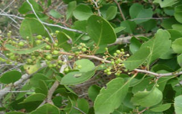
Figure 1: Maytenus emarginata
Botanical classification:
Botanical name: Maytenus emarginata (Willd.) Ding Hou
Synonyms:
Table 1. Various Phytochemical Constituents Reported in Maytenus emarginata
|
Sr. No |
Nature of Metabolite |
Identified Phytochemical |
|
1 |
Flavonoids |
Luteolin, Kaempferol, Quercitin |
|
2 |
Phytosterol |
β-sitosterol, Stigmasterol, Cholesterol, α-Amyrin acetate |
|
3 |
Triterpenoid |
Lupeol, Valemol |
|
4 |
Alkaloid |
Evonine |
|
5 |
Triterpene |
Squalene, Betulin, Friedelin, Lupenyl acetate, α-Caryophyllene, Isovellerdial, Germacrene A |
|
6 |
Triterpene glycoside |
Methyl commate C, Methyl commate A |
|
7 |
Alkane hydrocarbon |
Tridecane, Dodecane |
|
8 |
Alcohol |
Glycerin |
|
9 |
Aromatic compounds |
Myoinositol |
|
10 |
Fatty compounds |
Myristic acid, Pentadecanoic acid, Palmitoleic acid, Eicosanoic acid, α-Monostearin, n-Hexadecenoic acid, 9-Hexadecenoic acid, Octadecanoic acid, Linoleic acid |
MATERIALS AND METHODS
Extraction by maceration method
The leaves of Maytenus emarginata were collected from local area of Bhopal in the period of February, 2025
Collected leaves of Maytenus emarginata were cleaned properly and washed with distilled water to remove any kind of dust particles. Cleaned and dried plant materials were converted into moderately coarse powder in hand grinder. Powdered plant materials were weighed (50 gm) and packed in (250 ml) air tight glass Bottle. The plant materials were subjected to extraction by ethanol solvent as solvent for about 24 hrs59. The liquid extract was collected in a tarred conical flask. The solvent removed from the extract by evaporation method. The extracts obtained with ethanol solvent were weighed to a constant weight and percentage w/w basis was calculated.
Formulation of gel
Method of Preparation
In order to prepare the gel formulation, first dissolve carbopol 940 in distilled water. Next, add methyl paraben, propyl paraben, and glycerine, and let the mixture sit overnight. Consider the leaf extracts in propylene glycol, to which polymer dispersion was added later. After that, the remaining water was added, then triethanolamine was added while stirring constantly to get the pH down to 7.
Table No: 2: In vivo wound healing activity of gel
|
Sr. No. |
Ingredients |
F1 |
F2 |
|
1. |
Extract (%) |
1gm |
2gm |
|
2. |
Carbopol 934 (%) |
1gm |
1gm |
|
3. |
Propylene Glycol (ml) |
8ml |
8ml |
|
4. |
Methyl paraben (%) |
0.4gm |
0.4gm |
|
5. |
Propyl paraben (%) |
0.2gm |
0.2gm |
|
6. |
Triethanolamine (ml) |
qs |
qs |
|
7. |
Glycerin (ml) |
1ml |
1ml |
|
8. |
Distilled water (ml) |
Upto 100 ml |
Upto 100 ml |
Animals
Wistar rats weighing between 180-200 g with no prior treatment were used for the study. Animals were housed in polypropylene cages maintained at 22 ± 2? temperature with 12 h light and 12 h dark cycle. The animals were fed on a standard pelleted diet and had free access to water throughout the experiment. Animals that are described as fasting were deprived of food for at least 16 h but allowed free access to drinking water. The animal study was approved by the Institutional Animal Ethics Committee, as stated by prescribed guidelines of the Committee for the Purpose of Control and Supervision of Experiments on Animals (CPCSEA), No LNCP/IAEC/2025/036 Government of India.
Acute dermal toxicity
Acute dermal toxicity was determined, according to OECD 404, in rats62. Approximately 24 hours before the test, fur was removed by closely clipping the dorsal area of the
both flanks using a suitable depilatory material. Special attention was given not to abrade the skin. Skin was washed with sterile water a day before the toxicity test procedure. A limit test dose of 2,000 mg/Kg of the 10% (w/w) gel was applied on the shaved back of the rats within a range that was approximately 10% of the body surface area and held in contact to the skin with a gauze patch to facilitate good contact and uniform distribution of the test chemical on the skin for the duration of the exposure period (24 hours). The test was first conducted in a single rat for initial testing, and then after observing the response of the first rat for 24 hours a confirmatory test was done on another two rats simultaneously. At the end of the exposure period, the gel was removed and the animals were observed for 14 days for the development of any adverse skin reactions like inflammation, irritation, or redness. Behavior, general condition, posture and reflexes, attitude towards food, and water were also evaluated.
Experimental model
Male Sprague-Dawley rats were divided into four groups of six rats each and housed separately in individual cages. Before the creation of wounds, the animals were anaesthetised with ketamine (30 mg/kg, i.m.) and xylazine (3 mg/kg, i.m.). To create the wound, an electrical shaver was employed to shave the dorsal neck of each rat. A mark was made in the dorsal thoracic region, one centimetre away from the vertebral column and five centimetres from the ear. An excision wound was created by cutting out the full thickness of skin using a circular stamp with a diameter of two centimetres
Experimental designs
Wounds in the control group, designated as Group 1, were treated with normal saline (0.2 ml) twice daily. The reference standard control, labelled as Group 2, involved rats that were given 0.2 ml of Betadine twice daily. Rats in Groups 3 and 4 received Maytenus emarginata extract gel (1% and 2%, respectively) twice daily, and no dressing was applied to the wound site.
Assessment of degree of wound healing
Progressions of the wound healing were assessed on days 0, 3, 7, and 11 post-wound creation. Wound area was traced by a transparent tracing paper placing over the wound, which was then placed on a sheet of graph paper (2 mm2) to count the number of squares within the wound area64. The percentage of wound closure was measured using the following formula:
Wound Closure (%) = [(Wound Area at Day 0 - Wound Area at Later Day) / Wound Area at Day 0] X 100
Data Analysis
The data is expressed as mean ± Standard Deviation (SD). Results were analysed using one- way ANOVA followed by Dunnet’s test. Differences were considered statistically significant at P < 0.05 when compared with control.
RESULTS AND DISCUSSION
Extractive values of Maytenus emarginata
The yields were found to be 8.62% w/w of crude drug of ethanolic extract with dark black colour solid mass for Maytenus emarginata. Obtained results were recorded .
Table No: 3 Preliminary phytochemical screening of Maytenus emarginata
|
Sr. No. |
Phytoconstituents |
Test Name |
Ethanolic extract |
|
1. |
Alkaloids |
Mayer’s Test |
-ve |
|
Dragendorff’s Test |
-ve |
||
|
Wagner’s Test |
-ve |
||
|
Hager’s Test |
-ve |
||
|
2. |
Glycosides |
Raymond’s Test |
-ve |
|
Killer Killani Test |
-ve |
||
|
Legal Test |
+ve |
||
|
3. |
Carbohydrates |
Molisch’s Test |
-ve |
|
Fehling’s Test |
+ve |
||
|
Benedict’s Test |
-ve |
||
|
4. |
Tannins |
Vanillin- HCl Test |
-ve |
|
Gelatin Test |
+ve |
||
|
5. |
Flavonoids |
Lead acetate Test |
+ve |
|
Alkaline Reagent Test |
+ve |
||
|
6. |
Resins |
Turbidity Test |
-ve |
|
7. |
Steroids |
Libermann- Bur chard Test |
-ve |
|
Salkowski Reaction |
-ve |
||
|
8 |
Proteins & Amino acids |
Biuret Test |
+ve |
|
Precipitation test |
-ve |
||
|
9. |
Phenols |
Ferric chloride test |
+ve |
|
10. |
Saponin |
Froth test |
+ve |
|
11. |
Diterpenes |
Copper acetate Test |
+ve |
Estimation of Total Phenolic Content
Total phenolic content was estimated by gallic acid and expressed as milligrams of gallic acid equivalent (GAE). All the extracts contained a considerable amount of phenolic contents of mg/100mg of extract. The results were presented in
Calibration curves of Standard (Gallic acid) at 765nm Table No - 4
|
Standard |
Concentration (µg/ml) |
Mean absorbance |
|
Gallic acid |
5 |
0.201 |
|
10 |
0.358 |
|
|
15 |
0.543 |
|
|
20 |
0.712 |
|
|
25 |
0.856 |
(n=3)
Calibration curves of Standard (Gallic acid)
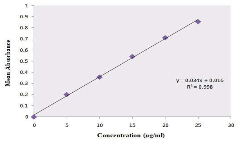
Total phenol content of ethanolic extract of Maytenus emarginata
Total phenol content of ethanolic extract of Maytenus emarginata Table No – 5
|
Sr. No. |
Extract |
Total phenol content (mg/100mg) |
|
1 |
Ethanolic extract |
0.562 |
Estimation of total flavonoids content
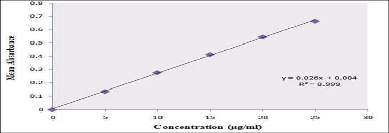
Flavonoid content was calculated from the regression equation of the standard plot (y = 0.026x + 0.004, R2 =0.999) and is expressed as quercetin equivalents (QE) (fig. 7.2).
Calibration curves of Standard (Quercetin) at 420nm Table No. 6
|
Standard |
Concentration (µg/ml) |
Mean absorbance |
|
Quercetin |
5 |
0.135 |
|
10 |
0.277 |
|
|
15 |
0.413 |
|
|
20 |
0.544 |
|
|
25 |
0.663 |
(n=3)
Total flavonoid content of ethanolic extract of Maytenus emarginata Table No - 7
|
Sr. No. |
Extract |
Total Flavonoid content (mg/100mg) |
|
1 |
Ethanolic extract |
0.785 |
The quantitative estimation of phytochemicals in the ethanolic extract of Maytenus emarginata revealed a moderate presence of total phenols and a relatively higher concentration of flavonoids. The total phenolic content was found to be 0.562 mg per 100 mg of extract, indicating the presence of compounds that may contribute to antioxidant and anti-inflammatory activities. More notably, the total flavonoid content was measured at 0.785 mg per 100 mg of extract, suggesting that flavonoids are a major class of secondary metabolites in this extract.
RESULTS OF EVALUATION OF IN VIVO WOUND HEALING ACTIVITY
Evaluation of wound healing activity of M. emarginata extract gel on rats Table No- 8
|
Groups |
Day 0 |
Day 3 |
Day 7 |
Day 11 |
|
I (Normal saline) |
0.0 ± 0.0 % |
18.5 ± 2.3 % |
42.7 ± 3.1 % |
61.4 ± 3.6 % |
|
II (Betadine) |
0.0 ± 0.0 % |
32.7 ± 2.5 % |
69.8 ± 2.8 % |
91.2 ± 2.4*** % |
|
III (M. emarginata extract gel 1%) |
0.0 ± 0.0 % |
25.1 ± 2.9 % |
57.3 ± 3.2 % |
79.6 ± 3.0*% |
|
IV (M. emarginata extract gel 2%) |
0.0 ± 0.0 % |
28.3 ± 2.7 % |
61.5 ± 3.5 % |
84.8 ± 2.9**% |
A one-way ANOVA was used for testing comparison with controls, and significance was assessed. Significance was defined as *P < 0.05, **P < 0.01 and ***P < 0.001.All values are expressed as mean ± SD.
It has been established that gel is an effective wound care product. As a positive control, betadine was chosen because it has been reported to have broad activity spectrum, the ability to penetrate biofilms, the absence of associated resistance, anti-inflammatory properties, low cytotoxicity, good tolerability, and no adverse effects on wound healing (Bigliardi et al., 2017).
Throughout the experiment, no sign of change in behavior of experimental animals were observed. Sprague-Dawley Rats were considered for wound healing studies because in comparison to other animals, rats are easy to handle and more economic than other laboratory animals. Wound healing is a very important and complex procedure linking many active processes and final stage is formation of scar tissue. In our study, the experimental animals were studied for 14 days. The final results of wound healing rate was noted till 11th day as more than 98% healing was obtained.
CONCLUSION
The results show that the gel made from Maytenus emarginata extract greatly helps wounds heal, and the more you use it, the better it works. Both the 1% and 2% versions of the gel helped close wounds more than the normal saline group. The 2% gel worked even better, almost matching or slightly beating the standard Betadine treatment by day 11. The quick healing seen with the 2% extract suggests that M. emarginata has strong wound healing abilities, probably because of its plant chemicals that fight inflammation and help tissues repair.
REFERENCES

Rani Munde, Nikhil Jadhav, In Vivo Wound Healing Activity of Maytenus Emarginata Gel Extract, Int. J. of Pharm. Sci., 2026, Vol 4, Issue 3, 2869-2877. https://doi.org/10.5281/zenodo.19198997
 10.5281/zenodo.19198997
