View Article

  • Development and Analytical Characterization of Liposomes: A Comprehensive Approach

  • 1Research & Development, Thermo Fisher Scientific, Greenville, NC 27834, USA.
    2Director, Product Development (CMC), Frontage Laboratories, Exton, PA 19341, USA.
    3Analytical Development & Quality Control, Navinta LLC, 1499 Ewing, NJ 08618, USA.

Abstract

Liposomes have emerged as versatile drug delivery systems, widely employed in pharmaceuticals due to their biocompatibility, controlled release properties, and ability to encapsulate both hydrophilic and lipophilic drugs. This review comprehensively explores the development and analytical characterization of liposomes, highlighting essential parameters such as morphology, particle size, zeta potential, and membrane fluidity. Advanced analytical techniques, including spectroscopy, chromatography, and microscopic imaging methods, are discussed for precise liposomal evaluation. Key approaches for assessing encapsulation efficiency, drug release kinetics, and stability are outlined, emphasizing their impact on formulation performance. Furthermore, the application of the Quality by Design (QbD) framework in liposomal development is detailed, with a focus on defining critical quality attributes (CQAs), material attributes, and process parameters to ensure robust product quality. Regulatory perspectives from the FDA, EMA, and ICH are highlighted to address standardization challenges in liposomal characterization. Emerging advancements such as smart liposomal systems, novel analytical tools, and scalable manufacturing strategies are also discussed. This review offers a comprehensive guide for researchers and pharmaceutical scientists seeking to improve liposomal formulation development and characterization techniques.

Keywords

Liposomes, Analytical Characterization, Drug Delivery Systems, Encapsulation Efficiency, Stability Studies, Quality by Design (QBD).

Introduction

Liposomes are self-assembled phospholipid vesicles comprising one or more bilayers encapsulating an aqueous core. Their amphiphilic nature allows for the encapsulation of both hydrophilic and hydrophobic drugs, making them ideal nanocarriers for pharmaceutical applications. Since their discovery in the 1960s, liposomes have gained prominence in drug delivery, gene therapy, vaccine formulations, and diagnostic imaging. The unique biocompatibility, biodegradability, and controlled release properties of liposomes enable site-specific drug delivery, enhancing therapeutic efficacy while minimizing systemic toxicity. Liposomes can be tailored by modifying lipid composition, surface charge, functionalization (e.g., PEGylation), and size, thus optimizing their pharmacokinetic and pharmacodynamic profiles. The flexibility in their structural design makes them suitable for various therapeutic applications, including: [1–3]

  • Cancer therapy – Encapsulation of chemotherapeutic agents (e.g., Doxil®, Onivyde®)
  • Antimicrobial delivery – Improved solubility and stability of antibiotics (e.g., AmBisome®)
  • Gene and RNA-based therapy – Lipid nanoparticles for siRNA and mRNA vaccines (e.g., Comirnaty™, Spikevax™)
  • Targeted drug delivery – Functionalized liposomes for site-specific action

Table 1 Advantages of Liposomes in Drug Delivery


Characteristic

Advantage

Application

Biocompatibility & Biodegradability

Minimal toxicity, safe for systemic administration

Cancer therapy, vaccines

Encapsulation of Hydrophilic & Hydrophobic Drugs

Versatile drug loading

Chemotherapeutics, antibiotics

Controlled & Sustained Drug Release

Prolonged therapeutic action, reduced dosing frequency

Long-acting injectables

Enhanced Permeability & Retention (EPR) Effect

Passive tumor targeting

Oncology

Surface Modifications (PEGylation, Ligand Attachment)

Active targeting, prolonged circulation time

Precision medicine

Significance of Analytical Characterization in Liposomal Development

Liposomal formulations exhibit complex physicochemical properties, requiring robust analytical techniques for precise characterization. The structural and functional attributes, such as particle size, surface charge, drug loading efficiency, encapsulation stability, and in vitro drug release, directly impact their bioavailability and therapeutic performance.

Analytical characterization ensures:

  • Batch-to-batch reproducibility and quality control
  • Optimization of formulation parameters (Critical Quality Attributes - CQAs)
  • Regulatory compliance with pharmaceutical guidelines (FDA, EMA, ICH)

Comprehensive characterization involves multiple analytical techniques, including dynamic light scattering (DLS) for size determination, high-performance liquid chromatography (HPLC) for drug content analysis, zeta potential analysis for stability assessment, and cryogenic transmission electron microscopy (Cryo-TEM) for morphological evaluation. [4–6]

Table 2 Key Analytical Parameters for Liposomal Characterization

Analytical Parameter

Importance

Common Techniques

Particle Size & Distribution

Influences biodistribution, drug release

DLS, NTA, AF4, TEM

Surface Charge (Zeta Potential)

Predicts colloidal stability

Electrophoretic Light Scattering (ELS)

Encapsulation Efficiency (EE%)

Determines drug-loading capacity

HPLC, UV-Vis Spectroscopy

Morphology & Lamellarity

Confirms vesicular integrity

Cryo-TEM, SEM

In Vitro Drug Release

Evaluates release kinetics

Dialysis, HPLC, UV Spectroscopy

Stability Studies

Ensures shelf-life and batch uniformity

Differential Scanning Calorimetry (DSC), DLS

Regulatory Perspectives and Market Trends

The commercialization of liposomal products necessitates adherence to global regulatory guidelines set by agencies such as the U.S. FDA, European Medicines Agency (EMA), and International Council for Harmonisation of Technical Requirements for Pharmaceuticals for Human Use (ICH). These agencies define critical quality attributes (CQAs) and establish standardized analytical methods to ensure efficacy, safety, and consistency in liposomal formulations.

Key regulatory challenges include:

  • Variability in characterization techniques across different research and industrial settings
  • Lack of globally harmonized standardization for liposome-based drug approval
  • Stringent requirements for demonstrating stability, biocompatibility, and clinical efficacy

Despite these challenges, the global liposomal drug delivery market has witnessed significant growth, driven by advancements in targeted therapies, vaccine technologies, and personalized medicine. The approval of lipid-based mRNA vaccines (Pfizer-BioNTech and Moderna) has further accelerated interest in lipid nanoparticles and liposomal formulations.

Table 3 Key Regulatory Guidelines for Liposomal Pharmaceuticals

Regulatory Agency

Guideline

Focus Area

FDA (USA)

Guidance for Industry: Liposome Drug Products

Manufacturing, pharmacokinetics, bioavailability

EMA (Europe)

Data Requirements for Intravenous Liposomal Products

Comparative studies, bioequivalence

ICH

Q8(R2) Pharmaceutical Development

Quality by Design (QbD) principles

WHO

Annex 9: Liposomal Drug Delivery Systems

Safety, efficacy, standardization

The future of liposomal drug delivery lies in the development of next-generation functionalized liposomes, including stimuli-responsive, ligand-targeted, and hybrid lipid-polymer nanoparticles. Addressing current analytical and regulatory challenges will be pivotal for ensuring consistent quality, reproducibility, and widespread clinical adoption.[7,8]

Physicochemical and Structural Attributes of Liposomes

Liposomes are complex nanoscale vesicular systems with distinct physicochemical and structural properties that influence their stability, encapsulation efficiency, biodistribution, and therapeutic efficacy. Understanding these properties is critical for optimizing liposomal formulations for pharmaceutical applications.

Morphology and Structural Organization

Liposomes exhibit diverse morphologies and internal structures, which can significantly affect drug loading, release kinetics, and biological interactions. The basic structural organization includes:

  1. Unilamellar Vesicles (ULVs): Single phospholipid bilayer surrounding an aqueous core
    • Small Unilamellar Vesicles (SUVs): 20–100 nm
    • Large Unilamellar Vesicles (LUVs): 100–1000 nm
  2. Multilamellar Vesicles (MLVs): Multiple concentric bilayers encapsulating aqueous compartments (~500 nm to several microns)
  3. Multivesicular Vesicles (MVVs): Large liposomes with multiple small vesicles enclosed within

The choice of morphology depends on the desired drug release profile, encapsulation efficiency, and stability.

Table 4 Structural Types of Liposomes and Their Characteristics

Type

Structure

Size Range

Applications

SUVs

Single bilayer

20–100 nm

Gene delivery, mRNA vaccines

LUVs

Single bilayer

100–1000 nm

Sustained drug release

MLVs

Multiple bilayers

500 nm–5 µm

Hydrophobic drug delivery

MVVs

Multivesicular structure

500 nm–5 µm

Long-acting injectables

Size Distribution and Polydispersity Index (PDI):

Particle Size and Its Significance

Particle size is a critical quality attribute (CQA) that affects cellular uptake, biodistribution, and clearance rates. Liposomes in the nano-range (50–200 nm) are optimal for tumor targeting via the Enhanced Permeability and Retention (EPR) effect, whereas larger liposomes (>400 nm) are useful for prolonged circulation and depot formulations.

Polydispersity Index (PDI) and Its Impact

  • PDI measures size uniformity: A low PDI (<0.3) indicates homogeneous liposomal dispersion, essential for reproducibility and stability.
  • High PDI (>0.5) suggests aggregation or instability, necessitating process optimization (e.g., extrusion, sonication).

Table 5 Liposomal Size Distribution and Applications

Size Range (nm)

Characteristics

Applications

Clearance Pathway

<50 nm

Rapid clearance

Short circulation time

Renal excretion

50–200 nm

Ideal for passive targeting

Cancer therapy

Prolonged circulation

200–500 nm

Controlled drug release

Sustained delivery

Hepatic clearance

>500 nm

Depot formulations

Long-acting injectables

Phagocytosis

Techniques for Size & PDI Analysis:

  • Dynamic Light Scattering (DLS) – Rapid and widely used
  • Nanoparticle Tracking Analysis (NTA) – Single-particle precision
  • Cryo-TEM & AFM – Structural and morphological insights
  • Asymmetrical Flow Field-Flow Fractionation (AF4) – High-resolution separation

Surface Charge (Zeta Potential) and Stability Considerations

The zeta potential represents the electrostatic charge on the liposomal surface, influencing colloidal stability, aggregation, and cellular interactions.

Key Roles of Zeta Potential:

  • High absolute values (±30 mV or more): Strong repulsion, stable dispersion
  • Low absolute values (<±10 mV): Increased aggregation, reduced stability
  • Charge-modified liposomes: Tailored for targeted delivery (cationic/anionic ligands)

Factors Influencing Zeta Potential:

  • Lipid composition (e.g., charged phospholipids)
  • pH and ionic strength of the dispersion medium
  • Surface modifications (e.g., PEGylation reduces charge and steric hindrance)

Table 6 Influence of Zeta Potential on Liposomal Stability

Zeta Potential (mV)

Stability Effect

Application

> +30 mV

Highly stable, cationic surface

Gene delivery (cationic liposomes)

+10 to +30 mV

Moderately stable

Targeted drug delivery

-10 to +10 mV

Poor stability, aggregation risk

Requires stabilization (e.g., PEGylation)

< -30 mV

Highly stable, anionic surface

Long-circulating liposomes

Analytical Methods for Zeta Potential

  • Electrophoretic Light Scattering (ELS)
  • Laser Doppler Velocimetry (LDV)
  • Streaming Potential Analysis

Bilayer Rigidity, Lamellarity, and Membrane Fluidity

Bilayer Rigidity and Its Significance

Bilayer rigidity dictates drug retention, release kinetics, and liposomal integrity. It is influenced by:

  • Lipid composition:
    • Saturated lipids (e.g., DPPC) → Rigid bilayers, slow release
    • Unsaturated lipids (e.g., DOPC) → Fluid bilayers, fast release
  • Cholesterol content: Enhances stability and reduces permeability

Lamellarity and Its Role in Drug Loading

  • Unilamellar liposomes (ULVs) – Efficient for small-molecule drugs
  • Multilamellar liposomes (MLVs) – Ideal for hydrophobic and sustained-release drugs

Membrane Fluidity and Drug Release

Membrane fluidity controls drug diffusion and release rates, determined by Differential Scanning Calorimetry (DSC) and Fluorescence Recovery After Photobleaching (FRAP) studies.

Table 7 Influence of Lipid Composition on Membrane Properties


Lipid Composition

Bilayer Rigidity

Drug Release Rate

Application

Saturated (DPPC, DSPC)

High

Slow

Sustained release

Unsaturated (DOPC, POPC)

Low

Fast

Gene delivery

Cholesterol-Enriched

Moderate

Controlled

Stability enhancement

PEGylated Liposomes

Reduced rigidity

Controlled

Long circulation

The physicochemical and structural attributes of liposomes play a crucial role in their stability, efficacy, and therapeutic application. Understanding and optimizing morphology, size, zeta potential, and membrane properties ensures the development of high-performance liposomal formulations for targeted drug delivery and controlled release applications.[9–11]

Analytical Techniques for Liposomal Characterization

Accurate characterization of liposomes is essential for ensuring reproducibility, stability, and efficacy in pharmaceutical applications. The complex structure of liposomes necessitates multi-faceted analytical approaches, including spectroscopy, chromatography, microscopy, scattering techniques, and fractionation methods. Each technique provides distinct yet complementary insights into liposomal properties such as size, morphology, charge, encapsulation efficiency, and drug release behavior.

Spectroscopic and Chromatographic Methods

UV-Vis Spectroscopy and Fluorescence Spectroscopy

Spectroscopic techniques are widely used for quantifying drug loading, lipid composition, and stability studies.

  • UV-Vis Spectroscopy:
    • Measures drug encapsulation efficiency by detecting absorption at specific wavelengths.
    • Non-invasive, rapid, and suitable for hydrophilic drugs.
    • Commonly used for doxorubicin-loaded liposomes (λmax ~ 480 nm).
  • Fluorescence Spectroscopy:
    • Useful for detecting fluorescently labeled lipids and encapsulated drugs.
    • Provides insights into membrane fluidity, phase transitions, and stability.
    • Applied for studying interactions with biological membranes.

Table 8 Spectroscopic Techniques for Liposomal Characterization

Technique

Principle

Key Applications

UV-Vis Spectroscopy

Absorbance of light at specific wavelengths

Drug quantification, Encapsulation Efficiency (EE%)

Fluorescence Spectroscopy

Emission of light from fluorophores

Membrane fluidity, Drug release studies

High-Performance Liquid Chromatography (HPLC) and Mass Spectrometry (MS)

HPLC and MS are essential for quantifying drug content, lipid composition, and degradation products.

  • HPLC:
    • Separates free drug from encapsulated drug for encapsulation efficiency (EE%) studies.
    • Reverse-phase HPLC (RP-HPLC) is commonly used for lipophilic drugs.
  • Mass Spectrometry (MS):
    • Identifies lipid degradation products and impurities.
    • Tandem MS (LC-MS/MS) provides high sensitivity for metabolite profiling.

Table 9 Chromatographic Techniques for Liposomal Characterization

Technique

Purpose

Example Applications

HPLC

Separation & quantification

Encapsulation efficiency, Drug release studies

LC-MS/MS

Structural identification

Lipid oxidation, Degradation analysis

Microscopic and Imaging Techniques

Transmission Electron Microscopy (TEM) and Cryo-TEM

  • TEM: Provides high-resolution images of liposome morphology and lamellarity.
  • Cryo-TEM: Preserves hydrated liposomes in their native state, avoiding artifacts from sample drying.

Table 10 TEM vs. Cryo-TEM for Liposomal Analysis

Feature

TEM

Cryo-TEM

Sample State

Dried

Hydrated

Resolution

High

Ultra-high

Artifacts

Possible

Minimal


Atomic Force Microscopy (AFM) and Scanning Electron Microscopy (SEM)

  • AFM: Measures surface roughness, mechanical properties, and 3D morphology at nanometer precision.
  • SEM: Provides topographical and morphological insights into liposome aggregation and surface modifications.

Table 11 Comparison of AFM and SEM for Liposome Analysis

Technique

Key Information Provided

Resolution

AFM

Surface roughness, mechanical properties

Atomic-level

SEM

Surface morphology, aggregation analysis

Nanometer

Field-Flow Fractionation and Nanoparticle Tracking Analysis

Field-Flow Fractionation (FFF) Techniques

FFF is a non-destructive technique that separates liposomes based on size, density, and charge without a stationary phase.

  • Asymmetrical Flow Field-Flow Fractionation (AF4):
    • Separates polydisperse liposomal populations.
    • Provides high-resolution size distribution profiles.
  • Hollow Fiber Flow Field-Flow Fractionation (HF5):
    • Miniaturized version of AF4, suitable for biological samples.

Nanoparticle Tracking Analysis (NTA)

NTA provides real-time size and concentration measurements of liposomes based on Brownian motion tracking.

  • Advantages:
    • Direct particle-by-particle visualization.
    • Measures liposomal concentration (particles/mL).
    • Suitable for low-concentration samples.

Table 12 Comparison of AF4 and NTA for Liposomal Analysis

Technique

Key Function

Best For

AF4

High-resolution fractionation

Polydisperse samples

NTA

Single-particle tracking

Particle concentration

A comprehensive analytical strategy integrating spectroscopy, chromatography, microscopy, scattering, and fractionation techniques is essential for precise liposomal characterization. These methods ensure quality, stability, and efficacy, aiding in the development of optimized liposomal formulations for pharmaceutical applications. [12–17]

Encapsulation Efficiency and Drug Release Profiling

Encapsulation efficiency (EE%) and drug release behavior are critical quality attributes (CQAs) in liposomal formulations, influencing therapeutic efficacy, bioavailability, and stability. A well-characterized encapsulation process ensures optimal drug loading, while a controlled release profile enhances targeted delivery and reduces systemic toxicity.

Quantification of Drug Loading and Encapsulation Efficiency

Encapsulation Efficiency (EE%) and Drug Loading (DL%)

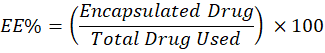
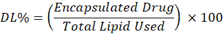
Encapsulation efficiency (EE%) measures the proportion of the total drug successfully entrapped within liposomes, while drug loading (DL%) represents the amount of drug per unit of liposome.

  • High EE% minimizes drug wastage and enhances therapeutic efficacy.
  • DL% depends on lipid composition, drug-lipid interactions, and preparation methods.

Mathematical Formulas:

EE%=Encapsulated Drug?Total Drug Used ×100

DL%=Encapsulated Drug?Total Lipid Used ×100

Methods for EE% and DL% Quantification

  • Separation of Free and Encapsulated Drug:
    • Ultracentrifugation – Isolates liposomes from free drug.
    • Size-Exclusion Chromatography (SEC) – Differentiates encapsulated and unencapsulated drug.
  • Drug Quantification Techniques:
    • UV-Vis Spectroscopy – Simple and non-destructive.
    • HPLC & LC-MS – Highly precise and sensitive.

Table 13 Methods for Determining Encapsulation Efficiency

Method

Principle

Applications

Ultracentrifugation

Differential sedimentation

Large-scale separation

Size-Exclusion Chromatography (SEC)

Size-based separation

Separation of free & loaded drug

HPLC/LC-MS

Chromatographic separation

Quantitative drug determination

In Vitro Release Kinetics and Mechanistic Models

Drug Release Mechanisms

Liposomal drug release can occur via:

  1. Diffusion-controlled release – Drug diffuses across the lipid bilayer.
  2. Degradation-mediated release – Lipid degradation triggers drug release.
  3. Trigger-induced release – External stimuli (pH, enzymes) modulate drug release.

Mathematical Models for Release Kinetics

  • Zero-Order Kinetics: Constant drug release over time.
  • First-Order Kinetics: Drug release dependent on concentration.
  • Higuchi Model: Drug diffusion from lipid matrix.
  • Korsmeyer-Peppas Model: Describes polymeric or lipid-based controlled release.

Table 14 Drug Release Kinetics Models for Liposomes

Model

Equation

Mechanism

Zero-Order

Qt = Q0 +k0t

Constant release

First-Order

Qt =Q0e-kt

Concentration-dependent

Higuchi

Qt =kHt1/2

Diffusion-based

Korsmeyer-Peppas

Qt / Q∞=ktn

Liposomal-controlled release

Controlled and Stimuli-Responsive Release Mechanisms

1. Passive Release

  • Dependent on membrane permeability and lipid composition.
  • Common for conventional liposomes (e.g., Doxil®).

2. Stimuli-Responsive Release

Triggered by internal (biological) or external (physical) stimuli:

  • pH-Responsive: Release triggered in acidic tumor microenvironments.
  • Temperature-Sensitive: Release at elevated temperatures (hyperthermia therapy).
  • Enzyme-Sensitive: Triggered by enzymes in diseased tissues.
  • Ultrasound, Light, or Magnetic Field-Triggered: Precise external control. [ 18–22]

Table 15 Stimuli-Responsive Drug Release Strategies

Stimulus

Mechanism

Application

pH-Sensitive

Lipid destabilization in acidic pH

Tumor targeting

Thermosensitive

Phase transition at elevated temperature

Hyperthermia-assisted release

Enzyme-Triggered

Degradation by specific enzymes

Inflammatory diseases

Magnetic/NIR Light

External activation

Site-specific release

Stability and Shelf-Life Assessment of Liposomes

Liposomal stability is critical for maintaining efficacy, safety, and regulatory compliance. Stability studies assess physicochemical changes that can compromise drug delivery performance.

Chemical and Physical Stability Evaluation

1. Chemical Stability:

  • Lipid Oxidation: Degradation of unsaturated lipids, causing vesicle instability.
  • Hydrolysis: Breakdown of ester bonds in phospholipids.
  • Drug Leakage: Loss of encapsulated drug due to membrane degradation.

2. Physical Stability:

  • Aggregation & Fusion: Uncontrolled vesicle growth leading to reduced efficacy.
  • Phase Separation: Bilayer instability at varying temperatures.

Stability Evaluation Methods:

  • Differential Scanning Calorimetry (DSC): Determines bilayer phase transitions.
  • Fourier-Transform Infrared Spectroscopy (FTIR): Detects chemical degradation.
  • Dynamic Light Scattering (DLS): Monitors size stability over time.

Table 16 Common Stability Issues in Liposomes

Instability Type

Cause

Impact on Drug Delivery

Oxidation

Lipid peroxidation

Reduced shelf-life

Hydrolysis

Ester bond cleavage

Membrane rupture

Aggregation

Vesicle fusion

Loss of uniformity

Drug Leakage

Increased permeability

Dose inaccuracy

Lyophilization and Storage Considerations

Lyophilization (Freeze-Drying) for Liposomal Stability

  • Converts liposomal dispersions into a dry powder for extended storage.
  • Prevents hydrolysis and aggregation.
  • Cryoprotectants (e.g., sucrose, trehalose) stabilize liposomes during freezing.

Key Steps in Lyophilization:

  1. Pre-Freezing: Freezing below lipid transition temperature.
  2. Primary Drying: Sublimation under reduced pressure.
  3. Secondary Drying: Removal of residual moisture.

Table 17 Lyophilization Advantages and Challenges

Aspect

Advantage

Challenge

Stability

Prolongs shelf-life

Potential drug leakage

Storage

Easy transport

Requires cryoprotectants

Reconstitution

Rapid rehydration

Aggregation risk

Impact of Lipid Composition on Long-Term Stability

Lipid composition affects:

  • Bilayer rigidity: Saturated lipids (e.g., DSPC) enhance stability.
  • Cholesterol content: Reduces membrane permeability.
  • Surface modifications: PEGylation increases circulation time and steric stability.

Table 18 Lipid Composition and Stability Correlation

Lipid Type

Effect on Stability

Example

Saturated (DSPC, DPPC)

High stability

Long-circulating liposomes

Unsaturated (DOPC, POPC)

Prone to oxidation

Fast-release formulations

Cholesterol-Enriched

Enhances rigidity

Controlled release liposomes

A robust understanding of encapsulation efficiency, drug release kinetics, and stability parameters is critical for optimizing liposomal formulations for pharmaceutical and clinical applications. [23,24]

Quality by Design (QBD) Approach in Liposomal Development

The Quality by Design (QBD) approach is a systematic, risk-based, and data-driven strategy for developing liposomal formulations with predefined quality, safety, and efficacy. It integrates scientific principles and regulatory expectations to enhance product robustness, manufacturing consistency, and regulatory compliance.

Defining Critical Quality Attributes (CQAs)

Critical Quality Attributes (CQAs) are measurable physical, chemical, biological, and microbiological properties that must be controlled to ensure product quality and performance.

CQAs for Liposomal Formulations

  • Physicochemical properties: Size, polydispersity index (PDI), zeta potential
  • Structural integrity: Lipid bilayer stability, lamellarity
  • Encapsulation efficiency & drug loading
  • Drug release kinetics (in vitro and in vivo)
  • Stability attributes: Oxidation, hydrolysis, aggregation

Table 19 Critical Quality Attributes (CQAs) and Their Impact on Liposomal Performance

CQA

Impact on Product Performance

Analytical Techniques

Particle Size & PDI

Affects bioavailability & circulation time

DLS, NTA, AF4

Zeta Potential

Governs colloidal stability & charge-based targeting

Electrophoretic Light Scattering

Encapsulation Efficiency

Influences drug dosage accuracy

HPLC, UV-Vis

Drug Release Rate

Determines therapeutic effectiveness

In vitro dissolution studies

Membrane Integrity

Prevents premature leakage

Cryo-TEM, DSC

Critical Material Attributes (CMAs) and Process Parameters

Critical Material Attributes (CMAs)

CMAs refer to properties of raw materials that significantly influence liposomal formulation quality.

Key CMAs in Liposome Development:

  • Lipid type and purity (Saturated lipids improve stability, unsaturated lipids enhance flexibility)
  • Cholesterol content (Regulates bilayer rigidity and permeability)
  • Surface modifications (PEGylation extends circulation time)
  • Solvent system (Affects particle formation and encapsulation efficiency)

Critical Process Parameters (CPPs)

CPPs are manufacturing process variables that must be controlled to ensure reproducibility and quality.

  • Hydration time and temperature (Affects vesicle formation)
  • Sonication/extrusion cycles (Controls liposome size and uniformity)
  • pH and ionic strength (Influences surface charge and drug loading)
  • Sterilization method (Autoclaving, filtration, or gamma irradiation)

Table 20 CMAs and CPPs Affecting Liposomal Quality

Factor

Influence on Liposomal Quality

Lipid Composition

Affects stability and drug encapsulation

Hydration Method

Determines vesicle formation and homogeneity

Sonication/Extrusion

Controls particle size and PDI

Sterilization

Prevents contamination without damaging vesicles

Risk Assessment and Design Space Optimization

Risk Assessment in QBD

Risk assessment ensures identification, evaluation, and mitigation of potential failure points in liposomal formulation. ICH Q9 recommends tools like:

  • Failure Mode and Effects Analysis (FMEA)
  • Ishikawa (Fishbone) Diagrams
  • Process Analytical Technology (PAT) for real-time monitoring

Design Space Optimization

ICH Q8(R2) defines the Design Space as the multi-dimensional combination of input variables that ensures quality. Design of Experiments (DoE) is used to optimize:

  • Factorial designs to study lipid ratios
  • Response Surface Methodology (RSM) to refine process conditions
  • Mixture designs for selecting excipients. [25,26]

Table 21 QBD Tools for Liposomal Development

Tool

Application

FMEA

Identifies formulation risks

DoE

Optimizes CMAs and CPPs

PAT

Real-time quality monitoring

Regulatory Guidelines and Standardization for Liposomal Products

Regulatory agencies (FDA, EMA, ICH) establish strict guidelines to ensure liposomal drug safety, efficacy, and quality. Standardization remains a challenge due to complex characterization techniques and batch-to-batch variability.

FDA, EMA, and ICH Guidelines for Liposomal Formulations

FDA Guidelines (USA)

  • "Guidance for Industry: Liposome Drug Products" (2018)
  • Covers CMC (Chemistry, Manufacturing, and Controls), bioavailability, labelling

EMA Guidelines (Europe)

  • "Data Requirements for Liposomal Products"
  • Emphasizes comparative studies with reference liposomal products

ICH Guidelines (International)

  • ICH Q8(R2): Pharmaceutical Development (QBD Principles)
  • ICH Q9: Quality Risk Management
  • ICH Q10: Pharmaceutical Quality System

Challenges in Standardizing Analytical Methods

Despite advancements, regulatory challenges persist in liposomal product evaluation.

  • Lack of globally harmonized standardization (Different agencies follow different protocols)
  • Complexity of liposomal characterization (multi-step analysis required)
  • Batch-to-batch variability (Difficult to ensure reproducibility)
  • Need for high-sensitivity techniques (TEM, AF4, DLS for in-depth analysis)

Harmonization of Global Regulatory Frameworks

Steps Toward Harmonization:

  • Standardized CQAs and analytical techniques across regulatory agencies
  • Development of international reference materials for liposome characterization
  • Adoption of real-time quality control methods (PAT, AI-driven QC monitoring)

Table 22 Approaches to Improve Liposomal Regulatory Standardization

Strategy

Impact

Global CQA Standardization

Reduces variability in quality evaluation

Regulatory Collaboration

Aligns FDA, EMA, and ICH expectations

PAT Implementation

Enables real-time monitoring

A QBD-driven approach, robust regulatory framework, and global harmonization are crucial for ensuring consistency, quality, and safety of liposomal drug products. By integrating risk assessment, analytical standardization, and advanced manufacturing controls, the pharmaceutical industry can streamline regulatory approvals and enhance liposomal drug delivery success. [ 27,28 ]

Advancements and Future Perspectives

The field of liposomal drug delivery is rapidly evolving, driven by technological advancements, innovative analytical techniques, and the demand for precision medicine. Future developments focus on enhanced characterization methods, intelligent multifunctional liposomes, and overcoming industrial scale-up challenges to ensure cost-effective, reproducible, and regulatory-compliant production.

Emerging Analytical Techniques for Next-Generation Liposomes

Traditional analytical methods such as Dynamic Light Scattering (DLS), Transmission Electron Microscopy (TEM), and High-Performance Liquid Chromatography (HPLC) have been instrumental in characterizing liposomes. However, next-generation liposomes require more advanced, high-resolution, and real-time characterization techniques to improve batch-to-batch consistency and regulatory compliance.

Recent Innovations in Liposomal Characterization

  • Super-Resolution Microscopy (SRM): Provides nanometer-scale imaging beyond TEM resolution.
  • Cryogenic Electron Tomography (Cryo-ET): Enables 3D reconstruction of liposomes in their native hydrated state.
  • Nanoparticle Tracking Analysis (NTA): Offers high-resolution particle-by-particle size distribution.
  • Microfluidic-Based Characterization: Real-time monitoring of liposome formation, stability, and drug encapsulation.
  • Surface Plasmon Resonance (SPR): Studies liposome-protein interactions for targeted drug delivery.

Table 23 Advanced Analytical Techniques for Liposomal Characterization


Technique

Principle

Key Advantage

Application

SRM

High-resolution fluorescence imaging

Nanometer precision

Structural studies

Cryo-ET

3D imaging of liposomes in native state

No dehydration artifacts

Morphology & bilayer studies

NTA

Tracking individual nanoparticles

Real-time size & concentration measurement

Batch uniformity testing

Microfluidics

Continuous-flow liposome analysis

On-demand, real-time monitoring

Process optimization

SPR

Optical detection of biomolecular interactions

Label-free detection

Drug-liposome binding studies

Smart and Multifunctional Liposomal Systems

Next-generation liposomes are being designed with intelligent functionalities, allowing for stimuli-responsive, targeted, and theranostic applications. These innovations include:

1. Stimuli-Responsive Liposomes

  • pH-Sensitive Liposomes: Drug release triggered in tumor microenvironments (acidic pH ~6.5).
  • Thermosensitive Liposomes: Release activated at fever-range temperatures (42°C) for localized delivery.
  • Redox-Responsive Liposomes: React to glutathione (GSH) gradients in cancer cells for intracellular drug delivery.

2. Ligand-Targeted Liposomes

  • Antibody-Conjugated Liposomes: Enhanced specificity via monoclonal antibodies (mAbs).
  • Peptide and Aptamer-Functionalized Liposomes: Improved targeting of cancer, infections, and brain disorders.

3. Theranostic Liposomes (Therapy +     Diagnostics)

  • Image-Guided Liposomal Therapy: MRI, PET, or fluorescence-labeled liposomes for precision medicine.
  • Hybrid Liposomes with Nanoparticles: Combination of lipid-based vesicles with magnetic, gold, or polymeric nanomaterials for multifunctional applications.

Table 24 Features of Smart and Multifunctional Liposomes


Liposome Type

Mechanism

Application

pH-Sensitive

Lipid destabilization at acidic pH

Tumor targeting

Thermosensitive

Drug release above transition temperature

Hyperthermia-assisted drug delivery

Redox-Responsive

Reacts to intracellular glutathione (GSH)

Cancer drug delivery

Antibody-Conjugated

Binds to specific cell surface markers

Monoclonal antibody-based therapies

Theranostic Liposomes

Dual function (drug + imaging)

Cancer diagnostics & treatment

Overcoming Challenges in Industrial Scale-Up

Despite significant advancements in liposome technology, industrial-scale manufacturing remains challenging due to:

1. Reproducibility & Batch Variability

  • Scaling up from small laboratory batches to large-scale production affects particle size, encapsulation efficiency, and stability.
  • Solution: Adoption of Microfluidics, High-Pressure Homogenization, and Continuous Manufacturing Systems.

2. Stability and Shelf-Life Issues

  • Liposomes are prone to oxidation, hydrolysis, and aggregation over time.
  • Solution: Improved lyophilization techniques, optimized lipid composition, and cryoprotectants.

3. Regulatory and Quality Control Challenges

  • Regulatory agencies require highly standardized analytical methods for batch consistency.
  • Solution: Implementation of Process Analytical Technology (PAT) for real-time monitoring. [29,30]

Table 25 Challenges and Solutions in Liposomal Scale-Up


Challenge

Impact on Manufacturing

Potential Solution

Batch Variability

Inconsistent liposome size & EE%

Microfluidic processing

Low Stability

Drug leakage & degradation

Lyophilization with cryoprotectants

High Production Costs

Expensive lipid materials

Optimization of lipid sourcing

Regulatory Compliance

Complex characterization

PAT & AI-based QC

CONCLUSION

Liposomal drug delivery has revolutionized modern therapeutics, offering enhanced solubility, targeted delivery, and controlled release. The next frontier in liposomal technology will focus on:

  • Integration of next-generation analytical tools (Cryo-ET, NTA, AI-assisted PAT).
  • Development of multifunctional & smart liposomes for precision medicine.
  • Adoption of microfluidics and continuous manufacturing for scalable production.
  • Regulatory harmonization & AI-driven quality control for batch consistency.

Future Directions:

  • AI-Driven Liposome Formulation: Machine learning for predicting lipid combinations.
  • 3D-Printed Liposomal Systems: Personalized nanomedicine for patient-specific dosing.
  • Next-Gen Theranostic Liposomes: Real-time disease monitoring and treatment in a single platform.
  • Green Chemistry Approaches: Sustainable lipid sourcing and solvent-free production methods.

The future of liposomal nanomedicine is bright, but success will depend on collaborative efforts between academia, industry, and regulatory bodies. By bridging the gap between laboratory innovation and large-scale manufacturing, liposomal therapeutics will continue to transform drug delivery and precision medicine.

Conflict of Interest

The author declares that there are no conflicts of interest regarding the publication of this article.

REFRENCES

        1. Düzgüne? N, Gregoriadis G. Introduction: The origins of liposomes: Alec Bangham at Babraham. Methods Enzymol. 2005;391(SPEC. ISS.). doi:10.1016/S0076-6879(05)91029-X
        2. Latrobdiba ZM, Fulyani F, Anjani G. Liposome optimisation for oral delivery of nutraceuticals in food: a review. Food Res. 2023;7(3). doi:10.26656/fr.2017.7(3).022
        3. Huang Z, Li X, Zhang T, et al. Progress involving new techniques for liposome preparation. Asian J Pharm Sci. 2014;9(4). doi:10.1016/j.ajps.2014.06.001
        4. Liu G, Hou S, Tong P, Li J. Liposomes: Preparation, Characteristics, and Application Strategies in Analytical Chemistry. Crit Rev Anal Chem. 2022;52(2). doi:10.1080/10408347.2020.1805293
        5. Fan Y, Marioli M, Zhang K. Journal of Pharmaceutical and Biomedical Analysis Analytical characterization of liposomes and other lipid nanoparticles for drug delivery. J Pharm Biomed Anal. 2021;192.
        6. Fan Y, Marioli M, Zhang K. Analytical characterization of liposomes and other lipid nanoparticles for drug delivery. J Pharm Biomed Anal. 2021;192. doi:10.1016/j.jpba.2020.113642
        7. Ayaz Ali B SY, Umair Aftaab S, Ayaz Ali S. Liposomes chemistry, manufacturing and control: Regulatory perspective. International Journal of Research and Analytical Reviews. 2021;(August).
        8. Liu P, Chen G, Zhang J. A Review of Liposomes as a Drug Delivery System: Current Status of Approved Products, Regulatory Environments, and Future Perspectives. Molecules. 2022;27(4). doi:10.3390/molecules27041372
        9. Koutsoulas C, Pippa N, Demetzos C, Zabka M. The role of ζ-potential on the stability of nanocolloidal systems. Pharmakeftiki. 2012;24(4).
        10. De Leo V, Milano F, Agostiano A, Catucci L. Recent advancements in polymer/liposome assembly for drug delivery: From surface modifications to hybrid vesicles. Polymers (Basel). 2021;13(7). doi:10.3390/polym13071027
        11. Matsuura-Sawada Y, Maeki M, Uno S, Wada K, Tokeshi M. Controlling lamellarity and physicochemical properties of liposomes prepared using a microfluidic device. Biomater Sci. 2023;11(7). doi:10.1039/d2bm01703b
        12. Yahata S, Hirose M, Ueno T, Nagumo H, Sakai-Kato K. Effect of sample concentration on nanoparticle tracking analysis of small extracellular vesicles and liposomes mimicking the physicochemical properties of exosomes. Chem Pharm Bull (Tokyo). 2021;69(11). doi:10.1248/cpb.c21-00452
        13. Sabeti B, bin Noordine MI, Javar HA, Davoudi ET, Kadivar A. Characterization of diclofenac liposomes formulated with palm oil fractions. Tropical Journal of Pharmaceutical Research. 2014;13(2). doi:10.4314/tjpr.v13i2.3
        14. Dave V, Gupta A, Singh P, Gupta C, Sadhu V, Reddy KR. Synthesis and characterization of celecoxib loaded PEGylated liposome nanoparticles for biomedical applications. Nano-Structures and Nano-Objects. 2019;18. doi:10.1016/j.nanoso.2019.100288
        15. Memete AR, Miere F, Laslo V, et al. An In Vitro Study of the Healing Potential of Black Mulberry (Morus nigra L.) Extract in a Liposomal Formulation. Applied Sciences (Switzerland). 2023;13(2). doi:10.3390/app13021041
        16. Shi Y, Li X. High-Performance Liquid Chromatography Coupled with Tandem Mass Spectrometry Method for the Identification and Quantification of Lipids in Liposomes. Methods Mol Biol. 2023;2622. doi:10.1007/978-1-0716-2954-3_20
        17. Siejak P, Smu?ek W, Nowak-Karnowska J, Dembska A, Neunert G, Polewski K. Bird Cherry (Prunus padus) Fruit Extracts Inhibit Lipid Peroxidation in PC Liposomes: Spectroscopic, HPLC, and GC–MS Studies. Applied Sciences (Switzerland). 2022;12(15). doi:10.3390/app12157820
        18. Agiba AM, Arreola-Ramírez JL, Carbajal V, Segura-Medina P. Light-Responsive and Dual-Targeting Liposomes: From Mechanisms to Targeting Strategies. Molecules. 2024;29(3). doi:10.3390/molecules29030636
        19. AlMajed Z, Salkho NM, Sulieman H, Husseini GA. Modeling of the In Vitro Release Kinetics of Sonosensitive Targeted Liposomes. Biomedicines. 2022;10(12). doi:10.3390/biomedicines10123139
        20. Jain A, Jain SK. In vitro release kinetics model fitting of liposomes: An insight. Chem Phys Lipids. 2016;201. doi:10.1016/j.chemphyslip.2016.10.005
        21. Badran MM, Alouny NN, Aldosari BN, Alhusaini AM, Abou El Ela AES. Transdermal Glipizide Delivery System Based on Chitosan-Coated Deformable Liposomes: Development, Ex Vivo, and In Vivo Studies. Pharmaceutics. 2022;14(4). doi:10.3390/pharmaceutics14040826
        22. Karn PR, Cho W, Park HJ, Park JS, Hwang SJ. Characterization and stability studies of a novel liposomal cyclosporin a prepared using the supercritical fluid method: Comparison with the modified conventional Bangham method. Int J Nanomedicine. 2013;8. doi:10.2147/IJN.S39025
        23. Pasarin D, Ghizdareanu AI, Enascuta CE, et al. Coating Materials to Increase the Stability of Liposomes. Polymers (Basel). 2023;15(3). doi:10.3390/polym15030782
        24. Tai K, Rappolt M, Mao L, Gao Y, Yuan F. Stability and release performance of curcumin-loaded liposomes with varying content of hydrogenated phospholipids. Food Chem. 2020;326. doi:10.1016/j.foodchem.2020.126973
        25. Xu X, Khan MA, Burgess DJ. A quality by design (QbD) case study on liposomes containing hydrophilic API: I. Formulation, processing design and risk assessment. Int J Pharm. 2011;419(1-2). doi:10.1016/j.ijpharm.2011.07.012
        26. Charankumar K, Bagasariya D, Jain N, et al. Quality by design (QBD) abetted development of pioglitazone incorporated liposomes-loaded hyaluronic acid-based in situ hydrogel for the management of melanoma. J Drug Deliv Sci Technol. 2023;84. doi:10.1016/j.jddst.2023.104453
        27. Chaurasiya A, Gorajiya A, Panchal K, Katke S, Singh AK. A review on multivesicular liposomes for pharmaceutical applications: preparation, characterization, and translational challenges. Drug Deliv Transl Res. 2022;12(7). doi:10.1007/s13346-021-01060-y
        28. Schlich M, Musazzi UM, Campani V, et al. Design and development of topical liposomal formulations in a regulatory perspective. Drug Deliv Transl Res. 2022;12(8). doi:10.1007/s13346-021-01089-z
        29. Wang J, Gong J, Wei Z. Strategies for Liposome Drug Delivery Systems to Improve Tumor Treatment Efficacy. AAPS PharmSciTech. 2022;23(1). doi:10.1208/s12249-021-02179-4
        30. Jiang Y, Li W, Wang Z, Lu J. Lipid-Based Nanotechnology: Liposome. Pharmaceutics. 2024;16(1). doi:10.3390/pharmaceutics16010034

Reference

  1. Düzgüne? N, Gregoriadis G. Introduction: The origins of liposomes: Alec Bangham at Babraham. Methods Enzymol. 2005;391(SPEC. ISS.). doi:10.1016/S0076-6879(05)91029-X
  2. Latrobdiba ZM, Fulyani F, Anjani G. Liposome optimisation for oral delivery of nutraceuticals in food: a review. Food Res. 2023;7(3). doi:10.26656/fr.2017.7(3).022
  3. Huang Z, Li X, Zhang T, et al. Progress involving new techniques for liposome preparation. Asian J Pharm Sci. 2014;9(4). doi:10.1016/j.ajps.2014.06.001
  4. Liu G, Hou S, Tong P, Li J. Liposomes: Preparation, Characteristics, and Application Strategies in Analytical Chemistry. Crit Rev Anal Chem. 2022;52(2). doi:10.1080/10408347.2020.1805293
  5. Fan Y, Marioli M, Zhang K. Journal of Pharmaceutical and Biomedical Analysis Analytical characterization of liposomes and other lipid nanoparticles for drug delivery. J Pharm Biomed Anal. 2021;192.
  6. Fan Y, Marioli M, Zhang K. Analytical characterization of liposomes and other lipid nanoparticles for drug delivery. J Pharm Biomed Anal. 2021;192. doi:10.1016/j.jpba.2020.113642
  7. Ayaz Ali B SY, Umair Aftaab S, Ayaz Ali S. Liposomes chemistry, manufacturing and control: Regulatory perspective. International Journal of Research and Analytical Reviews. 2021;(August).
  8. Liu P, Chen G, Zhang J. A Review of Liposomes as a Drug Delivery System: Current Status of Approved Products, Regulatory Environments, and Future Perspectives. Molecules. 2022;27(4). doi:10.3390/molecules27041372
  9. Koutsoulas C, Pippa N, Demetzos C, Zabka M. The role of ζ-potential on the stability of nanocolloidal systems. Pharmakeftiki. 2012;24(4).
  10. De Leo V, Milano F, Agostiano A, Catucci L. Recent advancements in polymer/liposome assembly for drug delivery: From surface modifications to hybrid vesicles. Polymers (Basel). 2021;13(7). doi:10.3390/polym13071027
  11. Matsuura-Sawada Y, Maeki M, Uno S, Wada K, Tokeshi M. Controlling lamellarity and physicochemical properties of liposomes prepared using a microfluidic device. Biomater Sci. 2023;11(7). doi:10.1039/d2bm01703b
  12. Yahata S, Hirose M, Ueno T, Nagumo H, Sakai-Kato K. Effect of sample concentration on nanoparticle tracking analysis of small extracellular vesicles and liposomes mimicking the physicochemical properties of exosomes. Chem Pharm Bull (Tokyo). 2021;69(11). doi:10.1248/cpb.c21-00452
  13. Sabeti B, bin Noordine MI, Javar HA, Davoudi ET, Kadivar A. Characterization of diclofenac liposomes formulated with palm oil fractions. Tropical Journal of Pharmaceutical Research. 2014;13(2). doi:10.4314/tjpr.v13i2.3
  14. Dave V, Gupta A, Singh P, Gupta C, Sadhu V, Reddy KR. Synthesis and characterization of celecoxib loaded PEGylated liposome nanoparticles for biomedical applications. Nano-Structures and Nano-Objects. 2019;18. doi:10.1016/j.nanoso.2019.100288
  15. Memete AR, Miere F, Laslo V, et al. An In Vitro Study of the Healing Potential of Black Mulberry (Morus nigra L.) Extract in a Liposomal Formulation. Applied Sciences (Switzerland). 2023;13(2). doi:10.3390/app13021041
  16. Shi Y, Li X. High-Performance Liquid Chromatography Coupled with Tandem Mass Spectrometry Method for the Identification and Quantification of Lipids in Liposomes. Methods Mol Biol. 2023;2622. doi:10.1007/978-1-0716-2954-3_20
  17. Siejak P, Smu?ek W, Nowak-Karnowska J, Dembska A, Neunert G, Polewski K. Bird Cherry (Prunus padus) Fruit Extracts Inhibit Lipid Peroxidation in PC Liposomes: Spectroscopic, HPLC, and GC–MS Studies. Applied Sciences (Switzerland). 2022;12(15). doi:10.3390/app12157820
  18. Agiba AM, Arreola-Ramírez JL, Carbajal V, Segura-Medina P. Light-Responsive and Dual-Targeting Liposomes: From Mechanisms to Targeting Strategies. Molecules. 2024;29(3). doi:10.3390/molecules29030636
  19. AlMajed Z, Salkho NM, Sulieman H, Husseini GA. Modeling of the In Vitro Release Kinetics of Sonosensitive Targeted Liposomes. Biomedicines. 2022;10(12). doi:10.3390/biomedicines10123139
  20. Jain A, Jain SK. In vitro release kinetics model fitting of liposomes: An insight. Chem Phys Lipids. 2016;201. doi:10.1016/j.chemphyslip.2016.10.005
  21. Badran MM, Alouny NN, Aldosari BN, Alhusaini AM, Abou El Ela AES. Transdermal Glipizide Delivery System Based on Chitosan-Coated Deformable Liposomes: Development, Ex Vivo, and In Vivo Studies. Pharmaceutics. 2022;14(4). doi:10.3390/pharmaceutics14040826
  22. Karn PR, Cho W, Park HJ, Park JS, Hwang SJ. Characterization and stability studies of a novel liposomal cyclosporin a prepared using the supercritical fluid method: Comparison with the modified conventional Bangham method. Int J Nanomedicine. 2013;8. doi:10.2147/IJN.S39025
  23. Pasarin D, Ghizdareanu AI, Enascuta CE, et al. Coating Materials to Increase the Stability of Liposomes. Polymers (Basel). 2023;15(3). doi:10.3390/polym15030782
  24. Tai K, Rappolt M, Mao L, Gao Y, Yuan F. Stability and release performance of curcumin-loaded liposomes with varying content of hydrogenated phospholipids. Food Chem. 2020;326. doi:10.1016/j.foodchem.2020.126973
  25. Xu X, Khan MA, Burgess DJ. A quality by design (QbD) case study on liposomes containing hydrophilic API: I. Formulation, processing design and risk assessment. Int J Pharm. 2011;419(1-2). doi:10.1016/j.ijpharm.2011.07.012
  26. Charankumar K, Bagasariya D, Jain N, et al. Quality by design (QBD) abetted development of pioglitazone incorporated liposomes-loaded hyaluronic acid-based in situ hydrogel for the management of melanoma. J Drug Deliv Sci Technol. 2023;84. doi:10.1016/j.jddst.2023.104453
  27. Chaurasiya A, Gorajiya A, Panchal K, Katke S, Singh AK. A review on multivesicular liposomes for pharmaceutical applications: preparation, characterization, and translational challenges. Drug Deliv Transl Res. 2022;12(7). doi:10.1007/s13346-021-01060-y
  28. Schlich M, Musazzi UM, Campani V, et al. Design and development of topical liposomal formulations in a regulatory perspective. Drug Deliv Transl Res. 2022;12(8). doi:10.1007/s13346-021-01089-z
  29. Wang J, Gong J, Wei Z. Strategies for Liposome Drug Delivery Systems to Improve Tumor Treatment Efficacy. AAPS PharmSciTech. 2022;23(1). doi:10.1208/s12249-021-02179-4
  30. Jiang Y, Li W, Wang Z, Lu J. Lipid-Based Nanotechnology: Liposome. Pharmaceutics. 2024;16(1). doi:10.3390/pharmaceutics16010034

Photo
Ravi Patel
Corresponding author

Research & Development, Thermo Fisher Scientific, Greenville, NC 27834, USA

Photo
Krupal Morker
Co-author

Director, Product Development (CMC), Frontage Laboratories, Exton, PA 19341, USA.

Photo
Dipen Purohit
Co-author

Analytical Development & Quality Control, Navinta LLC, 1499 Ewing, NJ 08618, USA.

Ravi Patel*, Krupal Morker, Dipen Purohit, Development and Analytical Characterization of Liposomes: A Comprehensive Approach, Int. J. of Pharm. Sci., 2025, Vol 3, Issue 3, 2005-2021. https://doi.org/10.5281/zenodo.15062406

More related articles
Formulation And Evaluation Of Nebivolol Transderma...
K. Mugilan, A. Vasanthan, Senthilkumar K. L., Karthick S., ...
Analyzing Antibacterial Drugs Floating Times for E...
Vadapalli Rama Rao, Gajula Meghamala, Darisi Saketh, Chowtipalli ...
A Review On Nano-Emulsion ...
Shaikh Arbaz , Shaikh Faizan , Syed Zaid , Qazi Majaz, ...
Recent Advances In Drug Delivery System...
Nandkumar M. chaudhari, Amrita M. Singh, Nitin S. Bharud, Dhananjay R. Chavhan, Rushikesh D. Kakade,...
Analyzing Antihypertensive Drugs Floating Times for Efficiency Evaluation: A Rev...
Dr. Rama Rao Vadapalli, Darisi Saketh, Gangolu Yohan, Gurram Lokeswari, Bodapati Meghana, Jayanth Ka...
Related Articles
Pharmaceutical Aspects Of Accelerated Stability Studies...
Ramya Teja Medarametla, K. Venkata Gopaiah, J. N. Suresh Kumar, . Lakshmi Pravallika4, P. Mouli, S. ...
Review Article on Novel Drug Delivery System ...
Dr. C. S. Parameswari, K.Mounika, B Manasa, N.Navya sree , M.Sreya, M.Rajeswari, E. Anusha, ...
Floating Microspheres in Diabetes Treatment: A Comprehensive Review ...
Janhavi Chaudhari, Dr. Prashant Malpure, Rahul Arote, Dr. Gokul Talale, ...
Pharmaceutical Aspects Of Accelerated Stability Studies...
Ramya Teja Medarametla, K. Venkata Gopaiah, J. N. Suresh Kumar, . Lakshmi Pravallika4, P. Mouli, S. ...
More related articles
Formulation And Evaluation Of Nebivolol Transdermal Drug Delivery System ...
K. Mugilan, A. Vasanthan, Senthilkumar K. L., Karthick S., ...
Analyzing Antibacterial Drugs Floating Times for Efficiency Evaluation: A Review...
Vadapalli Rama Rao, Gajula Meghamala, Darisi Saketh, Chowtipalli Pushpanjali, Mohammadh Rafivulla, G...