University Department of Chemical Technology, Dr. Babasaheb Ambedkar Marathwada University, Chhatrapati Sambhajinagar - 4310004 Maharashtra, India
Melittin, the main bioactive component of honey bee venom (HBV), demonstrates significant therapeutic promise, especially as an anticancer agent. This review explores the physicochemical and pharmacological properties of HBV, highlighting melittin's mechanisms of action against cancer cells, such as triggering apoptosis, inhibiting cell proliferation, and preventing metastasis. The effects of traditional collection methods and advanced extraction technologies, including electric shock devices, on venom composition are examined. The study delved deeper into advanced analytical methods such as SDS-PAGE, HPLC-MS, and LC-MS, which are vital to the separation and identification of melittin from venom of honey bees. This study examines these approaches I greater detail. It also looks at delivery systems mediated by nanotechnology as an innovative approach to maximize melittin's therapeutic effectiveness while lowering systemic toxicity. This review highlights melittin's potential as a cancer treatment choice in the future and encourages ongoing advancements in analytical and delivery systems to promote pharmacotherapies based on HBV.
Insect venom is one of the most common causes of anaphylaxis, along with medications and foods. Apidae—honeybees (Apis mellifera) and bumblebees (Bombus) are two groups of Hymenoptera that might trigger allergy reactions in the nations of Central Europe. The majority of cases lead to anaphylactic reactions after a honeybee sting, which is caused by the higher number of honeycomb strikes. Children and beekeepers are more likely than others to suffer from honeybee venom (HBV) allergy, which is the second most frequent type of Hymenoptera venom allergy. The proteins in honeybee venom have allergic properties that can result in a variety of symptoms, ranging from small reactions to potentially fatal systemic anaphylaxis (1).
Venom hypersensitivity may be caused by non-immunogenic as well as immunogenic causes, such as IgE or non-IgE-mediated venom allergy (2). One of the mechanisms used by animals to ensure survival is the use of venoms and toxins. The order of insects contains numerous venomous species. Such as the sting of a hymenopteran can be fatal to humans, inducing anaphylactic shock, and causes a systemic allergic reaction in prey or predators. A member of the Hymenoptera family that can be located practically everywhere in the world is the honeybee (Apis mellifera). When stung, venom of honeybee (HBV) poses a risk to humans, but it also offers medicinal benefits (3).
Thus, it is currently the topic of numerous studies. As a result, HBV has been used medicinally to assist in managing a number of ailments, such as multiple sclerosis, Parkinson’s diseases, cancer, liver fibrosis, skin conditions, and pain. Venom immunotherapy, which is intended to lessen the possibility of a systemic reaction in the case of stings by Hymenoptera, is the second method of administering HBV (4). Consequently, it is essential to understand and standardize HBV. In order to protect a colony from predators, HBV is produced in specialized glands (5).
Tumor is a solid mass of cells resulting from cancer, a condition in which one or more cells lose their ability to control their rate of growth. It is one of the major killers worldwide, and the main treatments include surgery, chemotherapy, and/or radiation therapy. Over 60% of cancer cases require radiation therapy and radiotherapy, despite the fact that there are no special medications made for it. Radiotherapy (RT) is the primary treatment to cure cancer. Chemotherapy Patient numbers have notably risen in recent decades (6).
Chemotherapy side effects are a major and frequently undetected clinical barrier to managing cancer. They may alter the treatment strategy and negatively affect the patient’s quality of life. The most commonly reported side effects, were exhaustion and feelings of nausea, vomiting. Notable side effects that are commonly seen include diarrhea, dry mouth, air loos, altered taste, and decreased appetite. Over half of the patients said they had at least one of these side effects. Additionally notable negative effects include diarrhea, tingling or numbness in the hands and/or feet, changes in the skin (such as dry skin, redness, or itching), fever, harm to the mouth mucosa, flu-like symptoms, allergic reaction, memory issues, impaired renal function, loss of hearing, and/or ringing in the ears (7). Peripheral neuropathy symptoms were thought to be the most prevalent symptoms that were being disregarded (6.7%) (8).
Surgery, chemotherapy and radiotherapy are conventional medical interventions for cancer treatment(9). Thus, new cutting-edge techniques to cancer treatment have been developed, incorporating radionics, chemodynamic therapy, nanoparticles, targeted therapy, natural antioxidants, ablation therapy, and ferroptosis-based therapy. The goal of modern oncology practices is to develop safe and efficient cancer nanomedicines. Treatment with stem cells that addresses both primary as well as metastatic cancer has shown impressive results in healing and restoring diseased or infected tissues (10). Natural antioxidants has the capacity to hunt down free radicals and mitigate their negative. Targeted therapy preserves healthy cells while halting the development and proliferation of certain cancer cells (11). Ablation treatment can be used to burn of freeze cancers without requiring open surgery, a recent development in minimally invasive surgery nanoparticles, loaded neosomes have introduced novel diagnostic and therapeutic possibilities which serves as an effective medication for cancer care(10).
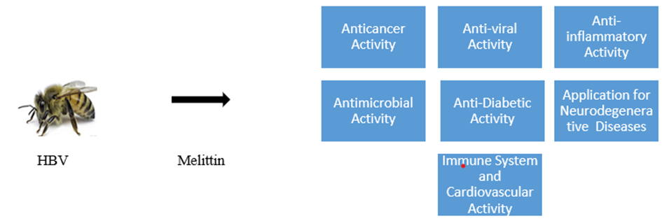
Different Activity of Melittin has been shown in figure no. 1
There are several natural substances which are used for cancer care due to their existing anti-cancer property. The primary ingredient in BV(Bee Venom), melittin (MEL), is thought to be an alternative therapy for a number of infections, antitum or/anticancer, anti-viral activity, anti-inflammatory activity, anti-microbial activity, anti-diabetic activity, immune system and cardiovascular activity and application for neurodegenerative diseases etc., Anti-tumor or antineoplasticity is one of the major properties of melittin (12), (13–16).
AIM AND OBJECTIVE
To study in detail analytical methods for characterization of melittin from honey bee venom.
The highest goal is to study about HBV (honey bee venom) and Hyphenated analytical techniques for characterization of melittin from HBV, a key ingredient of honey bee venom, for its antitumor activity.
Ancient cultures used bee products, and the Holy Qur'an, the Bible, and Vida all mention their healing efficacy (17). The treatment or prevention of disease is the focus of Apitherapy, which involves using bee-derived products like honey, pollen, propolis, royal jelly and benzoin. It is also known as "the science (and art) of employing honeybee products to maintain health and help people regain it when illness or accident interferes is another name for it (18).
Therefore, BV is the topic of several studies. A gland in the honey bees abdomen cavity produces its venom (19). It is produced by the honeybee venom glands associated with the sting apparatus of worker and queens, stored in the venom reservoir, and administered during the stinging procedure using the sting device (20). However, a bee’s venom sac contains approximately 300 µg of venom and in a single bite, it injects 50-10 µg of venom on average (21). It is used by adult honey bees for hive defense. In response to an attack, honey bees removes their stinger which has barbs, and their own venom sac from their body. After just one sting honey bees die (22).
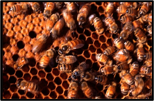
The Picture of Apis Mellifera has been shown in figure no. 2 (23)
Apitoxin is another name for bee venom, which is produced in bee venom glands as a protective mechanism. Humans have known about its properties since ancient times. Venom production occurs after two of three days of adulthood and decreases with age. Bee venom is a colourless, transparent fluid with a strong odour. The venom of honeybees is a clear liquid that can also turn yellow and it is odourless with an irritating (bitter taste) flavor. The pH of honeybee venom is about 4.5-5.5 (24).
When it arrives into contact with mucous membranes or the eyes, it produces a strong burning sensation and irritation. Because some of its protein have oxidized, the dried form is yellow, while some commercial preparations are brown in colour. A variety of volatile compounds included in honey bee venom are easily lost during the gathering process. The venom of honey bees is believed to be a good source of biogenic amines, peptides and enzymes (25). The bee venom gets rapidly dries and crystallizes into grayish-white crystals, when it comes into touch with air. The venom dissolves in water but does not dissolve in alcohol and ammonium sulphate (20).
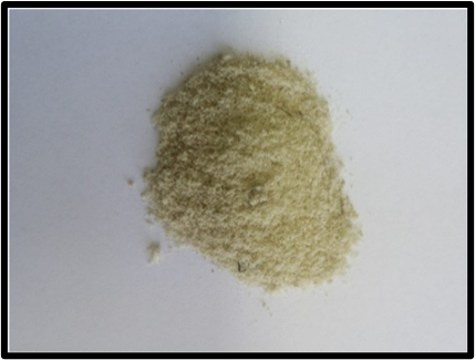
The Picture Bee Venom Powder has been shown in figure no.3 (26)
The vivo and in vitro studies are done to examined pharmacology of bee venom (27), (28), (29), (30), (31), (32) Bee venom has a wide range of pharmacological effects, including anti-mutagenic (32), radioprotective (32), anti-hepatotoxic (30), cytoprotective (30), anti-microbial (28), anti-viral (28), anti-inflammatory (28), (30), anti-oxidant (28), (30) anti-arthritic (27), anti-metastatic (29), and anti-tumor (29) effects.
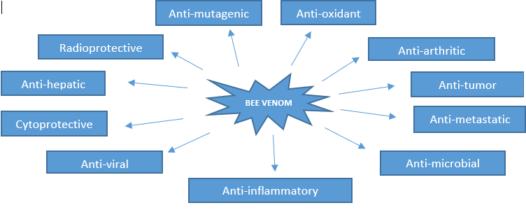
The Pharmacological Activities of Bee Venom has been shown in figure no. 4
The various extraction and collecting techniques have an impact on the composition of bee venom. Pence (1981) found from his research that the most effective venom seemed to be gathered under water to prevent the evaporation of several especially volatile components (33).The composition of the final products venom collected varies depending on the extraction or collection procedure used (20).
Variations in the composition of venom gland and venom sac production were discovered by surgically excising the poison gland and poison sac (reservoir) from worker honey bees of different Apis species (34). Early methods for gathering bee venom involved squeezing each bee separately to remove a drop from the sting’s tip or surgically excising the venom gland. The protein levels of venom extracted from surgically removed venom sacs different from those of venom extracted by electroshock method, which combined a chilling mechanism with the normal electro-shock collecting gear to retain more volatile chemicals(20).
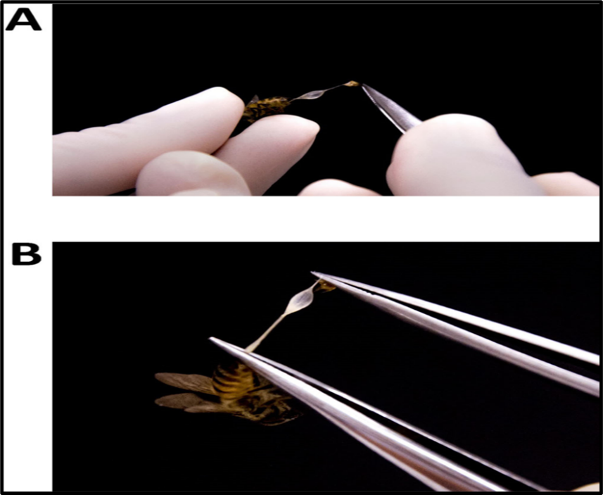
The Procedures for dissecting and separating the sting apparatus's venom reserves has been shown in figure no. 5 : (A) Use all-purpose fine-tipped tweezers to pull the venom reservoir and sting mechanism out of the bee's body; (B) After the two structures are fully extracted from the bee's body but remain together, use two tweezers (or one pair of micro scissors and one pair of tweezers) to separate them (35).
It was discovered that the Apis species venom gland and venom sac contents are significantly different in composition. Apart from acid phosphates and hexokinase, lipids, proteins, all other contents were concentrated in the venom sac as it houses both secretions from the venom gland and some of the venom sac’s secretory cells, hence enhancing the overall venom. Because the venom gland has a naturally low pH, leading to a higher presence of the acid phosphatase enzyme in that area. Hexokinase activity in the poison gland is higher, which indicates that more is secreted by the filamentous gland (the venom gland) than by the reservoir (the venom sac) (34).
Worker bee venom arbitrary sample was obtained from the hive. According to the findings of their experiments, Hsiang and Elliott (1975) came to the conclusion that the venom obtained by surgically extracting the venom sac had a different protein content than that obtained via electric shock. Gunnison (1966) applies a conventional electro-shock collecting equipment with a refrigeration mechanism to preserve the more volatile compounds (33). Since the early 1960s, After constant improvement, electro-shock extraction is now considered routine practice. It seems that the most potent venom is made underwater to prevent the evaporation of extremely volatile ingredients.
This technique, which has previously collected venom from solitary or small groups of bees using a variety of electrical methods, permitted to gather a gram of venom from around 20 colonies in a few of hours for the first time. This technology seems to work best for honey bees, while comparable electrical methods also work somewhat on other stinging insects. Obtaining venom from Africanized honeybees or the more defensive species is not recommended using the standard electroshock techniques (20). Galuszka (1972) proved that the electroshock approach was the most efficient collecting method, with a little adjustment comprising 15-minute stimulation at three-day intervals, followed by a two- to three-week repeat. This significantly boosted collection efficiency while leaving the hive undisturbed (33).
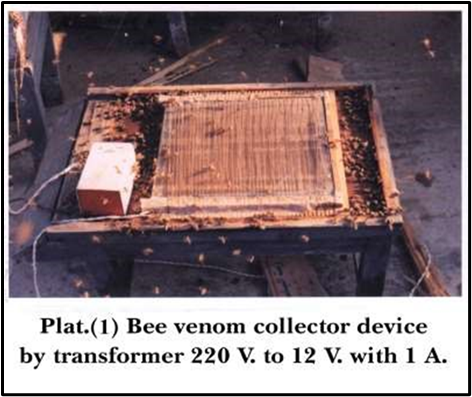
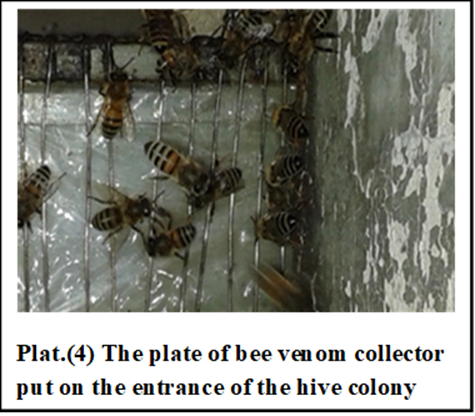
The collection of bee venom was scheduled as follows: Electric shock method design plat: 2 was used for bee venom collecting set on the colony entrance below the experiment production as Suggested by Benton et al. (1963), Nobre, (1990), Khattab, (1997), and El Ashhab (2002) (36), (37), (38), (39)
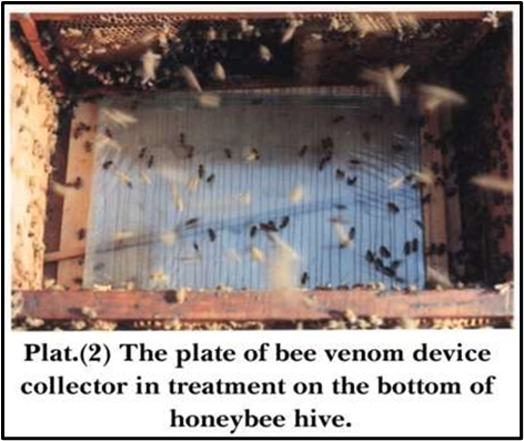
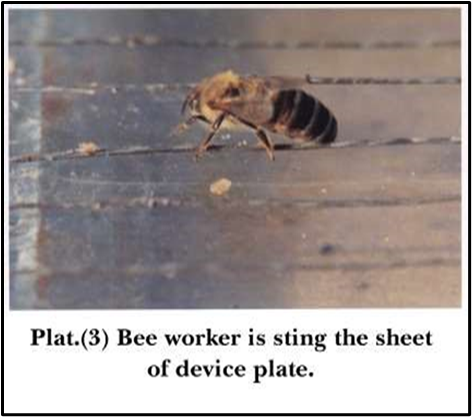
The equipment is based on the biology of honey bee workers and the intermitted pulse oscillation principle. The worker gets angry and releases venom from its sting onto the plastic sheet on the glass plate as a result of the electric shock, which has as effective voltage of 12 V. The entrance of the hive is where the functional electric wire is placed, and if a worker honey bee touches it, the venom is almost instantly ejected in an alternating pattern. The equipment is highly efficient and uses only a small amount of energy. The number of damaged bees is relatively minimal. The circuit is basic but smart.
The device is compact and straightforward to operate. The bees should not be dying from the electric shock. The following was the plan for the bee venom collection. Each of the three experimental colonies received three hours of treatment every ten days, with the electrical devices positioned at the entrance. Ten days a month, the collection procedures was carried out, and the amount of bee venom gathered was weighted (37).
According to Bogdanov (2017), devices for collecting bee venom have consistently developed and typically consist of four components:
The electrical stimulators should aim to prevent bees from becoming stuck and trapped between the glass slide and the active conductors. Since dried bee venom interferes with the device and is not electro-conductive, the active wires must be cleaned at the conclusion of each working day. The device ay lose up to 10% of its efficacy if its wires are not cleaned and maintained (36).
The Steps for Collection of Bee venom via Electric shock method has been shown in figure no. 6 (37)
The extraction apparatus used in these studies is powered by a 12VDC3 a continuous electric current Input 12 VDC3 Amperes (using either a battery or a 220-volt continuous current transformer). A standard 12-volt ignition coil is used to produce high voltage pulses. A contact breaker disrupts the coil's primary winding; high frequency energy is present in the secondary winding. In order to create a negative terminal and provide the bees with the required shock, the current from the ignition coil passes through a flexible glass plate. More than 20 plates can be powered by the current this system can provide. The glass plate (15 x 5.5 cm with a height of 0.2 cm); 14 x 7 cm with a height of 0.7 cm.
The glass plate put at the hive's entrance serves as a landing board for returning bees. The venom that had been deposited on the plate after drying was promptly removed and scraped off with a flexible blade. This procedure was carried out in a dark environment to avoid exposing the venom to direct sunlight for an extended period of time. The colonies were given pollen replacement along with sugar syrup (1:1) before the experiment started. The white plastic mesh screen has 1.5 mm square holes and is the same size as the typical inner hive cover. Horse leather mesh screen, 45 x 55 mm in size. Tiny holes in a histomorphic shape (as used to produce retinas). This data was compiled and subjected to statistical analysis. The impact of bee venom collection on the average amount of sealed brood produced.
• Composition of Honey Bee Venom
Bee venom contains roughly 18 physiologically active compounds, such as lipids, amino acids, enzymes, and polypeptides. Moreover, BV has a variety of peptides, such as melittin, adolapin, apamin and mast cell-degranulating peptide (MCDP) (40) As reported, venom of bees is a complex mixture of many compounds, containing smaller proteins and peptides like secapin, tertiapin, adolapin, melittin, and apamin, as well as proteins that function as enzymes like lysophospholipase, phosphatase, hyaluronidase, phospholipase A2, phospholipase B, and acid phosphomonoesterase. Phospholipids and biologically active amines like histamine, dopamine, and noradrenaline are other components. Minerals including calcium and magnesium, pheromones, carbohydrates like glucose and fructose, and amino acids are other constituents. But melittin, which has 26 amino acids, makes up the bulk of BV. It accounts up around 50% of the total BV dry weight (19).
The 22 amino acids make up the peptide, which accounts about 2% to 3% of the dry weight of BV. The enzymes in BV are hyaluronidase and phospholipase. These enzymes trigger immune responses and cause IgE reactions in susceptible individuals. Additionally, phospholipase A2 (PLA2), the main allergen in BV, makes up about 10–12% of its total weight (40).
Water makes about 88% of the bee venom. According to studies, melittin, a toxin found in bee venom, is primarily responsible for causing discomfort and itching in vertebrates. Histamine and other biogenic amines may also be involved. Venom has similar amounts of carbohydrates such as glucose, fructose, and phospholipids to those found in bee blood (41). The sticky polymer hyaluronic acid is hydrolyzed into non-sticky components by the enzyme hyaluronidase, commonly known as the "spreading factor," which is the second most significant allergen of BV. After the breakdown of extracellular chemicals, it also makes other toxins more active within the cells (42).
Apamin includes two disulfide bridges with 18 amino acids. Additionally, apamin is well-known for terminating potassium (K+) channels that have been activated by calcium ions (Ca2+) in a specific manner. Apamin causes the adrenal gland to produce more cortisol. The polypeptide adolapin is also a significant component of BV. It composed of 1% of the dry weight of BV and has 103 amino acids (43). Aside from carbohydrates, catecholamines, and minerals, BV also contains other low-molecular-weight substances such amino acids (40). Dopamine, norepinephrine, and histamine are among the amines found in BV. Histamine, which makes up the majority of an amine, helps substances penetrate deeper into blood vessels, causing an inflammatory response. Similar to catecholamines, norepinephrine, and dopamine, other substances such as them promote heartbeat, which facilitates the distribution and circulation of BV smallest neurotoxic of BV (42).
• Introduction of Melittin
Melittin is one of the main active pharmaceutical component of venom from honey bees. About 50% of the total BV dry weight is made up of melittin. Melittin has a molecular weight of around 2840 Da. They also discovered melittin, which contained 52% venom peptides (44). Melittin has potent anti-inflammatory properties and stimulates the body to produce cortisol. It is a desirable candidate for cancer therapy. In preclinical cell culture and animal model system this agent has demonstrated a wide range of anticancer actions. The peptide is water-soluble, heamolytic, amphipathic, cationic and linear. Its molecular formula is C131H229N39O31 and it includes 26 amino acids (45).
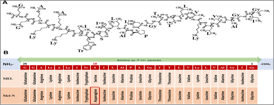
Chemical Composition of Melittin has been shown is figure no. 7: A) Molecular structure and B) Amino acid sequences of two melittin isoforms, Apis Mellifera and Apis Cerana. (45)
Melittin’s active form has polar characteristics, with the initial 20 amino acids are hydrophobic and the next six being hydrophilic. The complete sequence is as follows. NH2- Gly-Ile-Gly-Ala-Val-Leu-Lys-Val-Leu-Thr-Thr-Gly-Leu-Pro-Ala-Leu-Ile-Ser-Trp-Ile-Lys-Arg-Lys-Arg-Gln-Gln-CONH2. Melittin’s highly unique three-dimensional structure is the reason for this particular sequence. (44)At physiological pH, the N-terminal part of the molecule has +4 charges that are mainly hydrophobic, while the C-terminal part has +2 charges that are hydrophilic, for a total of +6 charges (45).
Recently, using apitoxin, especially its main component melittin, as a potential cancer therapeutic approaches has grown in significance. The peptide's lytic action is attributed to its hydrophilic carboxyl-terminal portion, whereas the amino-terminal part of its sequence is primarily hydrophobic and lacks lytic activity (31). Additionally, it makes it simple to introduce melittin into membranes by dissolving phospholipid bilayers that are both synthetic and natural. Prior research has demonstrated that melittin's mechanism of action in rupturing membranes is mediated by pore creation that non-selectively lyses both bacterial and eukaryotic cells (23).
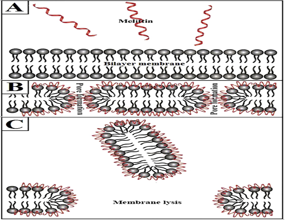
Diagrammatic representation of melittin’s lytic process has been shown in figure no. 8 (A) Melittin and membrane bilayer, (B) Pore formation, and (C) membrane lysis. (45)
• Melittin anticancer effects
According to studies, venom of bees is effective against melanocyte cancer cells, breast cancer, ovarian cancer cell, cervical cancer cells, lung cancer cells, hepatocellular cancer cells, prostate cancer cells, and leukemia (40). The synergistic action of MEL appears to be the reason for BV's effectiveness, and this anticancer peptide may be a better option than BV in its natural form. Melittin, a component of BV, prevents the spread of cancer cells by triggering apoptosis, which is a process that takes place in both healthy and diseased settings(46).
Two melittin (MEL) isoforms (Fig.7 ) have been demonstrated to have anti-cancer properties i) MEL derived from the venom of Apis mellifera and ii) MEL derived from the venom of Apis cerana (MEL-N).The most widely utilized pharmaceutical peptide in cancer research is MEL from A. mellifera, which has higher anti-cancer properties than MEL-N. However, a single study had shown that MEL-N inhibits cell proliferation and cytotoxicity in human ovarian cancer cells, specifically SKOV-3 and Pa-1. According to the literature currently available, which includes both in vitro and in vivo research, MEL influences signal transduction and regulatory pathways that result in a number of cancer death mechanisms, such as cell cycle arrest, angiogenesis inhibition, apoptosis induction, and inhibition of cancer motility, migration, metastasis and invasion etc (45).
Several studies have suggested that BV (melittin) is potential cancer agent against breast cancer (45). Melittin, the major component of honeybee venom, is efficient at causing cell death in aggressive breast cancer subtype like triple-negative and HER2-enriched. By preventing receptor phosphorylation at the plasma membrane, they block the activation of EGFR and HER2. According to a mutational analysis, the positively charged C-terminal sequence of melittin is essential for its antitumor activity and interaction with membranes (47).
In ovarian cancer, SKOV3 and PA-1 cells, MEL was demonstrated to activate a death receptor-induced apoptotic cell death pathway. This is accomplished by upregulating DR3 and DR6 transcripts and inhibiting the JAK2/STAT3 pathway(48). A research study indicated that BV triggers apoptosis in leukemia U937 cells by downregulating the ERK and Akt signaling pathways. With melittin-MEL-2 showing remarkable efficiency in promoting cytotoxicity in T and NK cells, the fusion protein greatly increased IFN-y synthesis in PBMCs (49).
MEL's impact on apoptosis regulation and the various variables that control apoptosis induction in a range of cancer types have been the subject of much research. MEL induced a rise in generated an increase in [Ca (2+)] within osteosarcoma MG63 cells by triggering Ca (2+) influx via L-type Ca (2+) channels, independently of the activity of phospholipase A(2) and protein kinase-C. This rise in [Ca (2+)] led to apoptosis (45).
Diagrammatic representation of melittin’s primary anti-cancer modes of action has been shown in figure no. 9 : (23), (12)
However, in cancer, this regulatory mechanism breaks down, leading to unchecked cell division and, eventually, tumor growth and advancement. There are several examples when MEL has been demonstrated to control the machinery involved in the cell cycle. The proliferation of HCC SMMC-7721 cells was efficiently inhibited by MEL via the downregulation of MeCP2 in vitro, the suppression of the signaling pathway, and the induction of G0/G1 cell cycle arrest. Furthermore, MEL was noted to significantly impact angiogenesis and tumor development, while also reducing the EGF-induced levels of HIF-1α protein. Moreover, the reduced half-life of MEL was assumed to be the cause of the suppression of the HIF-1α protein level (45).
Prostate cancer cell growth is inhibited by bee venom and melittin, its main ingredient. The anti-cancer effects may be mediated through the down-regulation of anti-apoptotic gene products such as cyclooxygenase-2 (COX-2), X-linked inhibitor of apoptosis (XIAP), inducible nitric oxide synthase (iNOS), and B-cell lymphoma 2 (Bcl-2) (45).
In melanoma, increased calcium levels and the activation of a caspase-independent mechanism cause apoptosis (50) Additionally, by activating the DR-induced apoptotic pathway and blocking NF-kB, BV suppressed the spread of cancer cells (51). In cervical cancer, melittin inhibits HIF-1a, thereby suppressing EGF-induced VEGF secretion and the formation of new blood vessels. Inhibition of the ERK and mTOR/p70S6K pathways in human cervical cancer cells results in this suppression (52).
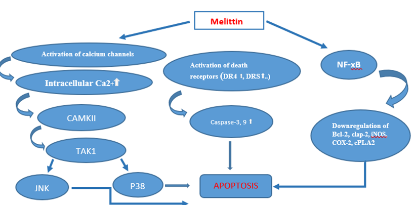
Bee venom and melittin demonstrate anti-tumor properties by activating both extrinsic and intrinsic apoptotic pathways. They enhance the expression of pro-apoptotic factors such as cytochrome C, p53, Bax, Bak, Caspase-3, Caspase-9 and death receptors, while simultaneously decreasing the levels of the anti-apoptotic protein Bcl-2. However, they inhibit NF-kB and Ca2+/CaM signaling pathways, which in turn suppresses proliferation, angiogenesis, invasive and metastasis. As a result, tumor growth and survival are significantly diminished. They also raise the amounts if AIF and EndoG, which promotes caspase-independent apoptosis (53).
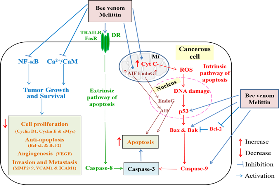
Diagram illustrating the suggested anti-cancer modes of action of melittin and bee venom has been shown in figure no. 10 : Examples of cytochrome C (Cyt C), nuclear factor-κB include reactive oxygen species (ROS), deoxyribonucleic acid (DNA), protein 53 (p53), B-cell lymphoma 2 (Bcl-2), Bcl-2-associated Xprotein (Bax), Bcl-2 homologues antagonist/killer (Bak), and death receptor (DR); MMP2/9, matrix metaloproteinase 2 and 9; cMyc, c-Myelocytomatosis; Bcl-xL, B-cell lymphoma extra-large; AIF, apoptosis-induced factor; EndoG, endonuclease G; TRAILR, TNF-related apoptosis-inducing ligand receptor; FasR, fas receptor; VEGF, vascular endothelial growth factor; VCAM1 vascular cell adhesion molecule 1; ICAM1, intercellular adhesion molecule 1; (53)
There are several methods for characterisation of Melittin form BV which consist of separation methods, chemical strategies based on common protein reactions, ad biological testing. Several aanalytical methods are employed to accelerate the eventual standardization of HBV. The primary means of separating, purifying and characterizing HBV peptides were achieved through gel chromatography and thin-layer chromatography. The identification of melittin may also be accomplished using capillary electrophoresis. In spite of this, HPLC remains the primary chromatographic method for peptides and proteins, whether for fingerprinting, product standardization, or quality control. Melittin, apamin, and tertiapin structural analyses, which include peptides in solution at various temperatures, have been the primary applications of spectroscopic methods (54).
To study the complex substances modern pharmaceutical analysis requires advanced analytical methods. Thus, researcher use “hyphenated techniques” which combine multiple analytical methods for better sensitivity, resolution and specificity (55). When two separate analytical methods are combined or coupled with the aid of a suitable interface, it is referred to as a hyphenated technique (56). By combining spectroscopic techniques like FTIR, PDA, MS and NMR with chromatographic methods like HPLC, GC, CE .Thus the hyphenated techniques integrate separation technologies with spectroscopic methods to enable both quantitative and qualitative analysis of complex samples (57).
The amount of melittin in bee venom may be measured using a variety of analytical methods, including reversed-phase high-performance liquid chromatography (RP-HPLC). Following RP-HPLC, the detection methods are diode array detector (DAD) (58), ultraviolet (UV) detector (59) , photodiode array detector (PDA), and tandem mass spectrometry (MS/MS) (60,61). Also, ultra-performance liquid chromatography-quadrupole time-of-flight mass spectrometry (UPLC-QqTOF-MS) is used for the quantitative measurement of melittin in Asian honeybee venom (Apis cerana) (62).
ANALYTICAL TECHNIQUES TO ACCESS MELITTIN FROM HBV HAS BEEN SHOWN IN TABLE NO. 1 (54).
|
Type |
Teechnique |
Component of HBV |
|
Electrophoresis |
SDS-Page |
Melittin |
|
Chromatography |
HPLC |
Melittin |
|
|
HPLC-MS /LC-MS |
|
HPLC-MS and LC-MS is one of the hyphenated technique used for characterisation of melittin. Spectroscopic techniques are typically paired with chromatographic procedures. Chemical components in a combination that were pure or almost pure were separated using chromatography, and spectroscopy generates specific data for identification through the use of library or standard spectra. The term "hyphenated techniques" refers to a variety of methods that combine separation and strategies for separation, separation-identification, and identification-identification (56).
MATERIAL AND METHOD
HPLC-grade acetonitrile and water, and phosphoric acid (85%) were from Merck (Darmstadt, Germany). Melittin (91.8% purity) was from Sigma (M2272, 119K4001) (63).
One mg of powdered venom of honey bee was transferred into a screw capped tube and dissolved in 10 mL of pure water by mixing with a vortex mixer for 3 min. following 5 min a centrifuging at 4000 rpm, 3 mL of supernatant was transferred to a 5 mL vial. This solution was filtered using a syringe filter with a 0.45 µm membrane for the HPLC analysis. 1gram of cream with one mg of honey bee venom and 10mL of 0.4% Phosphoric acid in water (elute A) were added in a screw-capped tube, heated for 5 minutes at 70oC and then mixed. The solution was centrifuged at 4000 rpm for 10 min at ambient temperature. After being filtered, the supernatant was combined with one milliliter of hexane to clarify it. For HPLC analysis, the aqueous solution was filtered using a 0.45 µm membrane 63).
The stock solution was made by dissolving the melittin in pure water to achieve the concentration of 1000 µg/mL. The stock solution was diluted with pure water and this diluted solution stored at 4°C in the dark before analysis to produce a 200 µg/mL working standard solution. The analyte level anticipated in the samples determined the concentration of the standard solutions. Melittin was calibrated using five concentrations (63).
The high-performance liquid chromatography (HPLC) was performed using the Knauer UHPC/PLC PLATINE system (Knauer, Berlin, Germany), which included a PDA detector, confined column chamber, and two pumps. The samples were separated at 25°C and chilled at a rate of 1mL/min using a Europa Protein 300 C18 column (250 x 4 mm, 5 m). The mobile phase consisted of acetonitrile (eluent A) and 0.4% phosphoric acid in water (eluent B), with a gradient elution that began at 5% to 52% A over 15 minutes, then rose to 80% A in 5 minutes, and ultimately reached 100% A in another 5 minutes. Every sample was examined three times after being injected with a volume of 20 μL. The PDA detector covered the wavelength range of 200 to 380 nm, with the detection wavelength set at 220 nm. By comparing the retention times and UV spectra of the analytes with known substances and published data, the analytes were identified (63).
Most components of honey bee venom can be separated from one another and identified using a technique called high performance liquid chromatography (HPLC). Melittin, tertiapin, MCDP, apamin and several enzymes (phospholipase A2 (PLA2), hyaluronidase) are the most significant substances found in the protein/peptide fraction. It is quite challenging to validate the results because there aren't any reference materials with certified values of the bee venom's constituent parts. For the purpose of removing the matrix effects and enhancing accuracy and precision, applying an internal standard is a definite requirement.
The following is the procedure:
Separation conditions are as follows: a linear gradient of 5% B to 80% B at 30 minutes; flow rate ¼ 1 ml/min; injection volume ¼ 40 ml; separation temperature ¼ 25 -C; and k ¼ 220 nm.
• Mass spectrometry (35)
High performance liquid chromatography-mass spectroscopy (HPLC-MS) or liquid chromatography-mass spectrometry (LC-MS) is the preferred technique for a thorough qualitative examination of the protein composition of bee venom. The wide variations in the concentrations of different proteins in the venom extracts, however, undermine the technique. In particular, tryptic digests of bee venom samples exhibit an overrepresentation of peptides derived from melittin and the different isoforms of phospholipase C. A number of techniques for pre-fractionating proteins before analysis are discussed in order to increase the dynamic range of proteomic analysis. Highly abundant proteins are usually removed using affinity reagents, such as cocktail sets of antibodies. We present the use of a combinatorial peptide library that is commercially accessible and serves as a "equalizer." Hexapeptides attached to beads make up this peptide library. During the washing processes, saturation causes excess, abundant protein to be removed.
Proteins recovered from the beads are separated by SDS-PAGE, cut into peptides and then analyzed via LC-MS or HPLC-MS. The peptide analysis can be performed using any high-resolution mass spectrometer (Orbitrap, QTOF) for proteomic analysis, but the use of FT-ICR-MS mass meters will be discussed later. It should be noted that quantitative data on variations in protein abundance are lost when employing the peptide library technique.
We explain how to utilize SDSPAGE on a 10% SDS-PAGE in this approach. For small proteins (less than 10 kDa), a Tris-tricine-SDS-PAGE (Sch€agger & VON Jagow, 1987) is the preferable method.
A commercial casting stand (small gel format) and frame can be used to cast a SDS-PAGE. Put two glass plates in the holder, spaced apart by a spacer, after assembling the casting stand. Make use of deionized water to check for leaks.
• Preparation for gel electrophoresis.
• Running of the gel electrophoresis.
• Liquid chromatography-mass spectrometry (35)
• Nanotechnology approach for bee venom and Melittin
MEL is considered an anticancer chemotherapeutic drug, but its ability to break down quickly and exhibit non-specific cellularlytic behavior poses significant challenges. The toxic effects of intravenous MEL therapy, such as hemolysis, (64) hinder its widespread adoption in cancer treatment. It has recently been clear that MEL and its conjugates may be utilized to treat certain cancer types by conjugating with hormone receptors, gene therapy, and nanoparticles (45). The toxicity, non-specificity, breakdown, low bioavailability, and hemolysis are all potential drawbacks of using melittin for cancer treatment (64). These are being overcome by means of methods such as nanotechnology, gene therapy, and immunoconjugation to improve efficacy in both in vitro and animal models (65).
Bee venom therapy can be done through electrophoresis, topical forms, injections, or direct stings. Traditional sting therapy is painful, has an inconsistent dosage, and needs to be administered repeatedly because of melittin's short half-life (66). Researchers are creating alternatives like polymer and (NPs) nanoparticle-based delivery methods in order to improve stability and control because of the short plasma half-life and dosage difficulties of bee venom (53).
Effect of Bee Venom Loaded on Nanoparticles (NPs) and Polymers
A sustained-release system is necessary to produce long-term therapeutic benefits because of the disadvantages of honeybee stings and venom injections. Recently, nanoparticles (NPs) that are effective carriers of bioactive chemicals like bee venom have been created using biodegradable polymers such as poly-D., L. and Glyc acid (PLGA), alginate, and chitosan. The delivery and release profile of bee venom is improved by loading it onto these polymer-based NPs, which lowers the frequency of administration and increases patient adherence. By modifying the polymer type, molecular weight, or nanoparticle architecture, it is possible to accurately change the breakdown rate of these nanostructures from days to years. This regulated release ensures that bee venom remains effective for a long period of time (66). It is also found that nano-fungal chitosan loaded with bee venom greatly enhanced anti-cancer action against cervical carcinoma (HeLa) cells, causing substantial apoptosis in a time- and dose-dependent manner (67).
Recent research has shown that delivery of nanovaccines targeting the LN leads to both humoral and cellular immune responses, which can help treat tumor (68). Due to the limited number and variable immunogenicity of tumor-associated antigens (TAAs), it is challenging to manufacture nano vaccines. Although neoantigens have potential, they need sophisticated prediction methods. On the other hand, whole-cell tumor antigens offer a wide range of antigens, which lowers the possibility of tumor escape and recurrence. Although the manufacturing procedure is time-consuming, Recently created chitosan nanoparticles packed with entire cell lysates to target dendritic cells in the lymph node (LN) (69). Methods for releasing antigens in situ, such as radiotherapy or oncolytic viruses, produce only little immune responses and require immunomodulators. As a result, there is a critical need for LN-targeted nanovaccines that can effectively activate antigen-presenting cells (APCs) and release whole-cell antigens (70). Melittin's lytic action is primarily attributed to its capacity to integrate into phospholipid bilayers and disrupt the integrity of cell membranes; however, when administered via nanoparticles, it may travel to intracellular membranes, activate the intrinsic pathway, and result in apoptosis (65).
Researchers developed α-peptide-NPs, a high-density lipoprotein-mimicking scaffold, and successfully loaded melittin to create ultrasmall α-melittin-NPs (10–20 nm) (71). These nanoparticles reduce melittin’s cytotoxicity to red blood cells while retaining its ability to induce tumor cell death and release whole-tumor antigens in situ. Their optimal size allows efficient lymph node (LN) targeting, enhancing melittin’s immunomodulatory effects. In vivo studies show that α-melittin-NPs rapidly drain to LNs, activate antigen-presenting cells (APCs), and trigger systemic immune responses. In a B16F10 tumor model, they eliminated 70% of primary and 50% of distant tumors, demonstrating strong potential as LN-targeted whole-cell nanovaccines for cancer immunotherapy. α-Melittin-NPs are LN-targeted whole-cell nanovaccines that efficiently slow tumor progression and elicit long-lasting, targeted, and strong systemic antitumor immunity. Most significantly, the necessary dose of melittin peptide is significantly reduced by the use of nanotechnology and intratumoral injection, resulting in additional cost savings (70).
Theoretically, Melittin (MLT) has a strong anticancer potential because of its ability to kill tumor cells (72), (73) It can control gene expression and alter immunity without causing drug resistance. However, its widespread cytolytic and hemolytic toxicity restricts its clinical application. Improvements in nanotechnology have made it possible to target MLT delivery using nanoparticles, lowering toxicity and increasing therapeutic effectiveness (74). Despite the availability of MLT-based therapies, more research is still needed on its antitumor processes and delivery techniques. Nanoparticles have benefits such as biocompatibility, adjustable pore size, and a large surface area. Researchers have enhanced functionalization strategies, such as bionic modifications, stimuli-responsive systems, and active targeting, by utilizing receptor variances and site-specific stimuli, resulting in increased bioavailability, safety, and delivery efficiency of melittin in vivo (75). The report summarizes recent advances and examines potential avenues for future study (75).
Melittin (MLT) was delivered effectively in vivo by perfluorocarbon nanocarriers with a PFOB core, resulting in tumor accumulation and considerable tumor growth retardation without toxicity (45). MLT also increases the bioavailability of orally administered drugs by crossing intestinal barriers, and targeted delivery via hemifusion reached endothelial and cancer cells(47). For stomach cancer, a legumain-activable MLT-decorated nanovehicle improved sorafenib delivery (76). Additionally, plasmid DNA encoding MLT is used in nanotech-based gene therapy, which has the potential for targeted therapy without any resource issues (75).
Research on cancer prevention and treatment has shown a great deal of interest in medication creation techniques mediated by nanotechnology. The unusual physicochemical characteristics of nanoparticles, such as nanometer size, high surface area-to-mass ratio, and effective interaction with cells, suggest that this technology has a bright future in the treatment of cancer. Numerous nanotechnology-mediated MEL conjugates have been successfully produced and evaluate in preclinical models for a wide variety of human cancers. In vitro tumor cell selectivity of sterically immunoliposomal peptides has been created and evaluated in BV (45).
CONCLUSION AND FUTURE PROSPECTS
Since both BV and its major peptide MEL are promising candidates for cancer treatment. The cumulative evidence thus far points towards the possibility of using BV and all of its components, especially MEL, as a cancer treatment. It's crucial to keep researching and developing MEL's potential as a cancer therapy option due to its anticancer properties. In areas where access to standard treatments is restricted, bee venom (BV) and melittin (MEL), its main peptide, demonstrate significant promise as inexpensive cancer cures. The effects of standard chemotherapeutic treatments are mirrored by the way that MEL causes apoptosis, impairs mitochondrial activity, and prevents angiogenesis and metastasis. Its membrane-lytic character and nonspecific toxicity, however, restrict its clinical application. By increasing targeted delivery, reducing hemolysis, and boosting treatment effectiveness, new strategies, especially nanotechnology, provide a viable solution.
By increasing the accessibility and efficacy of novel treatments, ongoing innovation in MEL-based nanocarriers has the potential to revolutionize cancer care. Due to its strong anticancer, anti-inflammatory, and antimicrobial characteristics, melittin, the major bioactive peptide in honey bee venom, is a viable option for therapeutic uses. The use of sophisticated analytical methods like SDS-PAGE, HPLC-MS, and LC-MS has allowed for accurate characterization and quantification of melittin, while nanotechnology-based delivery methods have greatly enhanced its stability, bioavailability, and targeted effectiveness. Despite its therapeutic promise, its clinical use is restricted by difficulties such hemolytic toxicity and a brief half-life in the plasma. To establish the safety and efficacy of melittin, future research should concentrate on improving nanocarrier systems, investigating gene therapy and immunoconjugates, and performing rigorous clinical trials. The use of melittin in personalized medicine and combination therapies may transform cancer treatment and expand its pharmacological applications.
We believe that a variety of further changes are still needed in order to increase the efficacy of BV and MEL in treating cancer. Although BV or MEL have not yet been tested as cancer therapies, we hope that ongoing research on this topic will eventually enable the assessment of these substances as potential anticancer treatments in the future. Thus, melittin should be characterized by use of hyphenated techniques to increase interest in research concerning the Bee venom and its potential cancer treatment.
ABBREVIATIONS
DECLARATIONS
Ethics approval and consent to participate
(Not Applicable) This work did not require formal ethics approval as it did not involve the use of honey bees practically.
Consent for publication
All authors have reviewed and agreed for publication.
Availability of data
No datasets are generated by any own assumption. Data was collected from different review and research papers.
Competing interests
The authors have no potential conflicts of interest regarding this publication.
Funding
There was no funding for this project.
Author’s Contributions
ACKNOWLEDGEMENTS
Not Applicable
REFERENCES


Nandita Tangale, Dr. Preeti Sable, Dr. Shushama Vaishnav, Dr. Praveen Wakte, Characterization of Melittin from Honey Bee Venom using Analytical Methods, Int. J. of Pharm. Sci., 2026, Vol 4, Issue 1, 915-939. https://doi.org/10.5281/zenodo.18207521
 10.5281/zenodo.18207521
