College of pharmaceutical sciences, Government medical college, Thiruvananthapuram.
Calcium Phosphate Microflowers are emerging as a novel category of hierarchically organized biomaterials that demonstrate significant promise for use in the biomedical field. These flower-like hybrid microparticles of calcium phosphate microflowers are produced by one-pot technique that combines ionotropic gelation with biomimetic mineralization. The formations are derived from chitosan, a natural biopolymer recognized for its compatibility with biological systems, natural degradability, and antibacterial properties. Extensive surface area and porosity of calcium phosphate microflowers, enhances their efficacy in various applications such as drug delivery, wound healing, antibacterial effects, and hemostasis. The hemostatic properties of calcium phosphate microflowers stem from their rapid water absorption and platelet aggregation without generating exothermic reactions. In the realm of bone regeneration, calcium phosphate microflowers facilitate early osteogenic differentiation via cellular uptake and upregulation.
Calcium phosphate heterogeneous microflowers (CM) represent a hierarchically porous material with micro/nanostructures that hold promise for biomedical applications. Chitosan (CS), a natural cationic polysaccharide obtained through the deacetylation of chitin, is considered one of the most suitable options for developing drug delivery systems due to its excellent biocompatibility, biodegradability, and strong ability to adsorb various pharmaceutical compounds. The structural features of biomaterials play a crucial role in hemostasis, antimicrobial activity, and tissue repair. However, chitosan can easily form different configurations during its preparation, such as nanoparticles, hydrogels, and microsphere. When compared to other inorganic materials, chitosan as a natural macromolecule, displays relatively poorer physical properties, including limited thermostability, low surface area, and mechanical strength, which hinder the drug delivery capabilities of pure chitosan.Therefore, to achieve better comprehensive performances, CS carriers often need further modification with other inorganic drug delivery system materials, such as silica, montmorillonite, graphene, calcium phosphate (CaP), and etc [1] . Its low production cost and natural biocompatibility and absorption, calcium phosphate (CaP) has found widespread use in drug delivery, being an inorganic material commonly found in nature [2]. Systems that utilize CaP nanoparticles for delivery have shown potential in preserving the integrity of polysaccharides and antigens while also initiating a humoral immune response, as indicated by an increasing body of research [3]. Additionally, the calcium (Ca) and phosphorus (P) present in CM are vital components involved in bone formation, which enhances CM’s utility in bone repair applications [4,5]. CS is quickly cross-linked with tripolyphosphate (TPP) through ionic gelation, resulting in nanocomplexes that exhibit notable improvements in stiffness, hardness, and surface characteristics [6]. In contrast to inorganic hemostatic agents like zeolite and kaolin, the layered porous architecture of CM features high porosity, a large specific surface area, and does not cause exothermic reactions when in contact with water [7].
Preparation of Calcium Phosphate Microflowers and drug loaded Calcium Phosphate Microflowers
A 1 weight percent acetic acid solution (2 mg mL−1) was used to dissolve chitosan (CS). After 10 minutes of stirring, 1.2 mL of 125 mg mL−1 TPP solution was added to 3 mL of 2 mg mL−1 CS solution. After stirring the mixture for ten more minutes at room temperature, 4.2 mL of 100 mM CaCl2 was added. After centrifugation and washing, the Calcium Phosphate Microflowerss were subsequently produced [3].

Figure 1. Preparation Calcium Phosphate Microflowers [3]
A 0.5 g Calcium Phosphate Microflowers was placed in 100 ml of 1 mg/ml drug solution. After stirring for 24 h, the drug loaded Calcium Phosphate Microflowers were separated out and dried at 40 °C. The The encapsulation efficiency (EE) of drug loaded Calcium Phosphate Microflowers were calculated by the following equation.
EE (%) M0-M1M0

where M0 is the total amount of added , M1 is the amount of unencapsulated free drug . Three replicate tests were performed in the drug loading studies [1].
Characterization of Calcium Phosphate Microflowers
Morphological assessments are typically carried out using Scanning Electron Microscopy (SEM) and Transmission Electron Microscopy (TEM), which verify the multi-layered flower-like structure. Fourier-Transform Infrared Spectroscopy (FTIR) is utilized to detect particular chemical interactions, such as amide and phosphate bonds, which indicate successful crosslinking. X-ray Diffraction (XRD) offers essential information regarding crystallinity and phase composition, especially in CMFs that include inorganic phases such as calcium phosphate. Thermal characteristics and stability are evaluated through Differential Scanning Calorimetry (DSC) and Thermogravimetric Analysis (TGA). Furthermore, Zeta potential measurements assess colloidal stability, while Brunauer–Emmett–Teller (BET) analysis measures surface area, which is directly linked to surface reactivity and adsorption capability. Together, these complementary methods provide a thorough understanding of CMF properties, enhancing their optimization for specific biomedical and industrial uses [1].
|
(d) |


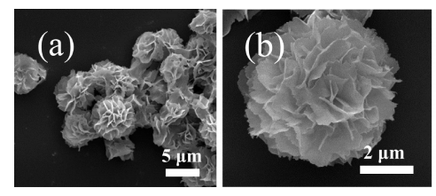
Figure 2. (a,b) SEM image ,(c) FTIR spectra, (d) TEM image ,(e) TGA and (f) DTG curves of the Calcium Phosphate Microflowers [1].
Applications of the Calcium Phosphate Microflowers
Dye Adsorption
Congo red (CR, Mw ≈ 696.7 Da, molecular charge: −2) is an azoanionic dye that is typically used as a model pollutant to assess the removal capacity of adsorbents. The synthesised microflowers were investigated for the removal of Congo red from water because of their porous and hierarchical structures, which may result in excellent adsorption properties [1]. The microflowers were treated with varying dosages, and their UV-vis absorption spectra were recorded [8]. The removal efficiency for Congo red increased as the microflowers dosage was increased .
Enzyme Immobilization
Microflowers made of chitosan can be employed as supports or carriers for enzyme immobilization. During the production of the microflowers, the catalase (CAT) was immobilized inside them by coprecipitation. To create the CAT-containing hybrid microflowers, a specified quantity of CAT was dissolved in the CaCl2 solution and then added to the chitosan and TPP mixture. This occurred as a result of CAT's (pI 5.4) electrostatic interaction with CS's amine groups and negative charge under neutral pH conditions, which affected the ionotropic gelation of CS-TPP nanocomplexes. Therefore, the inclusion of CAT lowered the effective nucleation sites for calcium pyrophosphate synthesis and hence the microflowers cannot be formed. That supported the creation mechanism of the microflowers, with the increase of CAT, the immobilization efficiency was lowered although the CAT coprecipitated in microflowers was increased. As compared with other nanoparticles, Calcium Phosphate Microflowerss are easily collected and more suitable for enezyme immobilization[1].
Drug Delivery Systems
Chitosan/calcium phosphate (CS/CaP) flower-shaped microparticles show great potential for drug delivery systems due to their complex structure and excellent physicochemical characteristics. These microflowers are synthesized using a one-pot method that merges ionotropic gelation of CS with biomimetic mineralization of CaP, resulting in formations made up of lamellar nanosheets measuring 2–5 nm in thickness and averaging 5–7 μm in diameter. The distinctive flower-like shape significantly enhances the specific surface area, improving drug adsorption and achieving an encapsulation efficiency of up to 80.77%. In vitro release experiments indicated remarkable sustained-release behavior under both acidic (pH 5.8) and neutral (pH 7.4) conditions, which were well-represented by the Korsmeyer-Peppas equation [1]. As a vehicle, the chitosan matrix acts as a structural template, while the mineralized CaP nanosheets bind pharmaceutical agents through electrostatic and hydrogen bonding interactions [9,10]. This design facilitates high drug loading and controlled release, especially in the pH conditions found in the intestine. Additionally, the CS/CaP microflowers demonstrate good cytocompatibility, exhibiting no considerable toxicity to Caco-2 intestinal epithelial cells at concentrations up to 500 μg/mL [11]. In summary, the flower-like microstructures effectively safeguard drug molecules, provide adjustable release profiles, and present significant potential for targeted and localized therapeutic delivery, especially for chronic diseases that require prolonged action.
Antibacterial and Wound Healing Properties
Chitosan microflowers (CMF) integrated into gelatin sponges display significant antibacterial and wound healing properties owing to their distinctive structure and biological characteristics [12]. CMF is recognized for its exceptional antibacterial and wound-healing abilities, which stem from its extensive surface area and unique interactions with pathogens[13]. The amino groups in chitosan, which carry a positive charge, engage with the negatively charged membranes of bacteria, disrupting their integrity and resulting in the leakage of their intracellular contents, ultimately leading to bacterial cell death [14]. Sponges loaded with CMF demonstrated a notable reduction in the growth of Pseudomonas aeruginosa, Klebsiella pneumoniae, Staphylococcus aureus, and Enterococcus faecium, showing enhanced antimicrobial activity at higher concentrations of CMF. Furthermore, Chitosan microflower loaded gelatin sponges efficiently inhibited biofilm formation, enhanced cell adhesion and proliferation, and decreased clotting times, thus facilitating tissue regeneration. These sponges also maintained over 90% viability of human skin fibroblast cells at concentrations below 100 μg/mL, confirming their excellent biocompatibility. The results highlight the potential of CMF as a versatile agent in sophisticated wound care, merging antimicrobial effectiveness with regenerative assistance [12].
Hemostatic properties
Polydopamine-coated chitosan/calcium pyrophosphate hybrid microflowers (PDA@CS-CaP) exhibit excellent hemostatic performance due to their porous and hierarchical structure, super hydrophilicity, and surface chemical functionality. The hybrid microflowers rapidly absorb water from blood, concentrating erythrocytes and platelets to accelerate clot formation. The presence of amino and phenol groups on the polydopamine coating enhances adhesion and stimulates the coagulation cascade. SEM imaging confirmed erythrocyte and platelet aggregation, with pseudopodia-like erythrocytes enhancing clot stabilization. The materials demonstrated significantly reduced clotting time in vitro (~115 s) and in vivo (85 ± 7.5 s in rat tail amputation model), outperforming zeolite and uncoated CS-CaP. Unlike traditional inorganic agents, PDA@CS-CaP does not induce exothermic reactions, ensuring safety by avoiding thermal tissue damage. These results underscore the hybrid microflowers' potential as a safe, efficient, and biocompatible hemostatic agent [15].
Bone Regeneration properties
Bone regeneration utilizing calcium phosphate (CaP) microflowers occurs through a complex mechanism that is based on the distinctive structure of the material and its biological interactions. The flower-shaped CaP microstructures, mainly consisting of dicalcium pyrophosphate and tricalcium phosphate, possess high porosity and an extensive surface area, which aids in cellular adhesion and infiltration. In vitro experiments demonstrated that at low concentrations (≤10 µg/mL), the microflowers are efficiently taken up by mesenchymal stem cells (MSCs), with uptake detected around the nuclei within 12 hours. This internalization triggers a series of osteogenic responses, particularly a notable increase in osteogenesis-related gene expression, including RUNX2 and BMP2 by day 3, and further elevation of OPN and BMP4 by day 14. This gene expression is associated with the early differentiation of osteoblasts and improved mineralization. In vivo, the implanted microflowers in rat calvarial defects exhibited quick and centripetal bone healing, with microCT revealing greater bone mineral density and ratios of bone volume to tissue volume compared to control groups. Histological evaluations further validated the development of trabecular bone and vascularization during the initial stages. Importantly, the bone regenerative effect was realized without the addition of cytokines or cells, highlighting the intrinsic bioactivity of the CaP microflower structure [5].
Vaccine adjuvant
Chitosan/calcium phosphate (CS/CaP) flower-like microparticles, especially when loaded with both ginsenoside Rb1 (GRb1) and IL-4, act as a potent immunoadjuvant system by boosting both humoral and cellular immune responses. These microparticles feature a structured “flower-like” formation with a textured surface that facilitates antigen and cytokine loading, achieving encapsulation efficiencies of 49.52% for GRb1 and 61.23% for IL-4. The GRb1/IL-4@CS/CaP system notably stimulated dendritic cells (DCs), as shown by the dose-dependent increase in MHC-II and CD80 expression, along with higher production of IL-1β and TNF-α. This stimulation is crucial because DCs are vital for antigen presentation and T-cell activation. In vaccinated chickens, the GRb1/IL-4@CS/CaP-IBDV formulation elicited significantly higher levels of IBDV-specific IgG as well as an increased IgG2a/IgG1 ratio, indicating a balanced but Th1-skewed immune response. Moreover, this adjuvant system raised serum concentrations of important cytokines such as IFN-γ, TNF-α, IL-4, IL-6, IL-1α, and IL-1β at both 28 and 42 days after immunization, underscoring its capacity to sustain and enhance immune activation. The chitosan microflower-based system also exhibited excellent biocompatibility within the relevant dosage range, minimal cytotoxicity towards dendritic cells, and a consistent zeta potential profile, establishing GRb1/IL-4@CS/CaP microparticles as a strong candidate for advanced vaccine delivery systems [2].
Advantages
Future Directions
In the future, research needs to concentrate on creating stimuli-responsive Calcium phospahte microflowers suited for precision medicine, enhancing their surfaces for targeted delivery, and combining them with advanced nanomaterials for integrated therapeutic and diagnostic uses. Calcium phospahte microflowers also offer promise in 3D bioprinting applications for regenerative engineering and environmental cleanup. Extensive long-term in vivo studies and regulatory approval will be essential for moving these materials from the laboratory to clinical and industrial applications.
CONCLUSION
Calcium phosphate microflowers (CMFs), particularly when paired with chitosan (CS), are a highly promising multifunctional biomaterial due to their complex porous structure, large surface area, and compatibility with biological systems. These composite materials show significant potential for applications in drug delivery, wound repair, enzyme immobilization, antibacterial therapies, bone regeneration, and as adjuvants for vaccines. Their capacity to encapsulate and release drugs in a controlled fashion, facilitate cellular interactions, and improve hemostatic properties establishes CMFs as a powerful foundation for advancements in biomedicine.
REFERENCES


Ramyasri M.*, Roma Mathew, Calcium Phosphate Microflowers: A Novel Platform for Biomedical Uses, Int. J. of Pharm. Sci., 2025, Vol 3, Issue 5, 4956-4963. https://doi.org/10.5281/zenodo.15554302
 10.5281/zenodo.15554302
