Department of Pharmaceutics JSPM Sudhakarrao Naik Institute of Pharmacy Pusad, India.
The benefits of the in-situ gel nasal drug delivery system based on nanoemulsions over oral administration in terms of systemic bioavailability are examined in detail in this review. A low- viscosity solution is injected into the nasal cavity using in-situ gel technology, a novel method of nasal drug delivery that turns into a gel when it comes into contact with the nasal mucosa. In comparison to traditional delivery methods, nasal drug delivery offers several advantages, such as avoiding first-pass metabolism, improving the permeability of some medications through the nasal epithelium, facilitating rapid absorption across the membrane, accelerating the onset of action, improving patient compliance and comfort, and extending the duration of drug release. Variations in temperature, pH, ion concentration, UV light, polymorphism, dissolution rate, solubility, viscosity, and osmolarity are some of the variables that variables that affect the gel formation process.
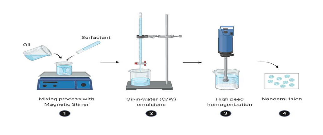
Nanoemulsions are colloidal systems containing submicron-sized particles, also known as submicron emulsions, ultrafine emulsions, or Mini emulsions. They generate homogeneous dispersions of two immiscible liquids, like water and oil, and are thought to be both kinetically and thermodynamically stable. An interfacial layer composed of surfactants and co-surfactants stabilizes these systems, ensuring a single-phase mixture. In nanoemulsions, a variety of surfactants with distinct characteristics both ionic and non-ionic are frequently used. In addition to anionic surfactants like potassium laurate and sodium lauryl sulfate, nonionic surfactants like sorbitan esters and polysorbates are also frequently used. Furthermore, zwitterionic and cationic surfactants such as quaternary ammonium halides are used. Oil-in-water (O/W) dispersions were the predominant form of nanoemulsions at first, with droplet sizes usually from 1-1000nm.The three primary categories in current classifications are water-in-oil (W/O), where water droplets disperse in an oil phase; oil-in-water (O/W), where oil droplets are dispersed in water; and bicontinuous systems, where water and oil form linked microdomains. It is easy to transition between these categories by changing the formulation components. Multiple emulsions, such as water-in-oil-in-water (W/O/W) or oil-in-water-in-oil (O/W/O) structures, are another type that involves protecting smaller droplets within larger ones. Oil-in-water (O/W) and water-in-oil (W/O) emulsions coexist in a single system in multiple emulsions, another type of nanoemulsion formulation. This dual-phase structure is stabilized by combining lipophilic and hydrophilic surfactants.
Compared to traditional dosage forms, these nanoemulsions offer a number of benefits, such as:
Components Of Nanoemulsions
Water, lipids, oils, surfactants, and water-soluble co-solvents are all components of nanoemulsion systems. Triglycerides such as tri-, di-, or mono-acylglycerols, vegetable oils, mineral oils, free fatty acids, etc., can be included in the oil phase of nanoemulsion formulation (3). The solubility of the medicine is typically the basis for selecting an oil. When developing nanoemulsions, oil phases with significant drug loading are typically used (6). Surfactants such as spans (sorbitan fatty acid esters), tweens (polyoxyethyl-ene (POE) derivatives of sorbitan fatty acid ester), Cremophor EL (polyoxyl-35 castor oil), lauroyl macro-golglycerides (Gelucire 44/14), polysaccharides (gum and starch derivatives), phospholipids (egg, soy, or dairy lecithin), and amphiphilic proteins (whey protein isolate and caseinate) are commonly used in nanoemulsions for drug delivery and food ingredients (7,8). The formation of nanoemulsions requires extremely low negative Interfacial tension. Co- surfactants or co-solvents are used along with a surfactant for this purpose. Polyethylene glycol, propylene glycol, ethanol, transcutol-P (diethylene glycol monoethyl ether), ethylene glycol, glycerin, and propanol are common co-surfactants or co-solvents used in the production of nanoemulsion systems (8,9).
The Preparation Method
A) The high-energy approach
B) Low-Energy Technique
The high energy approach, which uses strong mechanical forces to reduce bigger droplets to smaller sizes, is essential to the production of nanoemulsions (2). In order to produce stable and consistent nanoemulsions, this method uses devices including high-pressure homogenizers, microfludizers, and ultrasonicators to create powerful disruptive forces (3). The precise control that high-energy techniques provide control droplet size, formulation composition, and stability is one of their main benefits. These techniques affect the emulsions’ texture, color, and other rheological characteristics (4). High-energy nanoemulsion techniques in the food sector help preserve the integrity of nutrients and sensory attributes, reduce breakage, and increase shelf life without compromising food safety (5).
The smallest particle sizes are produced by high pressure homogenizers, which provide uniform flow and high energy. Thus, the most widely used technique of producing nanoemulsions is by using high-pressure homogenizers. Extremely very small particle size (up to 1 nm) nanoemulsions are formed by intensely disruptive forces generated by high-pressure homogenizers (10) With this technique, a mixture is forced through a small opening at a very high pressure, usually 500–5000 psi. Extreme agitation and hydraulic shear are produced during the process, which results in the formation of nanoemulsions with extremely small droplets. High pressure homogenization is considered to be one of the best methods to produce nanoemulsions. However, it has certain drawbacks, such as high energy usage and processing-induced emulsion temperature increases. Obtaining lower droplet sizes as well frequently calls for several cycles of homogenization. Using this technique, for example, phytosphingosine-based oil-in-water (O/W) nanoemulsions were effectively produced. It was also noted that the droplet size reduced during eight homogenization cycles, producing a stable nanoemulsion that lasted for more than six months (11).
II. Microfludization
In order to create extremely small particles in the sub-micron range, this method used a device known as a microfludizer, which uses a high-pressure positive displacement pump (500–20,000 psi) to push the final product out through the interaction chamber made of a stainless steel microchannel on the impact area. Until the desired particle size is reached, the mixture is passed through the microfluidizer several times. To create a homogeneous nanoemulsion and separate tiny droplets from bigger ones, the outcome is also run through a filter. Used a microfluidizer to create octadecane O/W nanoemulsions and found that the droplet size dropped as the total number of passes and homogenized pressure increased (12).produced tocotrienol-rich fraction nanoemulsions using two-step homogenization, in which a microfluidizer was used to further process an initial coarse emulsion that had been created with a stirrer. They noted that during ten homogenization cycles at elevated pressure, the droplet size decreased from 120 to 65.1 nm (13)
III. Ultasonication
When equipment relates to cleaning and operation, ultrasonication is superior to other high energy techniques (2,14). This method reduces the size of the droplets to nanodroplets by agitating a premixed emulsion at an ultrasonic frequency of 20 kHz. After that, the resulting emulsion is run through a high shear zone to create uniformly sized droplets. This method uses a water jacket to control the temperature. During ultrasonic emulsification, sonotrodes, often referred to as sonicator probes, used piezoelectric quartz crystals as energy sources. These sonotrodes expand and compress when an alternating electric voltage is applied. When the sonicator tip comes into contact with the liquid, mechanical vibrations are created. This causes cavitation, which causes the vapour cavities that have developed inside the liquid to collapse. When a droplet size of less than 0.2 µ is needed, this method is typically used. Shi et al. created a nanoemulsion filled with emodin. The mean diameter of the emodin-loaded nanoemulsion was determined to be between 10 and 30 nm utilizing the ultrasonic emulsification method at a frequency of 25 kHz (15).
B. Low-energy techniques
These techniques use little energy to create nanoemulsion systems. Since low-energy emulsification techniques use the systems’ intrinsic chemical energy and only need mild agitation to produce the nanoemulsions, they are more energy-efficient (16). Phase inversion emulsification and self-emulsification are common low-energy emulsification techniques. Low energy techniques are typically not taken into consideration for creating food-grade nanoemulsions because they require high surfactant concentrations, which have a negative impact on the safety and taste of food formulations (7). The method of phase inversion emulsification:
I. Phase inversion emulsification method
During the emulsification process, this method’s phase change is caused by the surfactant’s spontaneous curvature. Like at the HLB temperature in the PIT method, changes in change to zero cause changes in the surfactant’s spontaneous curvature. A lamellar or bi-continuous structure is created during this transition. The structures of the surfactant layer with zero curvature transform to having large positive curvature as more water is introduced, above the transition composition. Phase inversion and the production of nanoscale droplets are the results of this curvature shift. Phase inversion results from altering the system’s composition (16.17). Similar to this, additional compositional factors like salt addition and pH variations also result in nano-sized emulsion droplets by transitional phase inversion (18, 19, 20).
II. Self-nanoemulsification method
The self-emulsification approach produces nanoemulsions without altering the surfactant’s spontaneous curvature. Turbulence and nano-sized emulsion droplets are produced when surfactant and/or co-solvent molecules quickly move from the dispersed phase to the continuous phase. Another name for the self-emulsification process is the spontaneous emulsification method (16,21). Based on the self-emulsification phenomena, SNEDDS have a reduced lipid content and more hydrophilic surfactants or co-surfactants (co-solvents) (22). An isotropic combination of an oil, surfactant, co-surfactant, and medication is known as SNEDDS. Due to the moderate agitation caused by the stomach and intestine’s digestive motility, this mixture forms a thin and optically transparent O/W nanoemulsion when diluted by aqueous fluids in vivo (23, 24). Diffusion of the hydrophilic co-solvent or co-surfactant from the organic phase into the aqueous phase (16,25) and the formation of nanoemulsions with negative free energy at transient negative or ultra-low interfacial tension (27,28,29) are the two most frequently reported mechanisms of nanoemulsion formation from SNEDDS. For delivery of hydrophobic medications with low bioavailability, SNEDDS are also the most common and promising approach. Bioactive food ingredients have also been delivered via SNEDDS (30).
Nasal In Situ Gel
In situ gel is a new nasal drug delivery dosage type that recently became available. Compared to in situ gels, liquid nasal formulations are injected into the nasal cavity as low viscosity solutions. The polymer transforms into a gel upon contact with the nasal mucosa. In order to gradually release medications into the nasal cavity while also extending the duration of interaction between the drug and the absorption site. Cross-linking of polymer chains, which can be achieved by the production of covalent or non-covalent bonds, is how gel is formed. In situ gels were created using both synthetic and natural polymers. Plasma profiles with somewhat consistent sustained release can be produced by in situ gel systems.
The In Situ Gelling System Principle
The principle behind the in situ gelling system for solid nasal formulations is that, once administered, the nasal formulations absorb the nasal fluid and create a gel inside the cavity. The development of nasal gel inside the cavity helps prevent the sensation of a foreign body. The gel sticks to the nasal mucosa because it is bioadhesive. It functions as a prolonged medication delivery mechanism by acting as a matrix that controls release. The lowest layer of mucus in the nose comes and moves around the cilia, moving backward during the preparation phase and forward during the propulsion phase. The cilia parts scrape the top layer of mucus during their motion phase, penetrating it by approximately 0.5 mm. Zones of ciliary activity then appear at different times. Because cilia are positioned backward, any obstructions during the motion phase can be eliminated. Following gel formation, the mucociliary removal to the nasopharynx or disintegration take place. As a result, once the dosage form has run out of a medicine, it does not need to be removed (51,52).
Formation Methods for Nasal in Situ Gel:
Cold Method
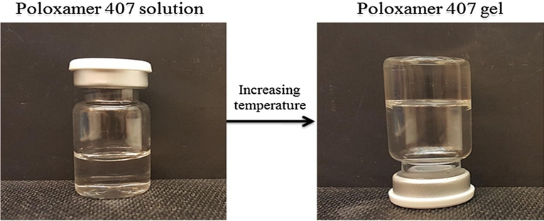
The product and a sample quantity of double-distilled water are combined in a refrigerator and kept at 4 °C overnight in this formulation procedure. The in situ gelling polymer is then gradually added while being continuously stirred. The dispersion is kept in a refrigerator until a clear solution is created and the volumes are changed. This approach is selected when formulation requires gelling polymers such as poloxamer, chitosan, or carbopol. Because the solubility of the propylene oxide chain in poloxamer decreases at high temperatures, causing precipitation or salting from the polymer, the polymeric dispersion of poloxamer remains a solution at lower temperatures and becomes concentrated in gel at higher nasal temperatures. Similarly, chitosan requires little ranges to remain a solution at room temperature, and as the temperature rises, its hydrophobicity increases (31).
Hot Method
When using pectin or gellan gum as a gelling polymer, this form Is recommended. Higher temperatures cause gellan chains to dissolve in water, develop a random coil shape with great segmental mobility, and proceed as a solution above those temperatures. When gellan gum solution is cooled, sol-gel transition takes place in the presence of ions like K or Ca2+. Additionally, pectin requires a higher temperature for demethoxylation, resulting in the formation of a solution or the dissolution of pectin (32).
Approaches Of Nasal in Situ Gel
A) In-Situ Gelling System That Responds to Stimuli
1. In-situ gel system induced by temperature.
2. In-situ gel systems induced by pH.
B) In-Situ Gelling System Induced by Osmoticism
C) In-Situ Gelling System Induced Chemically
1. Cross-linking of ions.
2. Cross-linking via enzymes.
3.Polymerization by photolysis.
D) In situ formation according to the physical process
A) In-Situ Gelling System That Responds to Stimuli
Small external changes in the environment can cause physical or chemical changes.
1. In situ gel induced by temperature
Certain polymers react to minor external changes in their ambient conditions by undergoing significant and unexpected physical and chemical changes. These polymers are known as Polymers that respond to stimuli. Other names use to describe them include intelligent, creative, stimuli-sensitive, and environmentally sensitive polymers. These polymers detect a stimulus as a signal, determine the signal’s strength, and then adjust their chain confirmation accordingly. The most researched class of environmentally responsive polymer systems in drug delivery is temperature-sensitive polymers. This is due to the fact that temperature can be easily controlled and applied both in vitro and in vivo. In this method, a change in temperature causes the solution to gel, maintaining the drug release. When these hydrogels come into contact with body fluids (35–37°C), immediately gel from their liquid state at ambient temperature (20–25°C).The use biomaterial whose transition from sol-gel is triggered by increase in temperature is an attractive way to approach in situ formation. The best critical temperature range for such systems is ambient and physiologic temperature, such that clinical manipulation is facilitated and no external source of heat other than that of body is required to trigger gelation(33,34).
Temperature sensitive polymers may be,
1. Positive thermosensitive gels: This system has an upper critical solution temperature (UCST), such Hydrogel contracts upon cooling UCST. E.g. Polymer networks of pol (acrylic acid)
2. Negative thermosensitive gels: This system have a lower critical solution temperature (LCST) and contract upon heating above the LCST. E.g. poly (N-isoprophylacrylamide)
3. Thermo reversible gels
Eg poloxamers/pluronics, tetronics
2. In situ gel induced by pH
This class of polymers has an acidic or basic group that, depending on the pH of the surrounding environment, either accepts or releases protons. These are known as pH-sensitive polymers as a result. The majority of anionic group-containing pH-sensitive polymers are based on PAA (Carbopol, Carbomer) and its derivatives. In these systems, a pH change induces the solution to gel transition. Additional acidic or basic groups included in all pH-sensitive polymers pick up or release protons in response to variations in the pH of their surroundings. Polymer electrolytes are polymers that include a large number of ionizable groups. The presence of polyelectrolytes in the formulation raises the pH outside, which causes the hydrogel to swell and create in-situ gel (16).
B) In-Situ Gelling System Induced by Osmoticism:
In this system, variations in ionic strength cause the fluid to gel. It is believed that the osmotic gradient across the gel’s surface determines the rate of gelation. When cations such as mono or divalent cations are present, the polymer’s aqueous solution turns into a transparent gel. The polymer exhibits osmotically triggered gelation, such as gellan gum and alginates.
C) In-Situ Gelling System Induced by Chemically
Ionic, enzymatic, and photo-polymerization are the chemical reactions that create in-situ gel systems.
1. Ionic crosslinking:
When different ions, such as Na, K, Ca, and Mg, are present, polymers may experience a phase transition. The ion-sensitive class includes a few of the polysaccharides (21). I-carrageenan primarily forms elastic gels in the presence of Ca 2, whereas k-carrageenan responds to modest levels of K by forming a hard and brittle gel. An anionic polymer called gellan gum gels in-situ when monovalent and divalent cations like Ca, Mg, K, and Na are present. Divalent cations, particularly Ca, can induce low methoxy pectin to gel. Similar to this, alginic acid gels in the presence of DGG Physiological feild situ gelation-polymeric generated by the interaction of the alginate chain with the glucuronic acid block when the tonic concentration of divalent polyvalent cations, such as Ca 2, changed (22). Various polymers are employed in the preparation of the in situ gelling system. Temperature-sensitive in situ gelling system polymers
Poloxamer:
It is a tri-block copolymer that dissolves in water. In an AIBA arrangement, it contains two polyethylene and polypropylene oxides (35). Also referred to as Pluronic, it possesses excellent drug resistance time and good thermal setting qualities. It serves as a solubilizing and gelling agent. It produces translucent, colorless gel. It comes in a wide range of molecular weights with various gelling qualities. The ratio and partition of the hydrophilic and hydrophobic chains determine the gelling characteristic. Essential polypropylene oxide is enclosed in polyethylene oxide to form the poloxamer. It functions as a viscous liquid at ambient temperature (25 °C) and changes into a translucent gel at 37 °C. At low temperatures, it forms a small micellar subunit in solution; as the temperature rises, the viscosity increases, causing swelling and the formation of a large micellar cross-linking network (36.37).
Chitosan
Alkaline acetylation of chitin, a naturally occurring substance found in shrimp and crab shells, yields chitosan. Chitosan contains a polycationic, thermosensitive, biodegradable polymer. Chitosan is a cationic polymer that depends on pH. The biocompatible quality of chitosan allows it to dissolve in aqueous solutions up to a pH of 6.2. A hydrated gel-like precipitate forms when an aqueous solution of chitosan is neutralized to a pH higher than 6.2. By adding polyol salts with a single anionic head, such as glycerol, sorbitol, fructose, or glucose phosphate salts, to chitosan aqueous solution, the pH gelling cationic polymer solution is converted to the thermally sensitive pH-dependent gel-forming aqueous solutions without the need for chemical changes (38, 39).
The pH in situ gelling system polymer:
Carbopol
The carbopol polymers' ability to absorb water is excellent. The polymers’ pka of 6.0 causes them to swell in water up to 1000 times their initial volume and 10 times their initial diameter, forming a gel until they are subjected to a pH between 4.0 and 6.0. In addition to having a large molecular weight and derivatives of cross-linked polyacrylic acid, carbopol polymer has potent mucoadhesive qualities. When cellulose is added, the concentration of polymers will decrease, concentration will increase, and gelling properties will improve. Carbopol 934 and Carbopol 841 are the gelling polymers that are most frequently employed (40).
The ion in situ gelling system polymer
Sodium Alginate
Alginic acid salts include sodium alginate. The source of it is brown algae. It is a linear block polysaccharide with residues of a-L glucuronic acid and two type monomers, B-D-mannuronic acid, linked by 1,4 glycosidic bonds. Because sodium alginate has a carboxylic group, it is non- toxic, biodegradable, and has good mucoadhesive properties. Alginate B-D-Mannuronic acid and a-L glucuronic acid monomers are arranged as M-M blocks with modifying sequences (M-G blocks) as the polymer’s mechanism. Development of a gel is the result of the interaction between the polymer’s G block and calcium. The type of crosslinker utilized, the concentration of alginate polymer solution, and the mechanical strength and porosity of hydrogel are all dependent on the G. M. ratio (42
Pectin
The polymer backbone of pectins, a family of polysaccharides, is primarily composed of residues of a-(1-4)-D galacturonic acid. H ions, the source of divalent ions, are necessary for pectin gelation to occur. Calcium ions are typically needed to create a gel that can be used as a drug delivery vehicle (43). Pectin’s primary benefit in these formulations is its water solubility, which removes the need for an organic solvent. When administered orally, the stomach’s divalent cations undergo a pectin-to-gel transition. To cause pectin gelation, formulations can contain complex forms of calcium ions (44) The majority of the calcium ions added to the formulation can form a compound with sodium citrate when added to the pectin solution. The majority of the calcium ions added to the formulation can form a compound with sodium citrate when added to the pectin solution. Because of this, the formulation can stay in a fluid state (sol) until the complex breaks down in the stomach’s acidic environment, when the release of calcium ions results in gelation (45).
Gum xanthan:
The gram-negative bacterium a species undergoes fermentation to create xanthan gum, a high molecular weight extracellular polymer. This naturally occurring cellulose derivative’s core structure consists of a cellulosic backbone (BD-glucose residues) and a trisaccharide side chain that is joined to the main chain’s alternating glucose residues by B-D-mannose, B-D-glucuronic acid, and a-D-mannose (46). Because the side chains of this polymer include both pyruvate and glucuronic acid groups, it has an anionic characteristic (47) Both hot and cold water, as well as alkaline and acidic environments, can dissolve xanthan gum. Has strong stability in alkaline environments.
Gellangum
One a-L rhamnose, one B-D-glucuronic acid, and two B-D-glucuronic acid residues compose a tetrasaccharide repeating unit of gellan gum polymer, an anionic deacetylated exocellular polysaccharide produced by Pseudomonas elodea (48). It tends to gelation that is cation-induced or temperature-dependent. A double helix junction zone is created during this gelation process, and then the double helix segments aggregate to form a three-dimensional network through complexation with cations and hydrogen bonding with water (49) By cross-linking negatively charged helices with monovalent or divalent cations (Na, Ca, Mg. K’), gellan gum produces cation-induced in-situ gelations (Ca, Mg). When it comes to inducing gelation, divalent ions exceed monovalent cations. Gelation improves the drug’s bioavailability and increases its residence time at the absorption site (50)
Marketed Product of Nasal in Situ Gel
|
Drug substance |
Indications |
Dosage form |
Manufacturer |
|
Fluconazole |
Used to prevent antifungal infection |
Solution (Spray) |
Pfizer limited |
|
Zinc gluconate Zinc acetate |
Used to prevent cold and relief from cold system such as sore, runny nose, cough and congestion |
Solution (Spray) |
Matrixx initiative Inc |
Fig: List of Marketed Product of Nasal in situ gel
Nanoemulsion Based Nasal in Situ Gelling System
|
Sr.no |
Stimulus responsive agent |
Triggering factor |
Drug |
Category |
Year |
Ref |
|
1 |
Poloxamer 407,Chitosan, Hpmck15 |
Temperature |
Ondansetron hydrochloride |
Antiemetic |
2018 |
53 |
|
2 |
Pluronic F127,F68 |
Temperature |
Clozapine |
Antischizop renic |
2021 |
54 |
|
3 |
Poloxamer 407,188 |
Temperature |
Temozolami de |
Antineoplas tic |
|
55 |
|
4 |
Pluronic F127 |
Temperature |
Buspirone |
Anti-anxiety |
|
56 |
|
5 |
Poloxamer 407,Hpmc |
Temperature |
Olanzepine |
Antischizop renic |
|
57 |
|
6 |
Gellangum |
Ion sensitive |
Disulfiram |
Antitumour |
|
58 |
|
7 |
Poloxamer 407 |
Temperature |
Tizanidine |
Spasticity |
|
59 |
|
8 |
Poloxamer 407,Carbopol 934 |
Temperature |
Dolutegravir |
Neurodegen erative CNS disorder |
|
60 |
|
9 |
Carbopol 934 |
PH |
Lasmiditan |
Antimigrane |
|
61 |
Fig: Nanoemulsion Based Nasal in Situ Gelling System
The Benefits of Nasal-Based Nanoemulsions in Situ Gel (53,54,55,56).
1. Improved Absorption of Drugs
Because of their small droplet size and surfactant action, nanoemulsions increase the solubility of lipophilic medicines and improve their penetration through the nasal mucosa.
2.Enhancement of Bioavailability
First-pass metabolism is avoided by the nasal route, and systemic drug availability is further enhanced by the use of nanoemulsions.
3.Uniform and Controlled Release
When in situ gels come into contact with the temperature-, pH-, or ion-sensitive nasal mucosa, they become stronger, facilitating gradual, controlled release.
4.The ability to adhere to mucosa
By extending nasal residence duration, polymers such as poloxamer or carbopol decrease mucociliary clearance.
5.Patient-friendly and non-invasive
a substitute for injections in the systemic distribution of proteins, peptides, and CNS-active medications.
6.Targeted Delivery to the Brain
Systems based on nanoemulsions can improve delivery from the nose to the brain by making it easier for substances to travel through the trigeminal and olfactory pathways.
Evaluation Parameters
1.Evaluation of Nanoemulsion (72)
• Measurement of droplet size, PDI
• Zeta Potential
• Phase Separation
2. Evaluation of Nasal In situ gel (71)
• Viscosity determination (67)
• Measurement of gel strength (69)
• pH determination
• In vitro diffusion study (45)
• Stability study
• Gelling capacity
• Determination of drug content (68)
CONCLUSION
Nasal in situ gels based on nanoemulsions are a new and promising method of medication delivery, especially when it comes to focusing on the central nervous system through the nose-to-brain connection. These systems combine the benefits of in situ gels, such as extended residence duration, mucoadhesion, and controlled drug release, with the advantages of nanoemulsions, such as improved solubility, stability, and bioavailability. This formulation method has been shown in numerous trials to have the potential to improve therapy outcomes for a variety of systemic diseases and CNS disorders. While promising preclinical results, additional investigation is necessary to address issues related to formulation scalability, long-term stability, mucosal toxicity, and clinical translation. It is expected that further developments in polymer science and nanotechnology will make it easier to develop safe, patient-friendly intranasal nanoemulsion- based medicines that use gels.
REFERENCES


Rani Chatte*, Dr. Ravikiran Wakade, Abhilash Deshmukh, Benefits of Nanoemulsion Based Nasal in Situ Gel: A Review, Int. J. of Pharm. Sci., 2025, Vol 3, Issue 7, 3095-3109. https://doi.org/10.5281/zenodo.16351929
 10.5281/zenodo.16351929
