Raffles University, Japanese Zone, NH-48, Neemrana-301705, District-Alwar, Rajasthan, INDIA
The study's primary goal is to assess the ethanolic extract of Leucas lavandulifolia roots' antioxidant and free radical scavenging properties utilizing a variety of in vitro assay techniques. It also aims to assess the phytochemical identification tests. On March 31, 2024, the roots of the Leucas lavandulifolia plant were gathered from Dolaigaon Road, New Colony, Natunpara, Bongaigaon, Assam, 783380, India. The antioxidant and free radical scavenging activity were estimated using standard procedures from the literature. Given that the obtained values are quite close to the utilized reference chemical, the results imply that the extracts can be regarded as a valuable source of antioxidant products. In this setting, antioxidant and free radical scavenging capabilities are novel to the literature. For future in vivo activities, this extension in vitro study of the chosen leucas species for antioxidant activity will be beneficial.
Leucas lavandulifolia roots exhibit a wide range of biological actions, such as analgesic, antioxidant, antibacterial, anti-inflammatory, and perhaps anticancer properties. To completely comprehend the medicinal properties of the roots and identify the chemical substances causing these benefits, more study is needed, especially clinical studies. It should be used with caution, as is the case with many conventional herbal therapies, particularly when used in conjunction with additional therapies or in patients who already have a medical condition [1]. There are 116 species in the broad genus Leucas, which is a member of the Lamiaceae (Labiatae) family. It is found in northeastern Australia and is widely dispersed throughout tropical Asia and Africa. Small shrubs, sub-shrubs, annual or perennial herbs, and plants with simple hairs, erect stems, and simple, opposite leaves are all members of the genus Leucas [2,3]. To cure a wide range of illnesses, including skin disorders, colds and coughs, snakebite, diarrhea, fever, STIs, wounds, ulcers, aphrodisiacs, rheumatic issues, jaundice, and more, various species of the genus Leucas are used extensively in traditional medicine [2,4]. Numerous biological activity, including antibacterial, hypoglycemic, anthelmintic [5], antioxidant, antitussive, anti-inflammatory[6], insecticidal, and cytotoxic properties, have been demonstrated by various extracts of the various plant sections . The Indian tradition system of medicine enlists a large number of plants for basic health care. Leucas lavandulifolia is mentioned in the ayurvedic medicinal system and also used among the folklores. The plant is used for the treatment of fever, asthma, psoriasis, dermatitis and healing snake bites. The scientific validation of the plant for their traditional use in different immune related disorders are yet to be explored [7]. In a study on rats with stomach ulcers caused by indomethacin, the L. lavandulifolia extract demonstrated dose-dependent ulcer prevention efficacy [8]. It has been discovered that both the methanolic and chloroform extract of L. lavandulifolia leaves exhibits antibacterial action against P. aeruginosa, S. aureus, B. subtilis, and E. coli [9]. It has been discovered that both the methanolic and By reducing gastrointestinal motility, the aerial portions of L. lavandulifolia at a dose of 400 mg kg-1p.o. greatly decreased the frequency of defecation, the wetness of the fecal droppings, and diarrhea.This herb can therefore be used as a general anti-diarrheal agent. Tannic acid and tannins may be the cause of the reaction because they denature proteins to create protein tannate, which increases the intestinal mucosa's resistance and decreases secretion [10,11]. Using codeine phosphate (10 mg kg-1), a prototypical antitussive drug, as a reference, the antitussive activity of the methanol extract of L. lavandulifolia was investigated in a cough model caused by sulfur dioxide gas in mice. When compared to the control, it showed notable effects, and the frequency of coughing was found to be dose-dependent.Within one hour of conducting the trial, the plant extract at doses of 100, 200, and 400 mg kg-1, p.o., demonstrated a 35.0, 51.9, and 56.5% inhibition of cough. These findings support the Tripuran tribal people's traditional use of the plant [12] The incision & excision wound model developed for albino rats was used to assess the wound healing activity of methanolic extract applied as an ointment and injected as powdered plant material.The extract formulation had activity similar to that of the common medication nitrofurazone and markedly improved the rate of wound contraction, duration of epithelialization, tensile strength, and restoration of tissues at the wound site.The findings support the traditional usage of L. lavandulifolia to treat skin conditions [13]. Using a variety of animal models, including behavior tests, sodium pentobarbitone sleep potentiation, exploratory behavior (Head dip test & Y-maze test), and relaxation action (Traction test, Rotarod test, and 30° inclined screen test) in mice and rats, the methanol extract of the plant was assessed for psychopharmacological profiles.In the Y-maze test, there was a notable reduction in the exploratory behavior of rats treated with extract at a dose of 100 mg kg™. Additionally, the extract significantly reduced the head dip responses in mice at doses of 100 mg kg-1and potentiated pentobarbitone-induced sleeping duration in mice at doses of 200 mg kg-1. The extract demonstrated exceptional motor coordination and muscle relaxation activity in tests pertaining to muscle relaxant action[14]. Ethanolic extract of plant aerial parts yielded flavones glycoside (Chrysoeriol-4"-O-α-L-rhamnopyranosyl (1>2) β-D glucopyrancside), which was tested for anti-inflammatory activity using carrageenan-induced paw edema in albino rats. After three hours, the extract at a dose of 300 mg kg™ demonstrated 62.5% inhibition of paw edema, which was equivalent to the standard medication, diclofenac[15,16]. The acetic acid-induced writhing model & hot plate techniques were used to examine the analgesic effects of the plant's ethyl acetate extract in rats. The extract (400 mg kg-1) dramatically decreased the number of writhings in the acetic acid-induced writing test. In comparison with the standard drug pentazocin, the hot plate test result showed a significant rise (p<0.001) in reaction time at 2 and 3 hours, but a smaller increase (p<0.05) at 1 hour. Bradykinin and prostaglandins were proposed to be key players in the pain process, and the extract may inhibit the production of chemicals that cause pain in the peripheral tissues. The flavonoid complex chrysoeriol-{OAC)-glucoside, which is contained in an extract of ethyl acetate of the herb's aerial portions, may be the cause of the action[17]. Rats with yeast-induced pyrexia were used to test the antipyretic effects of L. lavandulifolia methanol extract. Nineteen hours after delivery, a yeast suspension (10 mL kg-1, s.c.) raised the rectal temperature. The extract showed a substantial dose-dependent reduction in body temperature at dosages of 100, 200, and 400 mg kg -1 (i.p.); the antipyretic effect was similar to that of paracetamol, a common antipyretic medication[18]. Using glybenclamide as a reference chemical, the study examined the effects of the chloroform extract on L. lavendulifolia flowers upon blood glucose levels, glycosylated hemoglobin, and oral glucose tolerance in rats with alloxan-induced diabetes. Over the course of 30 days, oral administration of 0.15, 0.20, and 0.25 g kg-1 of L. lavenderifolia chloroform extract led to a significant decrease in blood sugar levels, glycosylated hemoglobin levels, and an increase in total hemoglobin; the effect was particularly significant for 0.25 g kg-1. Additionally, it prevented a decrease in body weight. When animals were given L. lavenderifolia flower extract, their glucose tolerance significantly improved[19]. Rats with streptozotocin-induced diabetes were given glybenclamide (1 mg kg-1) and methanolic plant extract at dosages of both 200 and 400 mg kg simultaneously. At a dose of 400 mg kg-1, the extract's efficacy peaked, resulting in a substantial 39.5% (p<0.001) drop in blood glucose levels when compared to control groups[20]. Rats with streptozotocin-induced diabetes were given glybenclamide (1 mg kg™) and methanolic plant extract at dosages of both 200 and 400 mg kg simultaneously. At a dose of 400 mg kg-1, the extract's efficacy peaked, resulting in a substantial 39.5% (p<0.001) drop in blood glucose levels when compared to control groups[21]. For 14 days, 200 and 400 mg kg™ of Chloroform extract of L. lavandulifolia aerial parts was taken orally as a fine solution in 0.3% sodium carboxymethyl cellulose. D (+) galactosamine was administered, causing liver injury. The serum levels of ASAT, ALAT, ALP, TB, LDH, and TC were significantly lower in the treated group than in the D (+) Gal N given group. Thus, it appears that this plant's chloroform extract has hepatoprotective properties in rats. Additional research is required to assess this extract's potential utility in clinical disorders related to liver injury [22].
MATERIALS AND METHOD:
Plant Collection:
The roots parts of Leucas lavandulaefolia were collected from North East states in india and its botanical identity was confirmed by Botanical survey of India. The roots of the plant Leucas lavandulifolia were collected in the 31st March of 2024 from Dolaigaon Rd, New Colony, Natunpara, Bongaigaon, Assam, 783380, India. Latitude and longitude of this places are respectively as follow: 26.482140 and 90.482140. The Leucas lavandulifolia roots were also collected from, New Colony, Mahabirstan, Bongaigaon, Assam, 783381, India. Latitude and longitude of this places are respectively as follow: 26.4821290 and 90.5358940. Leucas lavandulifolia was authenticated by Central National Herbalum, Botanical Survey of India, Howrah- 711103.
Extraction process:
Petroleum ether Extract:
At first take all the necessary items like leucus lavandulaefolia root , grinder, heating mantle , round bottom flask, extractor , condensers, pipe, continuous water supply, weight machine, petrolium ether , acetone , measuring cylinder etc. The root parts of the L.lavandulaefolia was shade-dried and Coarsely powdered by using grinder machine. Set up about two soxhlet extraction apparatus for the extraction .The coarse powder drug weighted by using weighing machine, take powdered drug about 108.830 gm from this total powdered take 50.010 gm in a thimble which is placed into solvent extractor and take 50.020 gm in another extractor. Next take 300 ml petroleum ether as a solvent which is known as defatting agent in two different round bottom flask. Then set up two Soxhlet apparatus and performed the extraction for 48 hrs .After the completion of extraction check the colour of solvent and concentrated it on water bath under 80?c. After this performed TLC of two different concentrated compound which is taken from two different Round Bottom Flask. At last calculate the Rf value[24].
Chloroform Extraction:
At first take all the necessary items like leucus lavandulaefolia root , grinder, heating mantle , round bottom flask, extractor , condensers, pipe, continuous water supply, weight machine, chloroform , acetone , measuring cylinder etc. The root parts of the L.lavandulaefolia was shade-dried and Coarsely powdered by using grinder machine. Set up about two soxhlet extraction apparatus for the extraction . The coarse powder drug weighted by using weighing machine, take powdered drug about 108.830 gm. From this total powdered take 49.080 gm in a thimble which is placed into solvent extractor and take 48.020 gm in another extractor.Next take 450 ml chloroform as a solvent in two different round bottom flask. Then set up two Soxhlet apparatus and performed the extraction for 24 hrs [23]. After 24 hrs check the solvent colour and take out it from the round bottom flask and put in a beaker. Next concentrate the solvent of two round bottom flask in the two different petridish by using water bath under 80?c temperature. After the concentration of the solvent, performed TLC of concentrated compound of two petridish. Next check the TLC plate under UV Spectrometer. At last measure the Rf value.
Ethyl Acetate Extraction:
At first take all the necessary items like leucus lavandulaefolia root , grinder, heating mantle , round bottom flask, extractor , condensers, pipe, Beaker, weight machine, Ethyl acetate, acetone , measuring cylinder etc. The root parts of the L.lavandulaefolia was shade-dried and Coarsely powdered by using grinder machine . Set up about two soxhlet extraction apparatus for the extraction . The coarsely powder drug weighted by using weighing machine, take powdered drug about 108.830 gm. From this total powdered take 49.080 gm in a thimble which is placed into solvent extractor and take 48.020 gmin another extractor. Next take 300ml Ethyl Acetate [23] as a solvent in two different round bottom flask. Then set up two Soxhlet apparatus and performed the extraction for 12 hrs . After the completion of extraction concentrate the solvent of two different round bottom flask in two different petridish. After this performed TLC of concentrated compound of two different petridish and check the plate under UV Spectrometer. Calculate the Rf value After 12 hrs of extraction we continued the process for 24 hrs. After this, concentrate the solvent in the same way. And perform TLC and calculate Rf value.
Ethanol Extraction:
At first take all the necessary items like Leucus lavandulaefolia root , grinder, heating mantle , round bottom flask, extractor , condensers, pipe, Beaker, weight machine, Ethyl acetate, Ethanol, acetone , measuring cylinder etc. The root parts of the L.lavandulaefolia was shade-dried and Coarsely powdered by using grinder machine . Set up about two soxhlet extraction apparatus for the extraction . The coarse powder drug weighted by using weighing machine, take powdered drug about 108.830 gm. From this total powdered take 49.080 gm in a thimble which is placed into solvent extractor and take 48.020 gm in another extractor. Next take 150 ml (50%) Ethyl acetate and 150 ml (50%) Ethanol as a solvent in a round bottom flask and take 300 ml (100%) Ethanol in another round bottom flask. Then set up two Soxhlet apparatus and continue the process for 6 hrs. After completion of the extraction , concentrate the solvent in two different petridish by using water bath under 80?c temperature. After concentration , perform the TLC and calculate Rf value.
Phytochemical Identification:
Mayer’s test: To a few drops of the Mayer’s reagent, 2 mg of ethanolic extract was added. Formation of white or pale yellow precipitate indicates the presence of alkaloids[25].
Wagner’s test: 2 mg of ethanolic extract was acidified with 1.5 % v/v of hydrochloric acid and a few drops of Wagner’s reagent was added. A yellow or brown ppt. indicates the presence of alkaloids [26].
Test for flavonoids:
Shinoda’s test: 2 mg of ethanolic extract was dissolved in 5ml of ethanol and to these 10 drops of dilute hydrochloric acid followed by a small piece of magnesium were added. Formation of pink or reddish-brown colour indicates the presence of flavonoids [27].
Test for saponins: In a test tube containing about 5 ml of an ethanolic extract, a drop of sodium bicarbonate solution was added. The test tube was shaken vigorously and left for 3 minutes. Formation of honeycomb like froth indicates the presence of saponins [28]
Test for tannins: To 1-2 ml of the ethanolic extract, few drops of 5% w/v FeCl3 solution was added. A green colour indicated the presence of Gallo tannins, while brown colour indicates the presence of pseudo tannins [29].
Test for Carbohydrates:
Molish’ s test: To 2 ml of plant sample extract, two drops of alcoholic solution of α- naphthol are added. The mixture is shaken well and few drops of concentrated sulphuric acid is added slowly along the sides of test tube. A violet ring indicates the presence of carbohydrates[29].
Benedict’s test: To 0.5 ml of filtrate, 0.5 ml of Benedict?s reagent is added. The mixture is heated on a boiling water bath for 2 minutes. A characteristic coloured precipitate indicates the presence of sugar[25].
Test for Fixed oils and Fats:
Spot test: A small quantity of extract is pressed between two filter papers. Oil stain on the paper indicates the presence of fixed oils[9].
Saponification test: A few drops of 0.5 N alcoholic potassium hydroxide solution is added to a small quantity of extract along with a drop of phenolphthalein. The mixture is heated on a water bath for 2 hours. Formation of soap or partial neutralization of alkali indicates the presence of fixed oils and fats.[9]
Test for Phenolic compounds:
Ferric Chloride test: The ethanolic extract is dissolved in 5 ml of distilled water. To this few drops of neutral 5% ferric chloride solution are added. A dark green colour indicates the presence of phenolic compound[30].
Alkaline reagent test: Add 5ml distilled water with ethanolic extract in a test tube. Then an aqueous solution of the extract is treated with 10% ammonium hydroxide solution. Formation of yellow fluorescence indicates the presence of flavonoids[25].
Test for Proteins: The extract is dissolved in 10 ml of distilled water and filtered through Whatmann No. 1 filter paper and the filtrate is subjected to test for proteins[26].
Millon’s test: To 3 ml of filtrate few drops of Millon?s reagent are added in a test tube. A white precipitate indicates the presence of proteins[26].
Antioxidant test using DPPH assay:
At first I took 100ml methanol in a conical flask. Then weighted 0.04 gm DPPH (2,2-diphenyl-1-picrylhydrazyl). Then the DPPH was added in the methanol and mixed well, a purple colour was formed.Wrapped the conical flask with an aluminium foil and placed it in a dark place for 30 minutes. Took 3 clean test tube. And took 1ml, 2ml & 3ml solution from ethanolic extract of Leucas Lavandulifolia roots in the 3 test tubes. Filled the test tube with methanol to make 10ml in each test tube and mixed well. Samples are ready. After 30 minutes. Took another 3 test tubes. Then took 1ml of sample from each test tube and put it into another test tubes. Then took 3 ml DPPH solution and put into the testubes then fill the test tubes with methanol upto 10 ml. Then the test tube’s mouth was sealed with cotton and placed in a dark place for 30 minutes for incubation. indicates the presence of antioxidant property. Prepared the UV Spectrophotometer to check the absorbance of the samples. Then set the wavelength to 517 nm. Then took methanol as a blank solution. Then checked the absorbance of 3 samples against the blank[31].
RESULT:
Petroleum ether Extract: As spot is not present in TLC plate so we can confirm that extraction of Petroleum Ether is successfully succeed.
Chloroform Extraction: As spot are present in TLC plate so we can confirm that extraction of Chloroform is successfully succeed
Ethyl Acetate Extraction: As spot is present in TLC plate so we have to continue this process with the same solvent.
Ethanol Extraction:Spot is present in TLC plate ,so we have to continue the process in the same way.
Phytochemical Identification:
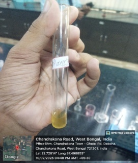
Mayer’s test: According to Mayer’s test, pale yellow precipitate is formed, so it is confirmed that alkaloid is present on the roots (Fig-1)
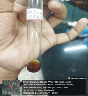
Wagner’s test: Brown precipitate is formed. A yellow or brown precipitate confirms the presence of alkaloids (Fig-2)
Test for flavonoids:
Shinoda’s test: Reddish brown colour is formed which indicates the presence of flavonoids in roots (Fig-3)
Test for saponins: Honeycomb like froth is formed which indicates that saponin is present. (Fig-4)
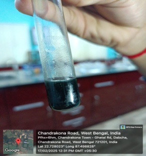
Test for tannins: Green colour is formed. It means roots contains Gallo tannins(Fig-5)
Test for Carbohydrates:
Molish’ s test: A violet ring is formed. Indicates the presence of carbohydrates. (Fig-6)
Benedict’ s test: A characteristic light blue coloured precipitate formed. Indicates the presence of sugar.(Fig-7)
Test for Fixed oils and Fats:
Spot test: No oil stain on the paper is formed. No indication of the presence of fixed oils.(Fig-9)
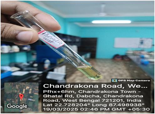
Saponification test: Partial neutralization of alkali is occur but no formation of soap. Indicates the presence of small amount of fixed oils and fats (Fig-8)
Test for Phenolic compounds:
Ferric Chloride test: A dark green colour is formed. Indicates the presence of phenolic compound.
Alkaline reagent test: Formation of yellow fluorescence. Indicates the presence of flavonoids (Fig-10)
Test for Proteins:
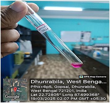
Millon’s test: White precipitate formed. Indicates the presence of proteins.(Fig-11)
|
|
|

|

|
|
Fig-1: Mayer’s Test |
Fig-2: Wagner’s Test |
Fig-3: Shinoda’s test |
Fig-4: saponins test |
|
|

|

|
|
|
Fig-5: Tannin Test |
Fig-6: Molish’ s test |
Fig-7: Benedict’s test |
Fig-8: Saponification test |
|

|
|

|
|
Fig-9: Spot Test |
Fig-10: Alkaline reagent test |
Fig-11: Millon’s test |
Antioxidant test using DPPH assay:
The radical scavenging activity of extract is evident in the DPPHassay, where it was seen that the purple color changed to yellow colour after the hydrogen donated by the antioxidant was accepted by the DPPH radical thereby changing the colour that was read at 517 nm. The extract showed a strong hydrogen donating capacity and can powerfully scavenge DPPH radical. The maximum free radical scavenging activity and potency was interpolated from figure to give results as shown intable 1.
|
Absorbance of Control |
Absorbance of Sample |
% Inhibition |
|
0.005 |
1.960 |
-39100 |
|
0.005 |
1.525 |
-30400 |
|
0.005 |
0.766 |
-15220 |
CONCLUSION:
The present study successfully demonstrated that the plant extract possesses significant antioxidant activity, as evidenced by its ability to scavenge DPPH free radicals in a concentration-dependent manner. The IC?? value indicated strong radical-scavenging potential, suggesting the presence of bioactive compounds with antioxidant properties. Phytochemical screening further revealed the presence of key secondary metabolites such as flavonoids, phenolics, tannins, and alkaloids, which are known to contribute to antioxidant effects. These findings support the potential therapeutic value of the plant extract and warrant further investigation into its active constituents and mechanisms of action for possible development into natural antioxidant agents.
Acknowledgement:
We are grateful to the P.G. Institute of Medical Science for providing the necessary resources and facility.
Funding:
This work was supported by the School of Pharmacy, Raffle University and P.G. Institute of Medical Science.
Data availability:
The data that support the findings of this study are available from the corresponding author, Nilanjan Adhikari, upon reasonable request..
Competing interests:
The authors declare that they have no competing interests.
REFERENCES


Nilanjan Adhikari*, Md. Shamsuzzaman, Antioxidant Study and Phytochemical Identification of Leucas Lavandulifolia Roots, Int. J. of Pharm. Sci., 2025, Vol 3, Issue 4, 2514-2523. https://doi.org/10.5281/zenodo.15258703
 10.5281/zenodo.15258703
