View Article

  • Anticancer Potential of Silver Nanoparticles by Using Lantana Camara Leaf
  • UG Scholar of Rashtriya College of Pharmacy Hatnoor, Tq. Kannad Dist. Chh. Sambhajinagar Maharashtra, India-431103.
     

Abstract

At present, green synthesized based delivery systems play a major role in the bio medical field. Lantana camara, also known as Unnichedi in Tamil, is an evergreen shrub that is involved in a variety of cancer diseases due to its pharmacological activity. In additions, the leaf extracts of Lantana camara was found to have excellent anti-cancer activity. The green synthesized AgNPs of Lantana camara leaves extract were more efficient against oral cancer. In the report, green synthesized nano-formulations in presence of AgNPs were formulated. The as prepared formulation was confirmed by UV spectroscopy, particle size and zeta potential, scanning Electron Microscopy (SEM). In UV spectroscopy the absorbance of the peak appears between the range of 200 to 300 nm corresponding to the formulation of AgNPs. The zeta potential shows high colloidal stability; the average potential value is -12.6Mv. The particle size results shown that the average size of AgNPs is 268.1 nm. The morphology and particle size determined using SEM analysis indicate spherical shaped particles. Further, the oral anti-cancer activity was evaluated on squamous cell cancer (SCC-25) CELL LINE. The cell line result shown is that the IC 50 Value for AgNPs was 39µg/ml and the plant extract shown 145µg/ml, this indicates that the silver nanoparticles have more potential when compared to the leaf extract

Keywords

AgNPs, Green Synthesis, Lantana camara, Oral Carcinoma, Plant Extract, SCC-25.

Introduction

Plants are a vital source of medicinal compounds. Also, many health issues were treated with medicinal herbs in antiquity. The examination of plants yields a wide range of bioactive compounds. Numerous plants have been studied and reported on for a variety of therapeutic properties.1 Lantana camara is also known as Lantana.2 it is a flowering ornamental plant. It belongs to the family: Verbenaceae, a kingdom:  planate, division: Magnoliopsida, Order: Lamiales, genus: Lantana, species: Lantana camaraLinn shown in Figure 1. It is commonly called as “unnichedi” in Tamil.3Lantana sp. (Verbenaceae) is a highly invasion tropical weed that attacks more than 60 % of forests worldwide.4 The genus harbors 150 species and is native to the tropical and subtropical areas of South America, Asia, and Africa. L.camarais the most dominant species.5 Although Lantana sp. Is used in many countries as decorative ornamental, the presence of pentacyclictriterpenoids, including lantadenes A and B in their leaves and seeds, has been correlated with the plants adverse effects, especially when ingested by animals, causing cholestasis, hepatotoxicity, and phototoxicity.

Taxonomic Classification6:

Kingdom : plantae

Subkingdom : Tracheobionta                           

Super division : Spermatophyta                                                                                                   

Division : Magnoliopdida.                               

Subclass : Asteridae                                    

Order : Lamiales.

       
            Lantana camara plant.png
       

Figure 1: Lantana camara plant

Family : Verbenaceae                                                    

Genus : Lantana                                                                        

Species : Camara.                                                                                                                   Vernacular names: English name - Wild sage; Hindi – caturang

Malayalam - Arippochedi; sanskrit – Caturangi, Vanacchedi; Kannada – Chitrangi.7

Nanoparticles:

       
            Synthesis of silver nanoparticles using fresh leaves of Lantana camara.png
       

Figure 2: Synthesis of silver nanoparticles using fresh leaves of Lantana camara

Nanopaticles represent a particle with a nanometer size of 1-100 nm. The nanoscale material has new, unique, and superior physical and chemical properties compared to its bulk structure, due to an increase in the ratio of the surface area per volume of the material/particle.8The most widely studied nanoparticle materials are metal nanoparticles because they are easier to synthesize. Moreover, these materials have a wide range of applications: detectors, catalysts, surface coating agents, and antibacterial/antimicrobials, among many others. Some of the most studied matallic nanoparticles include silver (Ag).9-10,gold (Au)11-12,platinum(Pt)13-14and palladium (Pd).15The nanoscale material due to an increase in the ratio of the surface area per volume of the material/particle. The material has unique, new and superior chemical and physical properties when compared to its bulk structure.16 Even though the methods involved in synthesis of nanoparticles results in different anticipated characteristics of a particles, the physical and chemical methods include lithography, ultrasonic fields, UV irradiation and photochemical reduction processes for the synthesis of nanoparticles have their own pitfalls while they are costly, labour-intensive, and toxic to both organisms and the environment.17-18

       
            Silver nanoparticles.png
       

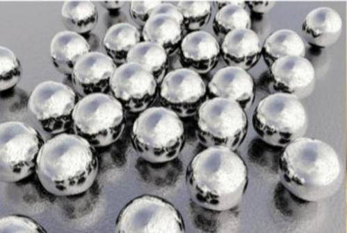
Figure 3 : Silver nanoparticles

  1. silver nanoparticles can be synthesized by several methods like chemical.   reduction. since the chemical reduction methods are economical and easier to  prepare these are most commonly used.19
  1. But the use of chemicals in the synthesis of Ag nanoparticles result in the adsorption of toxic chemicals on the surface of the material so that it will have adverse and harmful effects application.20

Cancer:

A cancer that start in the tissues of the mouthor throat is called oral cancer.21oral cancer is affectedby the various bio-environmental factors such as fatal illness, and in the mouth, it might result in plaques, foulbreath, or ulcers. Makeover, coughing and swallow lymph nodes in the neck are typical signs.22 The Lantana camara Linn plant is being studied for its potential as an oral anticancer because of its inherent ability to treat mouth ulcers. Squamous cell cancer makes up more than 90% of oral and oropharyngeal cancer cases. 23Cancer is a serious hazard to human health, and it causes death to millions of people’s worldwide annually. In Iraq, the cancer registry reported more than twenty thousand newly diagnosed cancer cases in

2012. 24 Hence, green synthesis of AgNPs using leaf extracts of Lantana camara might be an effective drug for oral cacer.The pharmacological activity of L. Camara is shown in Figure 4. 25

       
            Pharmacological activities of L. camara.png
       

Figure 4 :Pharmacological activities of L. camara.

Treatment Of Cancer :

Cancer is a life-threatening disease that is characterized by the continuous growth of cells. There are a number of drugs are available chemically for the treatment of cancer such as imatinib, gefitinib, rituximab, bevacizumab, lapatinib etc. Chemotherapeutic approach followed to kill cancerous cells by inhibiting  the process of cell division. But this approach is successful for highly developed stages of cancer because drugs can reach the site of cancer cells with reduced specifically. Hence the concept of nanoscale devices is designed to develop biodegradable self-assembled nanoparticles, which are used for targeting the delivery of anticancer drugs. 24Nanotechnology is gaining popularity in research that leads to the development of sophisticated, multifunctional, novel approaches which can recognize cancer cells and deliver drugs to target organ or tissue and help to prevent precancerous cells from becoming malignant. 26

Nanotechnology In Reduction of Obesity:

Nanotechnology can be used as a powerful public health tool for the provision of low calorie food which plays an important role in controlling obesity. Nanotechnology based food and products and food packaging materials are provided to consumers in some countries. 27

Nanotechnology In Diagnosis:

Carbon Nanotubes: carbon nanotubes are designed based on bisectors and employed for the detection of analytes in the Healthcare system. It’s also used for monitoring an detection of amino sugars, protein, albumin sugars, amino acids, immunoglobulin, neurotransmitters, insulin and human chronic gonadotropin etc. These are categorized into different types such as single walled, Double-walled and multi walled carbon nanotubes. SWNTs are characterized by strong covalency bonding, one – dimensional structure and nanometer size of 0.4-2 nm. The electrical and mechanical Properties of SWNTs may change due to breaking of C= C bond during chemical processes. DWNTs are made up of a pristine carbon nanotube core and chemically functionalities naotube shells. DWNTs in biological system are used as imaging and therapeutic agents. MWNTs consist of concentric tubes with multiple rolled layers of graphene. 28-29

Graphenes : It consists of thin layer of tightly packed carbon atoms and bonded all together in A hexagonal honeycomb. It is used in diagnostics nd biosensors due to its considerable Properties like high mechanical strength, good thermal conductivity,  elevated elasticity, and optical Properties. It is a transparent substance with a very low production cost and environmentally friendly mainly helpful for the identification of biological samples such s glucose, hemoglobin, cholesterol, dopamine, uric acid. 30-31

Quantum Dots : These sre inorganic nanocrystals which are prepared I. Between 3 and 15 nm and suitable for binding with specific biomolecules. They have unique optical Properties like narrow emission spctra, broad excitation, high photochemical stability, and less photo bleaching. It is used for the development of optical biosensors to identify organic compounds, ions, and biomolecules such as nucleic acids, amino acids, proteins, enzymes, sugars and neurotransmitters. 32-33

Phytochemical Analysis of Lantana Camara: 34

  1. Carbohydrates
  2. Saponin
  3. Tannins
  4. Glycosidic
  5. Flavonoids
  6. Phenols
  7. Proteins
  8. Triterpenoids
  9. Quinolones.

Nanotechnology In Drug Delivery System:

The drug delivery system significantly produces an impact on the use of drugs in patients. This system should minimize the side-effects and also reduce both the dose and frequency of dosage. Due to thir small size and large surface area there is a large saffinity for drugs and small molecules like antibodies or ligands for targeting specific diseases and releasing therapeutic agents at the controlled rate. 35-36

Emerging Scenario of Nanoparticles:

The advancement in naotechnology will lead to innovative synthetic routes along with new processing stratergies with economical manufacturing process. So the required for new drug development in the area of nanotechnology can be reduced which can save humn lives. 37-38  In coming year’s nanotechnology will play a significant role in the health care system to provide an innovative prospect for early detection in diseases, diagnostic and remedial measures to improve health condition and also enabling precise and effective therapy tailored to the patients. 39-40

Current And Future Development:

Currently various investigations are carried out on nanotechnologies to utilize its application in the field of therapeutic, diagnostic and drug delivery systems. In recent times, nano-based drug delivery systems are applied to facilitate the successful delivery of drugs into the target sites. Generally, the main targets in the body system are the receptors or proteins on cell membranes and cell surfaces respctively. Na notechnology will play a key role  to revolutionize medicine in future       developments. The nano robotic tools can be applicable in the treatment of various cardiovascular diseases and atherosclerosis by the year 2028. 41-42

Useful Parts of The Plant:

Lantana camara is ornamental plant, but due to the presence of various phytoconstituents, it has been used as a traditional medicine for an extended time. Generally, the parts use are leaves, flowers, Root, and Whole plant. 43

       
            fig-5,6,7,8.png
       

Phytochemistry Of the Drug:


Table 1 : phytochemistry of the drug44-45


Part

Chemical constituents

Seed

Organic matter, 95.2; crude protein,6.6; ether extract, 2.4; N-Free extract, 55.4; Crude fire,30.7; Total ash,4.8; total phenolic substances, 3.45%.

Aerial part

Triterpenoids, camarilic acid, camaracinic acid, lantadene A and ursolic, betulinic and oleanolic acids .

Leaves

Flavanol glycoside, camaraside, phenylpropanoid glyciside, lantanaside, lantoic acid, lantanilic acid, verbascoside. Lantadene A and B, steroid lancamarone, volatile oil lantanol

Flower

Volatile oil, anthocyanin

Dried flower

Volatile oil, 0.07%

Bark of stem and root

Quinine like alkaloid lantanine.


Traditional Uses:

  • Locally the traditional healers from Dakshin kannada district of karnataka use pounded leaves and leaf juice to manage cuts, ulcers, and swellings.
  • Taila is prepares by adding the decoction of both Lantana camara leaves, and Eupatorium odoratum is used for external application for the wound.
  • The decoction of leaves and fruits is used as a lotion for wounds.
  • Decoction of the leaves is used for the vitiated condition of vata and kapha.
  • In case of eczema and eruption, a decoction of the leaves is used internally in the dose of 10-20ml twice a day, along with an external application of leaf paste.
  • The leaf juice /decotion is used for gargling in mouth ulcers.
  • Malaria-leaf juice internally in the dose of 20ml twice daily.
  • For sparin and contusion, pounded fresh leaves are applied as a poultice.
  • For rheumatism, oil is spread on leaves, warmed, and applied to the affected part.46-47

Pharmacological Activites of Lantana Camara L.:

       
            Lantana camara and its compounds.png
       

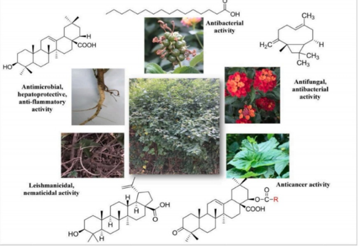
Figure 5 : Lantana camara and its compounds.

Antibacterial activity:

Ethanolic extracts of Lantana camara leaves and roots were reported for antibacterial activity. Microdilution method is performed for in-vitro antibacterial activity. The extracts exhibited antimicrobial activity against stephyloccus aureus, proteus vulgaris, pseudomonas aeruginosa, Escherichia coli, Vibrio cholareae and two multiresistant stains E.coli and S. aureus.

Antifungal activity:

The lantana camara extracts of hot water and ethanol is screened for its activity against wood destroying white and brown fungi. Through the both extracts resulted in efficient white and brown antifungal activity. But, the ethanol extract at very low concentration about 0.01% was showned to have highly potential antifungal activity.

Hemolytic activity:

The hemolytic activity of lantana camarais perfomed by using aqueous extract. The solvent fractions of different concentrations were taken [125,250,500,1000 µg/ml] using spectroscopic method. The aqueous extract and its solvent fractions exihibited very low hemolytic activity towards human erythrocytes. The haemolytic activity of the different extracts was found in order : chloroform fraction > hexane and ethyl acetate frection > aqueous extract > ethanol fraction  > methanol fraction.

Antimotility activity:

Methanol extract of Lantana. camara leaves was reported antimotility activity in mice. Intestinal motility assayed by charcoal meal test in mice. A dose of 1 g/kg body weight extract completely inhibited the transit of charcoal in mice. Intraperitoneal administration of 125mg/kg and 250 mg/kg body weight the extracts significantly reduced the fecal output in castor oil induced diarrhoea in mice.

Anti mutagenic activity:

22?-dimethylacryloyloxy and 22?- acidacetoloxy lantanolic acid from L. camara showed antimutagenic activity. The antimutagenicity test performed by micro-nucleus test in Swiss mice. High anti mutagenic activity is exhibited by both of the compounds in Mitomycin - C induced mutagenesis in mice.

Antioxidant activity:

The leaves of Lantana camara was reported by reducing power activity and 1,1- diphenyl-2- picrylhydrazyl by radical scavenging assay. The antioxidant activity is exhibited by the leaves. .younger leaves shown to exhibit stronger antioxidant activity when compared to matured or older leaves.

Antiurolithiatic activity:

The leaves of Lantana camara ethanolic extract was reported for antiurolithiatic activity against ammonium and chloride ethylene glycol induced calcium oxalate urolithiasis in male albino rats. Extract treatment significantly reduced the deposition of calcium; oxalate and also decreased urinary excretion of calcium; oxalate and creatinine.

Mosquito controlling activity:

Mosquito larvicidal activity of ethanol and methanol extracts of leaves and flowers of Lantana camara were reported against 3rd and 4th instar larvae of Ae. aegypti and Cx. quinquefasciatus mosquito. Both extracts exhibited significant larvicidal activity against both species of mosquitoes; however, at low concentrations extracts were highly active against Ae. aegypti than that of Cx. Quinquefasciatus.48

Toxicity Of Lantana Camara:

Lantana is one among the most toxic plants known so far possibly within top ten. Reports of lantana camara toxicity have been sated from India. America, Australia, New Zealand, South Africa. The consumption of high amount of plants material leads to toxicity. It is reported that goats, sheep and cattle are susceptible to lantadenes A,B,C and iatrogenic acid toxicity were as rats, horses, neonatal calves and lambs are not susceptible to lantadene A. The prominent clinical sigh of poisoning includes jaundice and photosensitization. Loss of appetite in poisoned animals occurs within 24-prominent clinical sign of in appetite is also observed, most severely poisoned animals die within 2-days of poisoning but usually death occurs after 1-3 weeks after poisoning. The kidneys are pale in colour and swollen, the gall bladder is grossly distended and the liver is enlarged. The oral toxic dose of lantadene A for sheep is 60mg/kg is toxic and 1-3 mg/kg by intravenous route.49-50

CONCLUSION:

Due to the advancement of science and technology, there is a tendency to ignore traditional knowledge and medicine. Documantation of this valuable traditional knowledge through ethnobotanical studies is essential for conserving natural resources. Lantana camara L. Belonging to the family Verbenaceae, is considered an invasive weed worldwide. It is quite obvious that from this review Lantana camara used for numerous therapeutic purposes different types of nanoparticles extracted from this even individual part have own therapeutic benefits such as cardiovascular diseases, atherosclerosis anti cancer, anti viral etc. Nanotechnology will play a key role to revolutionize medicine in further development.

REFERENCES

  1. Sanjeeb Kalita GK, Loganathan K, KokatiVenkataBhaskara R.A Review on Medicinal Properties ofLantanacamara Linn. J Pharm and Tech. 2012; 5(6):711-715.
  2. Sharma OP, et al. A review of the hepatotoxic plantLantanacamara .Crit Rev Toxicol. 2007; 37(4):313- 52. https://doi.org/10.1080/10408440601177863
  3. Ghisalberti EL, Lantana camara L. Verbenaceae. 2000; 71(5):467-486.https://doi.org/10.1016/S0367-326X(00)00202-1
  4. Sharma, G.P.; Raghubanshi, A.S.; Singh, J.S. Lantana invasion: An overview. Weed Biol. Manag. 2005, 5, 157–165. [CrossRef]
  5. Negi, G.C.S.; Sharma, S.; Vishvakarma, S.C.R.; Samant, S.S.; Maikhuri, R.K.; Prasad, R.C.; Palni, L.M.S. Ecology and Use of AlLantanacamara in India. Bot. Rev. 2019, 85, 109–130. [CrossRef]
  6. Mishra A: Allelopathic properties of Lantana camara, a review article.2014;32-52.
  7. Udayan PS & Indira Balachandran, Medicinal plants of Arya Vaidya Sala Herb Garden, Second impression; Department of Publication Arya Vaidya Sala Kottakkal, Kerala; 2011. P.222.
  8. B. L. Cushing, V. L. Kolesnichenko, and C. J. O’Connor, “Recent advances in the liquid-phase syntheses of inorganic nanoparticles,” Chemical Reviews, vol. 104, no. 9, pp. 3893– 3946, 2004.
  9. B. Ajitha, Y. Ashok Kumar Reddy, and P. Sreedhara Reddy, “Green synthesis and characterization of silver nanoparticles using Lantana camara leaf extract,” Materials Science and Engineering: C, vol. 49, pp. 373–381, 2015.
  10. A. Feng, S. Wu, S. Chen, H. Zhang, W. Shao, and Z. Xiao, “Synthesis of silver nanoparticles with tunable morphologies via a reverse nano-emulsion route,” Materials Transactions, vol. 54, no. 7, pp. 1145–1148, 2013
  11. M. Moreira dos Santos, M. JoãoQueiroz, and P. V. Baptista, “Enhancement of antibiotic effect via gold:silver-alloy nanoparticles,” Journal of Nanoparticle Research, vol. 14, no. 5, pp. 859–866, 2012.
  12. H. F. Aritonang, D. Onggo, C. Ciptati, and C. L. Radiman, “Synthesis of platinum nanoparticles from K2PtCl4 solution using bacterial cellulose matrix,” Journal of Nanoparticles, vol. 2014, Article ID 285954, 6 pages, 2014.
  13. H. F. Aritonang, D. Onggo, C. Ciptati, and C. L. Radiman, “Insertion of platinum particles in bacterial cellulose membranes from PtCl4 and H2PtCl6 precursors,” Macromolecular Symposia, vol. 353, no. 1, pp. 55-56, 2015.
  14. H. F. Aritonang, V. S. Kamu, C. Ciptati, D. Onggo, and C. L. Radiman, “Performance of platinum nanoparticles/ multiwalled carbon nanotubes/bacterial cellulose composite as anode catalyst for proton exchange membrane fuel cells,” Bulletin of Chemical Reaction Engineering & Catalysis, vol. 12, no. 2, pp. 287–292, 2017.
  15. R. W. Raut, A. S. M. Haroon, Y. S. Malaghe, B. T. Nikan, and S. B. Kashid, “Rapid biosynthesis of platinum and palladium Metal nanoparticles using root extract of Asparagus racemosus Linn.,” Advanced Materials Letters, vol. 4, no. 8, pp. 650–654, 2013.
  16. B. L. Cushing, V. L. Kolesnichenko, and C. J. O’Connor, “Recent advances in the liquid-phase syntheses of inorganic nanoparticles,” Chemical Reviews, vol. 104, no. 9, pp. 3893–3946, 2004
  17. E. McGillicuddy, I. Murray, S. Kavanagh et al., “Silver nanoparticles in the environment: sources, detection and ecotoxicology,” Science of the Total Environment, vol. 575, pp. 231–246, 2017
  18. M. Akter, M. T. Sikder, M. M. Rahman et al., “A systematic review on silver nanoparticles-induce cytotoxicity: physicochemical properties and perspectives,” Journal of Advanced Research, vol. 9, pp. 1–16, 2018.
  19. A. Zielinska, E. Skwarek, A. Zaleska, M. Gazda, and J. Hupka, “Preparation of silver nanoparticles with controlled particles size,” Procedia Chemistry, vol. 1, no. 2, pp. 1560–1566, 22009.
  20. A. Singh, D. Jain, M. K. Upadhyay, N. Khandelwal, and D. H. N. Verma, “Green synthesis of silver nanoparticles using Argenonemexicana leaf extract and their charactrization,” Digest Journal of Nanomaterials and Biostructures, vol. 6, no. 1, pp. 483–489, 2010.
  21. Montero PH, Patel SG. Cancer of the oral cavity. SurgOncolClin N Am. 2015; 24(3):491-508. https://doi.Org/10.1016/j.soc.2015.03.006
  22. D’Souza S, Addepalli V, et al. Preventive measures In oral cancer: An overview. Biomed Pharmacother. 2018; 107:72-80. https://doi.org/10.1016/j. Biopha.2018.07.114
  23. Wong T, Wiesenfeld D. Oral Cancer. Aust Dent J. 2018; 63(1):S91-S99. https://doi.org/10.1111/adj.12594
  24. N.A. Alwan, Breast cancer among Iraqi women: preliminaryFindings from a regional comparative Breast Cancer Research Project, J. Glob. Oncol. 2 (2016) 255e258
  25. D’Souza S, Addepalli V, et al. Preventive measures In oral cancer: An overview. Biomed Pharmacother. 2018; 107:72-80. https://doi.org/10.1016/j. Biopha.2018.07.114
  26. Boisselier E, Astruc D. Gold nanoparticles in nanomedicine: preparations, imaging, diagnostics, therapies and toxicity. Chem Soc Rev 2009; 38(6): 1759-82.
  27. Brigger I, Dubernet C, Couvreur P. Nanoparticles in cancer therapy and diagnosis. AdvDrugDelivRev2002;54(5):631-51.
  28. Jayaraj RL, Chandramohan V. Nanomedicines for Parkinson disease: current status and future perspective. Int J Pharm Bio Sci 2013; 4(1): 692-04.
  29. Lin JH, Lu AY. Role of pharmacokinetics and metabolism in drug discovery and development. Pharmacol Rev 1997; 49(4): 403-49. PMID: 9443165
  30. Li SD, Huang L. Pharmacokinetics and biodistribution of nanoparticles. Mol Pharm 2008; 5(4): 496-504.
  31. Mamo T, Moseman EA, Kolishetti N, et al. Emerging nanotechnology approaches for HIV/AIDS treatment and Prevention. Nanomedicine (Lond) 2010; 5(2):269-85.
  32. Mittal G, Kumar MN. Impact of polymeric nanoparticles on oral pharmacokinetics: a dosedependent case study With estradiol. J Pharm Sci 2009; 98(10): 3730-4.
  33. Moghimi SM, Hunter AC, Murray JC. Nanomedicine: current status and future prospects. FASEB J 2005; 19(3): 311-30.
  34. Biswa mohan sahool.B.V.V Ravikumar,ch.Niranjan patral, J.R Pandal, Bibhash C.Mohanta and Narahari N..Palei Nanotechnology:A Novel Approach For Drug Development In Health Care System Current Nanomaterials,(5)2020.
  35. Marshall, N. J., C. J. Goodwin, and S. J. Holt. “A critical assessment of the use of microculturetetrazolium Assays to measure cell growth and function.” Growth regulation 5, no. 2 (1995): 69-84.
  36. Pham CT. Nanotherapeutic approaches for the treatment of rheumatoid arthritis. Wiley Interdiscip RevNanomed Nanobiotechnology 2011; 3(6): 607-19.
  37. Sinha R, Kim GJ, Nie S, Shin DM. Nanotechnology in cancer therapeutics: bioconjugated nanoparticlesfor drug delivery. Mol Cancer Ther 2006; 5(8): 1909- 1DD
  38. Rosi NL, Mirkin CA. Nanostructures in biodiagnostics. Chem Rev 2005; 105(4): 1547-62.
  39. Deb S, Ghosh K, Shetty SD. Nanoimaging in cardiovascular diseases: Current state of the art. Indian JMedRes2015;141(3):285-98.
  40. Timko B. Advances in drug delivery. Annu Rev Mater Res 2011; 41: 1-20.
  41. Varshney HM, Rajnish K, Shailender M. Novel approaches for insulin delivery: current status. Int J of Therap Appl 2012; 7: 25-31.
  42. Vashist SK, Zheng D, Al-Rubeaan K, Luong JHT, Sheu FS. Advances in carbon nanotube based electrochemical sensors for bioanalytical applications. Biotechnol Adv 2011; 29(2): 169-88.
  43. Patil M, Mehta DS, Guvva S. Future impact of nanotechnology on medicine and dentistry. J Indian Soc Periodontol 2008; 12(2): 34-40.
  44. Warrier PK, Nambiar VPK, Ramankutty C, editors, Indian Medicinal Plants, vol-3, Orient Longman;1996. p. 300.
  45. Dr KS Krishnan Marg, Wealth of India second supplement series, National institute of science communication and information resources; 2007. p. 116.
  46. SN Yoga Narasimhan, Medicinal plants of India, Tamil Nadu, Vol-2, V. Srinivasan and N. Kosal Ram of CyberMedia, Bangalore; 2000. p. 315
  47. Warrier PK, Nambiar VPK, Ramankutty C, editors, Indian Medicinal Plants, vol-3, Orient Longman;1996. p. 300.
  48. DeFilipps RA, Maina SL, Crepin J. Medicinal Plants of the Guianas (Guyana, Surinam, French Guiana); Department of Botany, National Museum of Natural History, Smithsonian Institution: Washington, DC, USA, 2004.
  49. Sharma OP et al. A review of the toxicity of Lantana camara (Linn) in animals. Clinical Toxicology. 18 (9); 1981: 1077-1094.
  50. Sharma OP, Makkar HPS and Dawra RK. A review of the noxious plant Lantana camara. Toxicon. 26 (11); 1988: 975-987.

Reference

  1. Sanjeeb Kalita GK, Loganathan K, KokatiVenkataBhaskara R.A Review on Medicinal Properties ofLantanacamara Linn. J Pharm and Tech. 2012; 5(6):711-715.
  2. Sharma OP, et al. A review of the hepatotoxic plantLantanacamara .Crit Rev Toxicol. 2007; 37(4):313- 52. https://doi.org/10.1080/10408440601177863
  3. Ghisalberti EL, Lantana camara L. Verbenaceae. 2000; 71(5):467-486.https://doi.org/10.1016/S0367-326X(00)00202-1
  4. Sharma, G.P.; Raghubanshi, A.S.; Singh, J.S. Lantana invasion: An overview. Weed Biol. Manag. 2005, 5, 157–165. [CrossRef]
  5. Negi, G.C.S.; Sharma, S.; Vishvakarma, S.C.R.; Samant, S.S.; Maikhuri, R.K.; Prasad, R.C.; Palni, L.M.S. Ecology and Use of AlLantanacamara in India. Bot. Rev. 2019, 85, 109–130. [CrossRef]
  6. Mishra A: Allelopathic properties of Lantana camara, a review article.2014;32-52.
  7. Udayan PS & Indira Balachandran, Medicinal plants of Arya Vaidya Sala Herb Garden, Second impression; Department of Publication Arya Vaidya Sala Kottakkal, Kerala; 2011. P.222.
  8. B. L. Cushing, V. L. Kolesnichenko, and C. J. O’Connor, “Recent advances in the liquid-phase syntheses of inorganic nanoparticles,” Chemical Reviews, vol. 104, no. 9, pp. 3893– 3946, 2004.
  9. B. Ajitha, Y. Ashok Kumar Reddy, and P. Sreedhara Reddy, “Green synthesis and characterization of silver nanoparticles using Lantana camara leaf extract,” Materials Science and Engineering: C, vol. 49, pp. 373–381, 2015.
  10. A. Feng, S. Wu, S. Chen, H. Zhang, W. Shao, and Z. Xiao, “Synthesis of silver nanoparticles with tunable morphologies via a reverse nano-emulsion route,” Materials Transactions, vol. 54, no. 7, pp. 1145–1148, 2013
  11. M. Moreira dos Santos, M. JoãoQueiroz, and P. V. Baptista, “Enhancement of antibiotic effect via gold:silver-alloy nanoparticles,” Journal of Nanoparticle Research, vol. 14, no. 5, pp. 859–866, 2012.
  12. H. F. Aritonang, D. Onggo, C. Ciptati, and C. L. Radiman, “Synthesis of platinum nanoparticles from K2PtCl4 solution using bacterial cellulose matrix,” Journal of Nanoparticles, vol. 2014, Article ID 285954, 6 pages, 2014.
  13. H. F. Aritonang, D. Onggo, C. Ciptati, and C. L. Radiman, “Insertion of platinum particles in bacterial cellulose membranes from PtCl4 and H2PtCl6 precursors,” Macromolecular Symposia, vol. 353, no. 1, pp. 55-56, 2015.
  14. H. F. Aritonang, V. S. Kamu, C. Ciptati, D. Onggo, and C. L. Radiman, “Performance of platinum nanoparticles/ multiwalled carbon nanotubes/bacterial cellulose composite as anode catalyst for proton exchange membrane fuel cells,” Bulletin of Chemical Reaction Engineering & Catalysis, vol. 12, no. 2, pp. 287–292, 2017.
  15. R. W. Raut, A. S. M. Haroon, Y. S. Malaghe, B. T. Nikan, and S. B. Kashid, “Rapid biosynthesis of platinum and palladium Metal nanoparticles using root extract of Asparagus racemosus Linn.,” Advanced Materials Letters, vol. 4, no. 8, pp. 650–654, 2013.
  16. B. L. Cushing, V. L. Kolesnichenko, and C. J. O’Connor, “Recent advances in the liquid-phase syntheses of inorganic nanoparticles,” Chemical Reviews, vol. 104, no. 9, pp. 3893–3946, 2004
  17. E. McGillicuddy, I. Murray, S. Kavanagh et al., “Silver nanoparticles in the environment: sources, detection and ecotoxicology,” Science of the Total Environment, vol. 575, pp. 231–246, 2017
  18. M. Akter, M. T. Sikder, M. M. Rahman et al., “A systematic review on silver nanoparticles-induce cytotoxicity: physicochemical properties and perspectives,” Journal of Advanced Research, vol. 9, pp. 1–16, 2018.
  19. A. Zielinska, E. Skwarek, A. Zaleska, M. Gazda, and J. Hupka, “Preparation of silver nanoparticles with controlled particles size,” Procedia Chemistry, vol. 1, no. 2, pp. 1560–1566, 22009.
  20. A. Singh, D. Jain, M. K. Upadhyay, N. Khandelwal, and D. H. N. Verma, “Green synthesis of silver nanoparticles using Argenonemexicana leaf extract and their charactrization,” Digest Journal of Nanomaterials and Biostructures, vol. 6, no. 1, pp. 483–489, 2010.
  21. Montero PH, Patel SG. Cancer of the oral cavity. SurgOncolClin N Am. 2015; 24(3):491-508. https://doi.Org/10.1016/j.soc.2015.03.006
  22. D’Souza S, Addepalli V, et al. Preventive measures In oral cancer: An overview. Biomed Pharmacother. 2018; 107:72-80. https://doi.org/10.1016/j. Biopha.2018.07.114
  23. Wong T, Wiesenfeld D. Oral Cancer. Aust Dent J. 2018; 63(1):S91-S99. https://doi.org/10.1111/adj.12594
  24. N.A. Alwan, Breast cancer among Iraqi women: preliminaryFindings from a regional comparative Breast Cancer Research Project, J. Glob. Oncol. 2 (2016) 255e258
  25. D’Souza S, Addepalli V, et al. Preventive measures In oral cancer: An overview. Biomed Pharmacother. 2018; 107:72-80. https://doi.org/10.1016/j. Biopha.2018.07.114
  26. Boisselier E, Astruc D. Gold nanoparticles in nanomedicine: preparations, imaging, diagnostics, therapies and toxicity. Chem Soc Rev 2009; 38(6): 1759-82.
  27. Brigger I, Dubernet C, Couvreur P. Nanoparticles in cancer therapy and diagnosis. AdvDrugDelivRev2002;54(5):631-51.
  28. Jayaraj RL, Chandramohan V. Nanomedicines for Parkinson disease: current status and future perspective. Int J Pharm Bio Sci 2013; 4(1): 692-04.
  29. Lin JH, Lu AY. Role of pharmacokinetics and metabolism in drug discovery and development. Pharmacol Rev 1997; 49(4): 403-49. PMID: 9443165
  30. Li SD, Huang L. Pharmacokinetics and biodistribution of nanoparticles. Mol Pharm 2008; 5(4): 496-504.
  31. Mamo T, Moseman EA, Kolishetti N, et al. Emerging nanotechnology approaches for HIV/AIDS treatment and Prevention. Nanomedicine (Lond) 2010; 5(2):269-85.
  32. Mittal G, Kumar MN. Impact of polymeric nanoparticles on oral pharmacokinetics: a dosedependent case study With estradiol. J Pharm Sci 2009; 98(10): 3730-4.
  33. Moghimi SM, Hunter AC, Murray JC. Nanomedicine: current status and future prospects. FASEB J 2005; 19(3): 311-30.
  34. Biswa mohan sahool.B.V.V Ravikumar,ch.Niranjan patral, J.R Pandal, Bibhash C.Mohanta and Narahari N..Palei Nanotechnology:A Novel Approach For Drug Development In Health Care System Current Nanomaterials,(5)2020.
  35. Marshall, N. J., C. J. Goodwin, and S. J. Holt. “A critical assessment of the use of microculturetetrazolium Assays to measure cell growth and function.” Growth regulation 5, no. 2 (1995): 69-84.
  36. Pham CT. Nanotherapeutic approaches for the treatment of rheumatoid arthritis. Wiley Interdiscip RevNanomed Nanobiotechnology 2011; 3(6): 607-19.
  37. Sinha R, Kim GJ, Nie S, Shin DM. Nanotechnology in cancer therapeutics: bioconjugated nanoparticlesfor drug delivery. Mol Cancer Ther 2006; 5(8): 1909- 1DD
  38. Rosi NL, Mirkin CA. Nanostructures in biodiagnostics. Chem Rev 2005; 105(4): 1547-62.
  39. Deb S, Ghosh K, Shetty SD. Nanoimaging in cardiovascular diseases: Current state of the art. Indian JMedRes2015;141(3):285-98.
  40. Timko B. Advances in drug delivery. Annu Rev Mater Res 2011; 41: 1-20.
  41. Varshney HM, Rajnish K, Shailender M. Novel approaches for insulin delivery: current status. Int J of Therap Appl 2012; 7: 25-31.
  42. Vashist SK, Zheng D, Al-Rubeaan K, Luong JHT, Sheu FS. Advances in carbon nanotube based electrochemical sensors for bioanalytical applications. Biotechnol Adv 2011; 29(2): 169-88.
  43. Patil M, Mehta DS, Guvva S. Future impact of nanotechnology on medicine and dentistry. J Indian Soc Periodontol 2008; 12(2): 34-40.
  44. Warrier PK, Nambiar VPK, Ramankutty C, editors, Indian Medicinal Plants, vol-3, Orient Longman;1996. p. 300.
  45. Dr KS Krishnan Marg, Wealth of India second supplement series, National institute of science communication and information resources; 2007. p. 116.
  46. SN Yoga Narasimhan, Medicinal plants of India, Tamil Nadu, Vol-2, V. Srinivasan and N. Kosal Ram of CyberMedia, Bangalore; 2000. p. 315
  47. Warrier PK, Nambiar VPK, Ramankutty C, editors, Indian Medicinal Plants, vol-3, Orient Longman;1996. p. 300.
  48. DeFilipps RA, Maina SL, Crepin J. Medicinal Plants of the Guianas (Guyana, Surinam, French Guiana); Department of Botany, National Museum of Natural History, Smithsonian Institution: Washington, DC, USA, 2004.
  49. Sharma OP et al. A review of the toxicity of Lantana camara (Linn) in animals. Clinical Toxicology. 18 (9); 1981: 1077-1094.
  50. Sharma OP, Makkar HPS and Dawra RK. A review of the noxious plant Lantana camara. Toxicon. 26 (11); 1988: 975-987.

Photo
Komal Dumale
Corresponding author

UG Scholar of Rashtriya College of Pharmacy Hatnoor, Tq. Kannad Dist. Chh. Sambhajinagar Maharashtra, India-431103

Photo
Trupti Mokase
Co-author

UG Scholar of Rashtriya College of Pharmacy Hatnoor, Tq. Kannad Dist. Chh. Sambhajinagar Maharashtra, India-431103

Photo
Vaishnavi Raut
Co-author

UG Scholar of Rashtriya College of Pharmacy Hatnoor, Tq. Kannad Dist. Chh. Sambhajinagar Maharashtra, India-431103

Komal Dumale*, Trupti Mokase, Vaishnavi Raut, Anticancer Potential of Silver Nanoparticles by Using Lantana Camara Leaf, Int. J. of Pharm. Sci., 2025, Vol 3, Issue 1, 2047-2056. https://doi.org/10.5281/zenodo.14729430

More related articles
Comprehensive Review of Quality by Design (Qbd)...
Prajapati Rahul Vinitbhai, Devendrakumar D. Vaghela, Drashti C. P...
Design Development And Evaluation Of Floating Drug...
Shiv Sagar Mahapatra, Akanksha Patel, Manmath Purohit, ...
Formulation and Evaluation of Floating Microspheres Containing Non-Steroidal Ant...
Muggu Sankara bhavani, K. ChandraSekhar Gupta, A. Brahma Reddy, G. Venkata Vasu, ...
Therapeutic Applications of Corn Silk, a Versatile Natural Medicine: A Comprehe...
Amey Dongaonkar, Dadaso Mane, Gajanan Sormare, Milind Umekar, Nishant Awandekar , ...
Review on phytochemical and Pharmacological Investigation of Syzygium Guineense ...
Kokate Rohit, Kharbe Vitthal, Kirtankar Tejaswini, Lashkar Priti, Sumit S. Bodkhe, ...
Related Articles
A Detailed Review on The Use of Excipients in Drug Formulation...
Sakshi Wakchaure, Swapnil Kale, Shraddha Shelke , Sneha S. Kanase, Harshada Thombare , ...
Design And Characterization Of Polyherbal Single Use Soap For It’s Antibacteri...
Ankit P. Gondane, Samir R. Pakhidde, Sanket R. Ganvir , Tushar C. Bisen, Devesh R. Chourasia, ...
Formulation and Evaluation of Herbal Liquid Toner by Using Cymbopogon citratus a...
Deepa Chandrawanshi, Rutu Chaudhari , Tanu Hatel , Sayli Turkar , Swati Meshram , ...
Comprehensive Review of Quality by Design (Qbd)...
Prajapati Rahul Vinitbhai, Devendrakumar D. Vaghela, Drashti C. Patel, Dave Kunj Narendrabhai, Shah ...
More related articles
Comprehensive Review of Quality by Design (Qbd)...
Prajapati Rahul Vinitbhai, Devendrakumar D. Vaghela, Drashti C. Patel, Dave Kunj Narendrabhai, Shah ...
Design Development And Evaluation Of Floating Drug Delivery System For Lornoxica...
Shiv Sagar Mahapatra, Akanksha Patel, Manmath Purohit, ...
Comprehensive Review of Quality by Design (Qbd)...
Prajapati Rahul Vinitbhai, Devendrakumar D. Vaghela, Drashti C. Patel, Dave Kunj Narendrabhai, Shah ...
Design Development And Evaluation Of Floating Drug Delivery System For Lornoxica...
Shiv Sagar Mahapatra, Akanksha Patel, Manmath Purohit, ...