View Article

  • Antibacterial Evaluation of Jatropha spinosa Latex Extracts.

  • 1 Department of Pharmacognosy & Medicinal Chemistry, Faculty of Clinical Pharmacy, 21 September University of Medical and Applied Sciences, Sana’a, Yemen. 
    1,2,3,6,7,8,9 Department of Pharmacy -Faculty of Medical Science- Civilization University.
    2 Medicinal Chemistry and Drug Design, Department of Medicinal Chemistry, Faculty of Pharmacy, Sana'a University, Sana'a, Yemen.
    4 Department of Oral Biology, Faculty of Dental Medicine, Universitas Airlangga, Surabaya, Indonesia.
    5 Department of Oral Medicine, Faculty of Dental Medicine, Universitas Airlangga, Surabaya, Indonesia.

Abstract

This plant has been considered a medicinal plant in Yemen's traditional medicine, used as an antiseptic for wounds. This present study was carried out to evaluate the antibacterial properties of hexane, chloroform, butanol, and aqueous extracts of Jatropha spinosa latex. Agar disc diffusion tests were carried out to determine the antimicrobial effects of the Jatropha spinosa latex extracts against Gram-positive (B. cereus, S. aureus, and S. pyogenes), Gram-negative (Klebsiella, E. coli, and P. aeruginosa) bacterial strains. Among the various extracts, the hexane extract of Jatropha spinosa latex has the strongest antibacterial potency against Staphylococcus aureus (gram-positive), and the Butanol extract showed the highest effect on S. aureus, S. pyogenes, and Klebsiella at a higher concentration of 1mg/1ml with zones of inhibition 11.26mm, 11.53mm, and 11.6mm, respectively. Water extract showed the maximum (11.13± 1.02mm) inhibitory effect against Klebsiella at a higher concentration of 1mg/1ml, and no activity was observed at all concentrations against the Gram-positive S. pyogenes bacteria strain. The chloroform extract has potent antibacterial activity against the Gram-positive S. aureus and Gram-negative P. aeruginosa bacterial strains at a concentration 1mg\1ml with zone inhibition of 10.53mm and 10.83mm. The positive control Beta–sitosterol 0.25% W/W ointment did not show inhibitory effect against four tested bacteria strains (S. aureus, S. pyogenes, Klebsiella, and E. coli) at all concentrations. It has exhibited lesser inhibitory potency when compared with the Jatropha spinosa latex extracts. The present study has revealed that the J. spinosa latex extracts are a potential antimicrobial agent having a broad-spectrum activity.

Keywords

Jatropha spinosa, Antibacterial activity, Gram-positive bacteria, Gram-negative bacteria, Traditional medicine.

Introduction

Jatropha spinosa belongs to the family Euphorbiaceae, which comprises about 340 genera and more than 8000 species, mainly distributed in tropical regions across various habitats [1–4]. This family is considered one of the most important in Yemen, with over 106 species, including a high proportion of endemics [5,6]. Euphorbiaceae species have traditionally been used in Yemen for treating skin and infectious diseases [7,8], and in other regions of the world, they have also been applied in folk medicine for a wide range of disorders such as tumors, malaria, gastrointestinal and respiratory conditions, as well as renal, hepatic, rheumatic, and neurological ailments [9–10]. The genus Jatropha comprises about 175 species distributed in tropical regions worldwide [11]. The name Jatropha originates from the Greek words Jatros (physician) and trophe (food), reflecting its long-standing medicinal and nutritional value. The latex of several Jatropha species has been reported to possess pharmacological activities, including hemostatic, antibacterial, antifungal, antimalarial, antiparasitic, and insecticidal effects [12]. Traditionally, Jatropha trees have been utilized in Asia, Africa, and Latin America for the treatment of various ailments [13]. Their bioactivity is attributed to the presence of diverse phytochemicals such as terpenoids, tannins, and flavonoids, which contribute to their antimicrobial and therapeutic potential [14,15]. More broadly, medicinal plants have played a central role in human health care throughout history. They have been used for treating diseases, preventing epidemics, and preserving food [16]. Many modern drugs have been derived from these plants, and they continue to provide important leads for pharmacological development [17]. Previous research has shown that many medicinal plants have pharmacological qualities such as antibacterial, anti-inflammatory, anti-diarrheal, anti-ulcer, antioxidant, gastroprotective, and anti-constipation activities [18-22]. Millions of infectious or toxic diseases are caused by bacteria [23, 24], and approximately 80% of the global population still relies on plant-based traditional medicine for primary healthcare needs [25]. Therefore, the present study was designed to investigate the antibacterial activity of Jatropha spinosa latex extracts as part of ongoing research on Yemeni medicinal plants. This work aims to validate the traditional use of the species in folk medicine, assess its potential as a source of new antibacterial agents against resistant wound pathogens, and compare its efficacy with standard pharmaceutical drugs. Ultimately, the study seeks to contribute to the development of pharmaceutical formulations containing bioactive compounds from J. spinosa that may provide effective and competitive alternatives in the drug market.

MATERIALS AND METHODS

Plant material

The materials were collected from the Al-Wagah region at Taiz governorate, Yemen, especially in the rainy season. The plant sap (latex) collection was gathered by the aerial part of the stem injury using a sterile surgical blade, which was collected into a small glass bottle of about 40ml and transferred to the university laboratory.

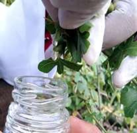
Figure 1. Extraction of J. spinosa latex.

Extraction

The 50ml of distilled water was added to the latex and put into a separating funnel, and 100ml of hexane solvent was added, and then shaken for 3 minutes, and the mixture was placed for 24 hours.

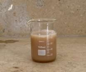
Figure 2. Distal water with latex.

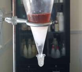
Figure 3. Hexane extraction.

Then, the hexane layer (lower layer) was separated from the other layers of the mixture. The 100ml of hexane solvent was again added to the mixture in a separating funnel and shaken for 3 minutes. After 24 hours, the hexane layer was separated from the other layers of the mixture. The 100ml of chloroform solvent was added to the mixture and shaken for 3 minutes, and the chloroform layer was separated after 24 hours. We added 75ml of butanol solvent, and the butanol layer was separated after 24 hours. This process was repeated three times to obtain a large amount of the extracts.

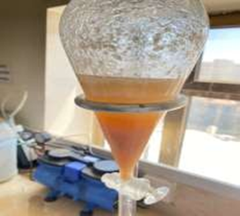
Figure 4. Chloroform extraction.

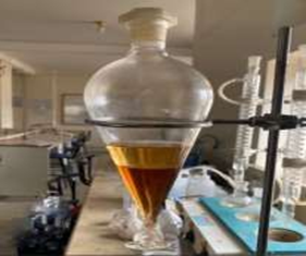
Figure 5. Butanol extraction.

All extracts were concentrated used rotary evaporator for antibacterial activity tests.

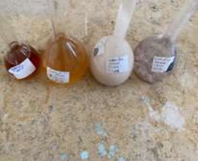
Figure 6. Extracts of J. spinosa

Disc diffusion method

Antibacterial activity of the tested plant extracts was carried out by the disc diffusion method [26]. First, the extracts of the plant tested were dissolved in DMSO at a concentration of 1mg/ml, 1mg/2ml, 1mg/3ml, and 1mg/4ml. Then, 100 µL of bacterial inoculums containing 10? CFU/ml were spread over plates containing Mueller Hinton agar, and discs (5 mm in diameter) impregnated with different concentrations of the plant extract solution as follows, were placed on the surface of the media. Also, we used reference four control discs containing DMSO and Meburn® (0.25 % disc) with different concentrations dissolved in DMSO: 1mg/ml, 2mg/ml, 3mg/ml, and 4mg/ml. The plates were incubated for 24 h at 37 ?. The diameters of the inhibition zones were measured, and antibacterial activity was assessed based on the inhibition zone diameters in mm [27]. This was applied three times for G+ve bacteria S. aureus, S. pyogenes, and B. cereus, and for G-ve bacteria Klebsiella, E. coli, and P. aeruginosa.

RESULTS AND DISCUSSION

The antimicrobial activities of Jatropha spinosa latex (hexane, chloroform, butanol, and water) extracts against the microorganisms examined in the present study, and their potency, were tested by the presence or absence of inhibition zones and zone diameter, and the results are given in Tables 1-4.

The results of Jatropha spinosa latex extracts showed increased activity with increasing concentration against all strains of Gram-positive and Gram-negative bacteria. The data indicated that S. aureus was the most sensitive strain tested with the hexane extract of Jatropha spinosa latex, which produced the largest inhibition zone, 14.3 mm at 1mg?1ml Table 1.     

Table 1. Zone inhibition of the Hexane extract

Zone of Inhibitor (mm)

Microorganism

Hexane Extracts

1mg\1ml

1mg\2ml

1mg\3ml

1mg\4ml

S. aureus

14.3±1.52

8.6±0.57

5.56±0.51

-

S. pyogene

10.66±1.52

10.40±0.60

-

-

Klebsiella

11.46±0.50

7.46±0.5

5.33±0.57

-

P. aeruginosa

8.76±0.20

6.1±0.15

-

-

E. coli

10.50±0.50

7.2±1.05

-

-

B. cereus

7.83±0.15

5.7±0.64

-

-

The highest activity of Jatropha spinosa water extract was observed against Klebsiella, with an inhibition zone of 11.13 mm at 1mg?1ml Table 2.

Table 2. Zone inhibition of the Water extract.

Zone of Inhibitor (mm)

Microorganism

Water Extracts

1mg\1ml

1mg\2ml

1mg\3ml

1mg\4ml

S. aureus

9.5±0.5

5.7±0.4

-

-

S. pyogene

-

-

-

-

Klebsiella

11.13±1.02

9.0±0.20

6.03±0.57

-

P. aeruginosa

9.6±0.52

7.4±0.5

6.1±0.5

-

E. coli

9.13±0.15

5.1± 0.17

-

-

B. cereus

9.73±0.30

5.4±0.51

-

-

Jatropha spinosa chloroform latex extract also displayed a variable degree of inhibition against the investigated bacterial strains. The maximum activity of chloroform extract was observed against S. aureus with an inhibition zone of 10.53 mm at 1mg?1ml Table 3.

Table 3. Zone inhibition of the Chloroform extract.

Zone of Inhibitor (mm)

Microorganism

Chloroform Extracts

1mg\1ml

1mg\2ml

1mg\3ml

1mg\4ml

S. aureus

10.53±.0.50

6.9±1.01

-

-

S. pyogene

8.56±0.51

5.33±0.57

-

-

Klebsiella

9.66±2.30

6.66±0.05

-

-

P. aeruginosa

10.83±0.76

8±1.0

6.0±0.11

-

E. coli

8.26±1.10

-

-

-

B. cereus

9.5±0.76

6.3±0.57

-

-

Also, the Klebsiella, found to be the most sensitive bacteria among the tested bacteria with butanol extract of Jatropha spinosa latex, which demonstrated an inhibition zone of 11.6 mm at 1mg?1ml, Table 4.

Table 4. Zone inhibition of the Butanol extract.

Zone of Inhibitor (mm)

Microorganism

Butanol Extracts

1mg\1ml

1mg\2ml

1mg\3ml

1mg\4ml

S. aureus

11.26±0.64

6.9±0.26

-

-

S. pyogene

11.53±0.5

8.23±0.20

-

-

Klebsiella

11.6±1.52

8.0±0.20

6.8±1.05

-

P. aeruginosa

9.86±0.15

8.1±0.57

5.9±0.17

-

E. coli

10.86±0.20

7.50±0.45

5.1±0.17

-

B. cereus

10.03±0.05

7.56±0.51

6.4±0.52

-

Based on these results, the hexane extract exhibited stronger and broader antimicrobial activity than the chloroform, butanol, and water extracts. Yemeni people have a tradition of using several plant species for the treatment of infectious diseases and various ailments, and they used J. spinosa as an antiseptic and wound healing [28]. Methanolic stem extract and the fresh stem juice of J. spinosa demonstrated activity against E. coli, S. aureus, and P. aeruginosa [29], and as a bio-guided study for drug discovery, our fractionation of the crude extract into different extracts by several solvents may lead to determining the extract that has the strongest active compounds against bacterial strains. The phytochemical evaluation of the aerial part of J. spinosa showed the presence of secondary plant metabolites, including flavonoids, carbohydrates, tannins, sterols, and triterpenes [30]. Whereas several studies have confirmed that some Jatropha genus plants contain many compounds that have a wound healing effect. The phytochemical investigation of Jatropha curcas, including Beta–sitosterol as an identified compound, which is an ingredient in wound healing ointment [31]. Several studies have been reported on the antibacterial activity of the plant extracts, which may be due to the presence of the natural compounds [32-36].

Significantly, in this study, it was found that (hexane, chloroform, butanol, and water) extracts of J. spinosa latex exhibited more antibacterial activity on tested bacteria compared to the Beta–sitosterol 0.25% W/W ointment, which is used as an ointment drug for wound healing. In contrast, the Beta–sitosterol 0.25% W/W ointment showed no antimicrobial activity against the bacterial strains S. aureus, S. pyogenes, Klebsiella, and E. coli at all concentrations Table 5.

Table 5. Zone inhibition of Beta-sitosterol 0.25 % W/W.

Zone of Inhibitor (mm)

Microorganism

Beta – sitosterol 0.25% W/W

1mg\1ml

1mg\2ml

1mg\3ml

1mg\4ml

S. aureus

-

-

-

-

S. pyogenes

-

-

-

-

Klebsiella

-

-

-

-

P. aeruginosa

5.9±0.11

7.6±0.35

9.96±0.57

12.96±1.00

E. coli

-

-

-

-

B. cereus

7.13±0.23

8.76±0.68

10.56±0.51

11.96±1.0

It has been reported that the Jatropha curcas contains flavonoids which play a role not only inhibit bacterial growth but also inhibit the formation of prostaglandins and stimulate blood cells to accelerate the granulation of tissues in the body [37]. Saponins can increase the optimal supply of oxygen and nutrients and increase the new blood vessel formation needed in wound healing components (angiogenesis) [38]. The flavonoids and saponins contained in the Jatropha curcas sap stimulate the formation of new epithelial cells and support the epithelialization process, so that a reduction in wound size has a positive correlation with the re-epithelialization process. The faster the re-epithelialization process, the smaller the wound size will be, thus shortening the wound healing process [39]. The present study revealed the potential antimicrobial activity of the J. spinosa latex extracts, which could be attributed to the presence of secondary metabolite products that may be similar or close to Jatropha curcas latex constituents. To the best of our knowledge, this is the first antibacterial study report on the different extracts of the latex from Jatropha spinosa. More studies should be carried out to determine the active ingredients of the latex extracts to discover new compounds that have an antibacterial effect against resistant bacteria. Also, to make an ointment formulation from the latex of Jatropha spinosa.

CONCLUSION

This study aimed to find effective plants to treat infections caused by Gram-positive bacteria S. aureus, B. cereus, S. pyogenes, and Gram-negative bacteria E. coli, Klebsiella, and P. aeruginosa. Based on the results of the study, the Jatropha spinosa latex extracts had antibacterial activity. The findings of the study support the view that certain medicinal plants are promising sources of potential antibacterial compounds and may be effective as a preventive pathogenesis of some diseases.

REFERENCES

  1. Webster GL. (1994). Synopsis of the genera and suprageneric taxa of Euphorbiaceae. Annals of Missouri Botanical Garden, 81(1), 33-144.
  2. Radcliffe-Smith A. (2001). Genera Euphorbiacearum. Royal Botanical Gardens, Kew. 464p.
  3. Wurdarck KJ, Hoffmann P, Samuel R, Bruijn A, Van Der Bank M, Chase MW. (2004). Molecular Phylogenetic analysis of Phyllanthaceae (Phyllanthoideae Pro Parte, Euphorbiaceae s.1.) using plastid rbcl DNA sequences. American Journal of Botany, 91(11), 1882-1900.
  4. Wurdack KJ, Hoffmann P, Chase MW. (2005). Molecular phylogenetic analysis of uniovulate Euphorbiaceae (Euphorbiaceae sensu stricto) using plastid rbcL and trnL-F sequence. American Journal of Botany, 92(8), 1397-1420.
  5. Wood JRI. (1997). A Handbook of the Yemen Flora (1st ed.). UK: Royal Botanic Gardens, Kew. p 434.
  6. Al-khulaidi AWA. (2000). The Flora of Yemen. Sustainable Environmental Management Programme YEM/97/100. Subprogram II Advice & Information of land Resources Utilization at (AREA).
  7. Fleurentin J, Pelt JM. (1982). Repertory of Drugs and Medicinal Plants of Yemen. J Ethnopharmacology, 6(1), 85-108.
  8. Fleurentin J, Pelt JM. (1983). Additional Formation for the Repertory of Drugs and Medicinal Plants of Yemen. Journal of Ethnopharmacology, 8: 237-243.
  9. Bussmann RW, Sharon D. (2006). Traditional Medicinal plant use in Northern Peru: tracking two thousand years of healing culture. Journal of Ethnobiology and Ethnomedicine, 2, 47.
  10. Garavito G, Rincón J, Arteaga L, Hata Y, Bourdy G, Gimenez A, et al. (2006). Antimalarial Activity of some Colombian Medicinal Plants. Journal of Ethnopharmacology, 107(3), 460-462.
  11. Webster GL. (1994). Synopsis of the genera and suprageneric taxa of Euphorbiaceae. Annals of the Missouri Botanical Garden, 81(1), 33-144.
  12. Bullangpoti V, Khumrungsee N, Pluempanupat W, Kainoh Y, Saguanpong U. (2011). Toxicity of ethyl acetate extract and ricinine from Jatropha gossypifolia senescent leaves against Spodoptera exigua hubner (Lepidoptera: Noctuidae). Journal of Pesticide Science, 36(2), 260-263.
  13. Aiyelaagbe OO, Adeniyi BA, Fatunsin OF, Arimah BD. (2007). In vitro antimicrobial activity and phytochemical analysis of Jatropha curcas roots. International Journal of Pharmacology, 3(1), 106-110.
  14. Bhamburdekar SB. (2015). Evaluation of antimicrobial potential and Phytochemical Constituents in leaf extracts of Jatropha Curcas L. World Journal Pharmaceutical Research, 4(3), 1430-1436.
  15. Rani B, Bhagat DK. (2013). Phytochemical Evaluation and In vitro Antimicrobial Activity of Jatropha glandulifera Roxb. (Family Euphorbiaceae). International Journal of Pharmacy and Life Sciences, 4(12), 3178-3184.
  16. Dar RA, Shahnawaz M, Qazi PH. (2017). General overview of medicinal plants: A review. The Journal of Phytopharmacology, 6(6), 349-351.
  17. Ramawat KG, Dass S, Mathur M. (2009). The Chemical Diversity of Bioactive Molecules and Therapeutic Potential of Medicinal Plants. In KG Ramawat (Ed.), Herbal Drugs: Ethnomedicine to Modern Medicine (pp. 7-32). Berlin, Heidelberg: Springer-Verlag.
  18. Noman L, Zellagui A, Hallis Y, Yaglioglu AS, Demirtas I, Gherraf N, et al. (2015). Antioxidant and antimicrobial activities of an endemic desert species Thymelaea microphylla Coss. et Dur. Pharmacognosy Journal, 7(6), 351–356.
  19. Noman L, Zellagui A, Yaglioglu AS, Demirtas I, Salah R. (2015). Antimicrobial activity of essential oil components and antiproliferative activity of trans-tiliroside compound from an endemic desert species Thymelaea microphylla Coss. et Dur. Journal of Essential Oil Research, 27(5), 428–434.
  20. Labib SN, Amar Z, Khaled M, Noureddine G, Mesbah L, Salah R. (2010). Essential oil composition of Thymelea microphylla Coss. et Dur. Chemistry of Natural Compounds, 46(6), 971–972.
  21. Oke-Altuntas F, Noman L, Zellagui A, Demirtas I, Salah R. (2018). Isochamaejasmin and other flavonoids isolated from an endemic Algerian desert species. Journal of the Turkish Chemical Society Section A: Chemistry, 5(2), 347–354.
  22. Abdulrasol S, Labib R, Alyousofi D, Abdelsamad I. (2025). Yemeni medicinal plants for respiratory health: Traditional uses, bioactive compounds, and anti-inflammatory mechanisms. African Journal of Biological Sciences. 7(9):283-305.
  23. Labib R, Irmawati A, Ridwan RD, Saskianti T, Noman L, Ahmad Y. (2024). Antibiotic Resistance in humans due to Microbes. International Journal of Advanced Multidisciplinary Research and Studies, 4(6), 624-627.
  24. Balqis NF, Labib R, Irmawati A, Ridwan RD, Saskianti T, Noman L, et al. (2024). Spread Bacterial Toxins through Food. International Journal of Advanced Multidisciplinary Research and Studies, 4(6), 620-623.
  25. Farnsworth NR, Akerele O, Bingel AS, Soejarto DD, Guo Z. (1985). Medicinal Plants in Therapy. Bulletin of the World Health Organization, 63(6), 965-981.
  26. Gulluce M, Sahin F, Sokmen M, Ozer H, Daferera D, Sokmen A, et al. (2007). Antimicrobial and antioxidant properties of the essential oils and methanol extract from Mentha longifolia L. ssp. Longifolia. Food Chemistry, 103(4), 1449-1456.
  27. Rahmoun NM, Ziane H, Boucherit-Otmani Z. (2014). Antibacterial and antifungal screening of four medicinal plants. Journal of Coastal Life Medicine, 2(12), 975-979.
  28. Fleurentin J, Pelt JM. (1983). Additional Information for the Repertory of Drugs and Medicinal Plants of Yemen. Journal of Ethnopharmacology, 8(2), 237-243.
  29. Subhash CP. (2012). Textbook of Microbiology and Immunology (1st ed., pp. 73-318). India: Elsevier.
  30. Algfri SK, Ali KS, Naser GA, Bin Shuaib A. (2025). Physicochemical and Phytochemical Analysis, Antioxidant and Antimicrobial Evaluation of Aerial Parts of Jatropha spinosa, Jatropha variegata and Euphorbia milii. Electronic Journal of University of Aden for Basic and Applied Sciences, 6(1), 1-10.
  31. Hufford CD, Oguntimein BO. (1978). Non-polar constituents of Jatropha curcas L. Lloydia, 41(2), 161-165.
  32. Lemoui R, Benyahia S, Noman L, Bencherchar I, Oke-Altuntas F, Rebbas K, et al. (2018). Isolation of phytoconstituents and evaluation of biological potentials of Berberis hispanica from Algeria. Bangladesh Journal of Pharmacology, 13(2), 179-186.
  33. Lemoui R, Cheriet T, Kahlouche F, Noman L, Seghiri R, Abudunia A. (2023). LC-MS profile, in vitro acetylcholinesterase inhibitory, antibacterial and hemostatic properties of Ranunculus bullatus extract. Natural Product Research, 37(17), 2911-2915.
  34. Mezrag A, Meriem A, Noman L, Abderrahmane B, Ahmed AM, Boukeloua A. (2024). Analysis of polyphenolic compounds and biological activities evaluation of extracts from Centranthus ruber. African Journal of Biological Sciences, 6(14), 9989-10004.
  35. Irmawati A, Iqbal M, Ahmad AY, Faizah BN, Muntadir L, Rashad QH, Labib R. (2025). Zahabba Patch: Innovation of Gingival Mucoadhesion Patch Combination of Ginger (Zingiber Officinale) And Black Cubit (Nigella Sativa) As Gingivitis Treatment. International Journal, 5(1), 211-217.
  36. Irmawati A, Mega SJ, Ummah NI, Ayna VKP, Zakia F, Azzaim YA, Alqhtani AS, Labib R. (2025). The effectiveness of Ocimum sanctum extract application as an Antimicrobial in the Supportive Therapy of Periodontal Disease. Research Journal of Pharmacy and Technology, 18(1), 177-184.37.
  37. Agyare C, Boakye YD, Bekoe EO, Hensel A, Dapaah SO, Appiah T. (2016). Review: African medicinal plants with wound healing properties. Journal of Ethnopharmacology, 177, 85-100.
  38. Balqis U, Darmawi, Iskandar CD, Salim MN. (2018). Angiogenesis activity of Jatropha curcas Linn. latex in cream formulation on wound healing in mice. Veterinary World, 11(7), 939-943.
  39. Salim MN, Silvia M, Daud R, Aliza D, Masyitha D, Iskandar CD, et al. (2020). Efficacy of Jatropha curcas latex cream in the epithelialization phase of wound healing in mice skin. E3S Web of Conferences, 151, 01038.   

Reference

  1. Webster GL. (1994). Synopsis of the genera and suprageneric taxa of Euphorbiaceae. Annals of Missouri Botanical Garden, 81(1), 33-144.
  2. Radcliffe-Smith A. (2001). Genera Euphorbiacearum. Royal Botanical Gardens, Kew. 464p.
  3. Wurdarck KJ, Hoffmann P, Samuel R, Bruijn A, Van Der Bank M, Chase MW. (2004). Molecular Phylogenetic analysis of Phyllanthaceae (Phyllanthoideae Pro Parte, Euphorbiaceae s.1.) using plastid rbcl DNA sequences. American Journal of Botany, 91(11), 1882-1900.
  4. Wurdack KJ, Hoffmann P, Chase MW. (2005). Molecular phylogenetic analysis of uniovulate Euphorbiaceae (Euphorbiaceae sensu stricto) using plastid rbcL and trnL-F sequence. American Journal of Botany, 92(8), 1397-1420.
  5. Wood JRI. (1997). A Handbook of the Yemen Flora (1st ed.). UK: Royal Botanic Gardens, Kew. p 434.
  6. Al-khulaidi AWA. (2000). The Flora of Yemen. Sustainable Environmental Management Programme YEM/97/100. Subprogram II Advice & Information of land Resources Utilization at (AREA).
  7. Fleurentin J, Pelt JM. (1982). Repertory of Drugs and Medicinal Plants of Yemen. J Ethnopharmacology, 6(1), 85-108.
  8. Fleurentin J, Pelt JM. (1983). Additional Formation for the Repertory of Drugs and Medicinal Plants of Yemen. Journal of Ethnopharmacology, 8: 237-243.
  9. Bussmann RW, Sharon D. (2006). Traditional Medicinal plant use in Northern Peru: tracking two thousand years of healing culture. Journal of Ethnobiology and Ethnomedicine, 2, 47.
  10. Garavito G, Rincón J, Arteaga L, Hata Y, Bourdy G, Gimenez A, et al. (2006). Antimalarial Activity of some Colombian Medicinal Plants. Journal of Ethnopharmacology, 107(3), 460-462.
  11. Webster GL. (1994). Synopsis of the genera and suprageneric taxa of Euphorbiaceae. Annals of the Missouri Botanical Garden, 81(1), 33-144.
  12. Bullangpoti V, Khumrungsee N, Pluempanupat W, Kainoh Y, Saguanpong U. (2011). Toxicity of ethyl acetate extract and ricinine from Jatropha gossypifolia senescent leaves against Spodoptera exigua hubner (Lepidoptera: Noctuidae). Journal of Pesticide Science, 36(2), 260-263.
  13. Aiyelaagbe OO, Adeniyi BA, Fatunsin OF, Arimah BD. (2007). In vitro antimicrobial activity and phytochemical analysis of Jatropha curcas roots. International Journal of Pharmacology, 3(1), 106-110.
  14. Bhamburdekar SB. (2015). Evaluation of antimicrobial potential and Phytochemical Constituents in leaf extracts of Jatropha Curcas L. World Journal Pharmaceutical Research, 4(3), 1430-1436.
  15. Rani B, Bhagat DK. (2013). Phytochemical Evaluation and In vitro Antimicrobial Activity of Jatropha glandulifera Roxb. (Family Euphorbiaceae). International Journal of Pharmacy and Life Sciences, 4(12), 3178-3184.
  16. Dar RA, Shahnawaz M, Qazi PH. (2017). General overview of medicinal plants: A review. The Journal of Phytopharmacology, 6(6), 349-351.
  17. Ramawat KG, Dass S, Mathur M. (2009). The Chemical Diversity of Bioactive Molecules and Therapeutic Potential of Medicinal Plants. In KG Ramawat (Ed.), Herbal Drugs: Ethnomedicine to Modern Medicine (pp. 7-32). Berlin, Heidelberg: Springer-Verlag.
  18. Noman L, Zellagui A, Hallis Y, Yaglioglu AS, Demirtas I, Gherraf N, et al. (2015). Antioxidant and antimicrobial activities of an endemic desert species Thymelaea microphylla Coss. et Dur. Pharmacognosy Journal, 7(6), 351–356.
  19. Noman L, Zellagui A, Yaglioglu AS, Demirtas I, Salah R. (2015). Antimicrobial activity of essential oil components and antiproliferative activity of trans-tiliroside compound from an endemic desert species Thymelaea microphylla Coss. et Dur. Journal of Essential Oil Research, 27(5), 428–434.
  20. Labib SN, Amar Z, Khaled M, Noureddine G, Mesbah L, Salah R. (2010). Essential oil composition of Thymelea microphylla Coss. et Dur. Chemistry of Natural Compounds, 46(6), 971–972.
  21. Oke-Altuntas F, Noman L, Zellagui A, Demirtas I, Salah R. (2018). Isochamaejasmin and other flavonoids isolated from an endemic Algerian desert species. Journal of the Turkish Chemical Society Section A: Chemistry, 5(2), 347–354.
  22. Abdulrasol S, Labib R, Alyousofi D, Abdelsamad I. (2025). Yemeni medicinal plants for respiratory health: Traditional uses, bioactive compounds, and anti-inflammatory mechanisms. African Journal of Biological Sciences. 7(9):283-305.
  23. Labib R, Irmawati A, Ridwan RD, Saskianti T, Noman L, Ahmad Y. (2024). Antibiotic Resistance in humans due to Microbes. International Journal of Advanced Multidisciplinary Research and Studies, 4(6), 624-627.
  24. Balqis NF, Labib R, Irmawati A, Ridwan RD, Saskianti T, Noman L, et al. (2024). Spread Bacterial Toxins through Food. International Journal of Advanced Multidisciplinary Research and Studies, 4(6), 620-623.
  25. Farnsworth NR, Akerele O, Bingel AS, Soejarto DD, Guo Z. (1985). Medicinal Plants in Therapy. Bulletin of the World Health Organization, 63(6), 965-981.
  26. Gulluce M, Sahin F, Sokmen M, Ozer H, Daferera D, Sokmen A, et al. (2007). Antimicrobial and antioxidant properties of the essential oils and methanol extract from Mentha longifolia L. ssp. Longifolia. Food Chemistry, 103(4), 1449-1456.
  27. Rahmoun NM, Ziane H, Boucherit-Otmani Z. (2014). Antibacterial and antifungal screening of four medicinal plants. Journal of Coastal Life Medicine, 2(12), 975-979.
  28. Fleurentin J, Pelt JM. (1983). Additional Information for the Repertory of Drugs and Medicinal Plants of Yemen. Journal of Ethnopharmacology, 8(2), 237-243.
  29. Subhash CP. (2012). Textbook of Microbiology and Immunology (1st ed., pp. 73-318). India: Elsevier.
  30. Algfri SK, Ali KS, Naser GA, Bin Shuaib A. (2025). Physicochemical and Phytochemical Analysis, Antioxidant and Antimicrobial Evaluation of Aerial Parts of Jatropha spinosa, Jatropha variegata and Euphorbia milii. Electronic Journal of University of Aden for Basic and Applied Sciences, 6(1), 1-10.
  31. Hufford CD, Oguntimein BO. (1978). Non-polar constituents of Jatropha curcas L. Lloydia, 41(2), 161-165.
  32. Lemoui R, Benyahia S, Noman L, Bencherchar I, Oke-Altuntas F, Rebbas K, et al. (2018). Isolation of phytoconstituents and evaluation of biological potentials of Berberis hispanica from Algeria. Bangladesh Journal of Pharmacology, 13(2), 179-186.
  33. Lemoui R, Cheriet T, Kahlouche F, Noman L, Seghiri R, Abudunia A. (2023). LC-MS profile, in vitro acetylcholinesterase inhibitory, antibacterial and hemostatic properties of Ranunculus bullatus extract. Natural Product Research, 37(17), 2911-2915.
  34. Mezrag A, Meriem A, Noman L, Abderrahmane B, Ahmed AM, Boukeloua A. (2024). Analysis of polyphenolic compounds and biological activities evaluation of extracts from Centranthus ruber. African Journal of Biological Sciences, 6(14), 9989-10004.
  35. Irmawati A, Iqbal M, Ahmad AY, Faizah BN, Muntadir L, Rashad QH, Labib R. (2025). Zahabba Patch: Innovation of Gingival Mucoadhesion Patch Combination of Ginger (Zingiber Officinale) And Black Cubit (Nigella Sativa) As Gingivitis Treatment. International Journal, 5(1), 211-217.
  36. Irmawati A, Mega SJ, Ummah NI, Ayna VKP, Zakia F, Azzaim YA, Alqhtani AS, Labib R. (2025). The effectiveness of Ocimum sanctum extract application as an Antimicrobial in the Supportive Therapy of Periodontal Disease. Research Journal of Pharmacy and Technology, 18(1), 177-184.37.
  37. Agyare C, Boakye YD, Bekoe EO, Hensel A, Dapaah SO, Appiah T. (2016). Review: African medicinal plants with wound healing properties. Journal of Ethnopharmacology, 177, 85-100.
  38. Balqis U, Darmawi, Iskandar CD, Salim MN. (2018). Angiogenesis activity of Jatropha curcas Linn. latex in cream formulation on wound healing in mice. Veterinary World, 11(7), 939-943.
  39. Salim MN, Silvia M, Daud R, Aliza D, Masyitha D, Iskandar CD, et al. (2020). Efficacy of Jatropha curcas latex cream in the epithelialization phase of wound healing in mice skin. E3S Web of Conferences, 151, 01038.   

Photo
Labib Noman
Corresponding author

Department of Pharmacognosy & Medicinal Chemistry, Faculty of Clinical Pharmacy, 21 September University for Medical and Applied Sciences, Sana’a, Yemen. Department of Pharmacy -Faculty of Medical Science- Civilization University.

Photo
Jalal H. Abdullah
Co-author

Department of Pharmacy -Faculty of Medical Science- Civilization University. Medicinal Chemistry and Drug Design, Department of Medicinal Chemistry, Faculty of Pharmacy, Sana'a University, Sana'a, Yemen.

Photo
Ebtesam Al-Ansi
Co-author

Department of Pharmacy -Faculty of Medical Science- Civilization University.

Photo
Anis Irmawati
Co-author

Department of Oral Biology, Faculty of Dental Medicine, Universitas Airlangga, Surabaya, Indonesia.

Photo
Raed Labib
Co-author

Department of Oral Medicine, Faculty of Dental Medicine, Universitas Airlangga, Surabaya, Indonesia.

Photo
Maher Al-Maeedhi
Co-author

Department of Pharmacy -Faculty of Medical Science- Civilization University.

Photo
Rabea Al-Haresi
Co-author

Department of Pharmacy -Faculty of Medical Science- Civilization University.

Photo
Seddeq Al-Magashy
Co-author

Department of Pharmacy -Faculty of Medical Science- Civilization University.

Photo
Khaled Al-Emad
Co-author

Department of Pharmacy -Faculty of Medical Science- Civilization University.

Labib Noman, Jalal H. Abdullah, Ebtesam Al-Ansi, Anis Irmawati, Raed Labib, Maher Al-Maeedhi, Rabea Al-Haresi, Seddeq Al-Magashy, Khaled Al-Emad, Antibacterial Evaluation of Jatropha spinosa Latex Extracts, Int. J. of Pharm. Sci., 2026, Vol 4, Issue 3, 187-195. https://doi.org/10.5281/zenodo.18856621

More related articles
In Vitro Anti-Oxidant and Hypolipidemic Activity o...
Meghana B. P., Harshitha S., Spandana A. R., Shilpa R., Abhishek ...
Gallic Acid Nanofiber Loaded in Polymer PVP And Pe...
Dr. Omprakash Bhusnure , Rajashree Biradar, Shivam Vyavahare , Ma...
Design, Synthesis, Characterisation and Biological...
S. Jothilingam, N. Irfan, L. Gopi, ...
Tamsulosin (Urimax) In Female Patient: A Case of Recurrent UTI and Left Pelvic C...
Dr. Ramadevi Pemmereddy, Ambala Syama , Mogilicharla Archana, Jarupla Mahika, ...
Formulation and evaluation of microemulsion based delivery system for the Co- en...
Kumari Varsha, Tanvi Kapil, Mohit Sharma, Dr. Sanjay Kumar, ...
A Comprehensive Review of Risk Factor and Management of Hyperlipidemia ...
Bhakti Nagare, Dr. Anjali Wankhade, Dr Vivek Paithankar, ...
Related Articles
Pharmacogenetics of intravenous fentanyl for postoperative analgesia in south In...
Omkar Parab , Santoshi kagne, Chaitrali Varunkar, Sujata Patil, Pranjali Raorane , Manasi karpe, ...
To Study Invitro Anticancer activity Brine shrimp Lethality and Anticoagulant Ac...
Kaufiya D Sayyad, Ashpak M. Tamboli, Manoj S. Patil, ...
Biomimetic Design and Bioengineering of Artificial Blood Substitutes: Recent Adv...
Shivanand Gavaroji, Shankar Gavaroji, Raj Kishore, Anoop Kumar, Narendra Mandoria, Harjeet Singh, Va...
In Vitro Anti-Oxidant and Hypolipidemic Activity of Ethanolic Extract of Mimosa ...
Meghana B. P., Harshitha S., Spandana A. R., Shilpa R., Abhishek K. S., ...
More related articles
In Vitro Anti-Oxidant and Hypolipidemic Activity of Ethanolic Extract of Mimosa ...
Meghana B. P., Harshitha S., Spandana A. R., Shilpa R., Abhishek K. S., ...
Gallic Acid Nanofiber Loaded in Polymer PVP And Peg by Electrospun...
Dr. Omprakash Bhusnure , Rajashree Biradar, Shivam Vyavahare , Mani Ganesh, Vijayendra Swammy , Hyun...