View Article

Abstract

Urolithiasis is the formation of calcium oxalate crystals within the urinary tract and kidney. The use of plant-based (natural) therapies for the treatment of kidney stones that exhibit few or less severe side effects than standard therapies. Background: The aim of this study was to evaluate the anti-urolithiasis potential of the aqueous and ethanolic extract of Catharanthus roseus leaves compared to the standard drug, using an in-vitro calcium oxalate crystallisation method. Methods: Leaves of the Catharanthus roseus plant were shade dried, powdered, and then extracted using aqueous and ethanolic solvents according to standard extraction techniques documented in the published literature. Preliminary phytochemical screening of the extracts was conducted to determine the major bioactive components of the extracts. The ability of each extract to prevent the formation of calcium oxalate crystals was determined using the egg membrane pouch method, and then determined using a modified KMnO4 titration method. Results: The aqueous extract of Catharanthus roseus inhibited the formation of calcium oxalate crystals by 64.5% while the ethanolic extract produced 61% inhibition, and the standard drug Neeri produced 70% inhibition of calcium oxalate crystal formation. These findings suggest that both aqueous and ethanolic extracts of Catharanthus roseus leaves exhibit significant anti-urolithiasis activity. Conclusions: The results of this study show that Catharanthus roseus leaf extract shows an effective anti-urolithiatic agent by reducing the formation of calcium oxalate crystals. There is a need for additional in-vivo and clinical research in order to determine its specific effectiveness in humans.

Keywords

Catharanthus roseus, Anti-urolithiatic activity, Calcium oxalate crystallization, In-vitro study, KMnO? titration method.

Introduction

    1. Global Burden, Epidemiology and Pathophysiological Basis:

Urolithiasis, commonly known as kidney stone disease, is a widespread and recurrent urological disorder affecting millions worldwide. The lifetime prevalence ranges from 1-20% globally, with India reporting approximately 7-8% [1]. Certain “stone belt” regions show even higher incidence due to hot climate, dehydration, and dietary habits rich in oxalate-containing foods. A major concern is recurrence, about 50% of patients develop another stone within 10 years [2].

The disease creates not only physical suffering but also economic burden due to hospital admissions, surgical procedures, and loss of productivity. Long-term complications include hypertension and chronic kidney disease. Risk factors include low water intake, high sodium consumption, excessive oxalate-rich foods, obesity, diabetes, and metabolic syndrome [3].

Stone formation is a complex physicochemical process. It begins with super-saturation of urine with calcium and oxalate ions, followed by nucleation, crystal growth, aggregation, and finally retention within renal tubules. Calcium oxalate stones account for nearly 75-80% of cases. Crystal adhesion to damaged renal epithelial cells plays a crucial role in stone development. An imbalance between promoters (calcium, oxalate, uric acid) and inhibitors (citrate, magnesium, glycoproteins) determines stone formation [3, 4].

    1. Clinical Presentation and Current Treatment Limitations:

Clinically, urolithiasis presents with severe flank pain radiating to the groin (renal colic), hematuria, nausea, and vomiting. Diagnosis is mainly confirmed by non-contrast CT scan, ultrasonography, and urine biochemical analysis [5].

Current treatments include medical expulsive therapy using alpha-blockers, extracorporeal shock wave lithotripsy (ESWL), ureteroscopy, and percutaneous nephrolithotomy (PCNL). Though effective in removing stones, these methods do not correct underlying metabolic abnormalities, leading to high recurrence rates [6].

Preventive pharmacotherapy, such as potassium citrate, thiazide diuretics, and allopurinol, is used, but long-term compliance is often poor due to cost and side effects. Hence, there is growing interest in herbal medicines that are cost-effective, safer, and suitable for long-term use [7, 8].

    1. Phytotherapy and Rationale for Catharanthus roseus:

Medicinal plants have been traditionally used for kidney stone management in Ayurveda and other systems of medicine. Many herbs exhibit diuretic, antioxidant, anti-inflammatory, and crystal-inhibiting properties [9].

Catharanthus roseus (family Apocynaceae), commonly known as Madagascar periwinkle, is widely cultivated in India. It is rich in bioactive compounds such as indole alkaloids (vindoline, vincristine, vinblastine), flavonoids (quercetin, kaempferol), triterpenoids (ursolic acid, oleanolic acid), phenolic acids, and saponins [10].

These phytochemicals exhibit multiple pharmacological actions relevant to urolithiasis:

  • Antioxidant activity protects renal epithelial cells from oxalate-induced damage.
  • Anti-inflammatory effects reduce cytokine-mediated crystal adhesion.
  • Diuretic action increases urine output, reducing supersaturation.
  • Crystal growth and aggregation inhibition through calcium chelation and surface adsorption [10, 11].

In vitro studies have demonstrated significant inhibition of calcium oxalate nucleation and aggregation by C. roseus leaf extract, suggesting strong anti-urolithiatic potential. Additionally, its reported antidiabetic and antihypertensive properties may help manage metabolic conditions associated with stone formation [12, 13].

    1. Research Rationale and Scientific Significance:

Despite promising preliminary evidence, systematic evaluation of C. roseus leaf extract in controlled experimental models remains limited. There is a need for standardised phytochemical profiling and mechanistic investigation under laboratory conditions [14].

The present study aims to evaluate the anti-urolithiatic activity of aqueous leaf extract of Catharanthus roseus using in-vitro calcium oxalate crystallisation models. The study focuses on assessing inhibition of nucleation and aggregation processes, which are critical stages in stone formation [15].

Scientific validation of this plant may provide a cost-effective, natural alternative or adjunct to conventional therapy. Given the high recurrence rate and economic burden of urolithiasis in India, the development of plant-based preventive strategies holds significant public health importance [16].

Thus, Catharanthus roseus represents a promising candidate for anti-urolithiatic research due to its rich phytochemical composition, multiple pharmacological mechanisms, and traditional therapeutic relevance [17].

  1. Plant Profile:

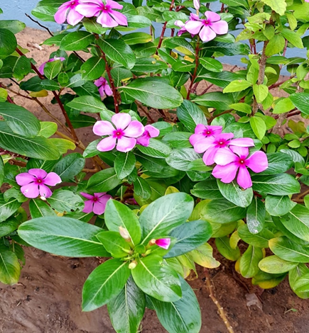
Fig 1: Catharanthus roseus

      1. Scientific Classification:

Kingdom: Plantae

Family: Apocynaceae

Genus: Catharanthus

Species: C. roseus [18].

      1. Common Names:

Madagascar periwinkle

Sadabahar

Vinca

Nithya Kalyani [19].

      1. Geographical Distribution:

Native to Madagascar

Widely cultivated in India, Africa, Southeast Asia, and tropical regions [20].

2.2. Morphological Characteristics:

2.2.1. Habit:

  • Evergreen perennial herb
  • Grows up to 30-100 cm tall [21].

2.2.2 Root:

  • Tap root system [22].

2.2.3. Stem:

  • Green, smooth, branched, and contains milky latex [23].

2.2.4. Leaves:

  • Simple, opposite
  • Elliptical or oblong
  • Dark green, glossy surface [24].

2.2.5. Flowers:

  • Pink, purple, or white
  • Five petals
  • Axillary position [25].

2.2.6. Fruit:

  • Pair of cylindrical follicles
  • Contains numerous small black seeds [26].

2.3. Chemical Constituents:

  • Vincristine
  • Vinblastine
  • Ajmalicine
  • Reserpine
  • These are important anticancer alkaloids [27].

2.4. Medicinal Uses:

  • Cancer treatment – Source of important anticancer drugs like vincristine and vinblastine.
  • Diabetes management – Leaf extract helps in lowering blood sugar levels.
  • Kidney stones – May help in reducing the formation of calcium oxalate crystals.
  • High blood pressure – Shows a mild antihypertensive effect.
  • Wound healing – Used traditionally for minor cuts and skin problems.
  • Antimicrobial action – Active against some bacteria and fungi.
  • Anti-inflammatory – Helps in reducing swelling and pain.
  • Antioxidant property – Protects cells from damage by free radicals [28, 29, 30].
  1. MATERIALS AND METHODS:
    1. Chemicals used:

Calcium chloride, Sodium oxalate, Potassium permanganate [KMnO4], Sulphuric acid [H2SO4], Tris buffer, Calcium chloride, Hydrochloric acid [HCl], Neeri [48, 49].

3.2. Collecting and Identifying the Plant Material:

 Fresh leaves of the plant Catharanthus roseus were collected from the local area of Nellore, Andhra Pradesh. They were authenticated as well as thoroughly cleaned using distilled water to remove dust and impurities. The leaves were then shade-dried at ambient temperature for several days before being ground to a coarse powder suitable for extraction using a mechanical grinder.

3.3. Preparation of Plant Extract:

The leaves were shade-dried and powdered. The crude plant extract was prepared by the Soxhlet extraction method. 50g of powdered plant material was extracted with 500 ml of ethanol and water individually. The process of extraction was carried out up to 6 cycles, till the solvent in the siphon tube of the extractor became colourless. The two extracts were filtered separately. Further, the dried extracts were maintained in a refrigerator at 4 °C for further antiurolithiatic activity [49].

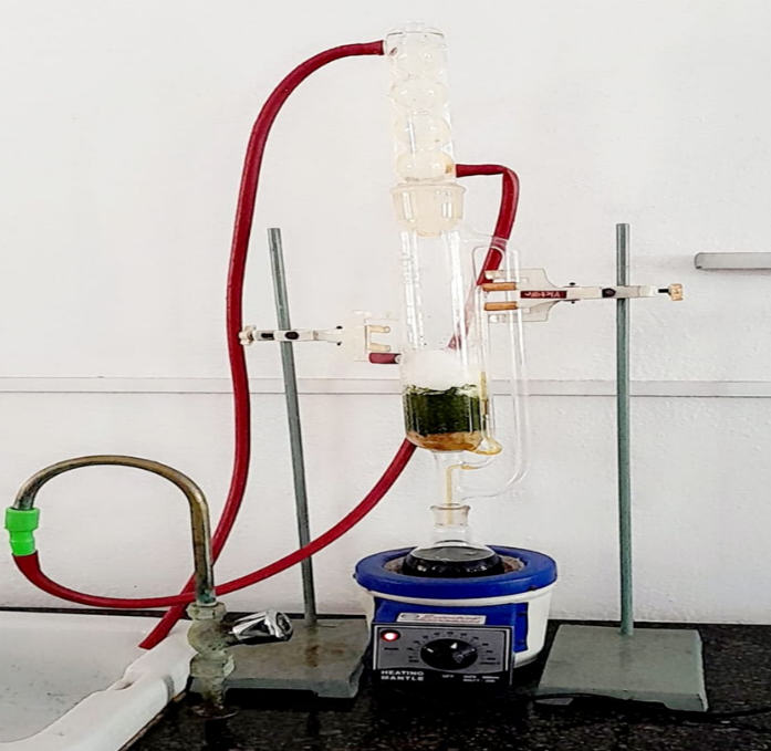
Fig 2: Preparation of plant extract using Soxhlet Apparatus.

3.4. Conducting Preliminary Phytochemical Screening:

The Aqueous and ethanolic Extract were screened qualitatively for the presence of major bioactive components, such as alkaloids; flavonoids; tannins; saponins; glycosides; and phenolic compounds through standard chemical tests.

3.5. Preparation of calcium oxalate crystals:

To prepare calcium oxalate, a 0.1 M calcium chloride solution is prepared by dissolving 1.47 g of CaCl?, 2H?O in 100 mL of distilled water. Similarly, 0.1 M sodium oxalate solution is prepared by dissolving 1.26 g of Na?C?O? in 100 mL of distilled water. The pH of both solutions is maintained between 5.5 and 6.5 [50].

The calcium chloride solution is placed on a magnetic stirrer, and the sodium oxalate solution is added slowly dropwise while stirring. This reaction leads to the formation of calcium oxalate precipitate. The mixture is stirred for 15-20 minutes and then allowed to stand for 30 minutes for proper crystal formation [51].

The precipitate is filtered, washed with distilled water, and dried at 40-50 °C. The dried crystals are then stored in a desiccator for further experimental use [52].

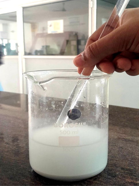
Fig 3: Preparation of calcium oxalate precipitate.

3.6. Preparation of a Semi-Permeable Membrane from Eggs:

Eggshell decalcification is performed to obtain the inner semi-permeable membrane, which can be used in in-vitro anti-urolithiatic studies. Eggshells are mainly composed of calcium carbonate (CaCO?). When exposed to white vinegar, which contains about 4-5% acetic acid, the calcium carbonate reacts with the acid to form soluble calcium acetate, carbon dioxide, and water. This reaction gradually dissolves the hard shell and leaves behind a thin membrane [53].

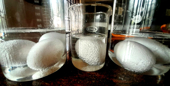
Fig 4: Decalcification of egg shells.

For the procedure, fresh chicken eggs are first washed with distilled water to remove impurities. The clean eggs are then placed in a glass container and completely submerged in white vinegar. They are left undisturbed for about 24-48 hours at room temperature. During this period, bubbles appear on the shell surface due to carbon dioxide release. After the shell dissolves, the soft decalcified egg is removed and washed with distilled water. A small opening is made to remove the egg contents, and the remaining membrane is rinsed and used for further experimental use [54].

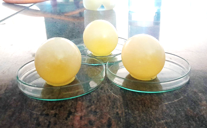
Fig 5: Decalcified eggs.

3.7. Preparation of 0.1M Tris Buffer:

Tris buffer is used to maintain a stable pH in biochemical experiments. To prepare a 0.1 M Tris buffer, 12.1 g of Tris (Tris (hydroxymethyl) aminomethane) is dissolved in about 800 mL of distilled water with continuous stirring [55]. The pH of the solution is checked and adjusted to pH 7.0-7.4 by adding dilute hydrochloric acid dropwise. Finally, the volume is made up to 1000 mL with distilled water, mixed well, and stored in a clean container for laboratory use [56].

3.8. Preparation of Egg Membrane Pouches Containing Calcium Oxalate and Extracts:

In this experiment, an egg membrane pouch model was used to evaluate the anti-urolithiatic activity of plant extracts. Previously prepared decalcified eggshell membranes were used as semi-permeable pouches. A total of four egg membranes were selected and carefully cleaned with distilled water to remove any remaining impurities [57].

Each egg membrane was filled with different contents to study their effect on calcium oxalate crystals. The first egg pouch contained only 1 g of calcium oxalate crystals and served as the control. The second pouch contained a mixture of 1 g of calcium oxalate and 1ml of ethanolic extract of the plant sample [58]. The third pouch was filled with 1ml of calcium oxalate along with the 1ml of aqueous extract of the plant material. The fourth pouch contained 1 g of calcium oxalate combined with the standard anti-urolithiatic drug Neeri, which was used for comparison with the plant extracts [59].

After filling, the open ends of the egg membranes were carefully tied to form sealed pouches. These prepared egg pouches were then placed in separate containers, each containing 100ml of Tris buffer solution [60]. The samples were incubated under controlled conditions for a specific period. This setup allowed interaction between the extracts and calcium oxalate crystals through the semipermeable membrane, which helps in evaluating the potential anti-urolithiatic activity [61].

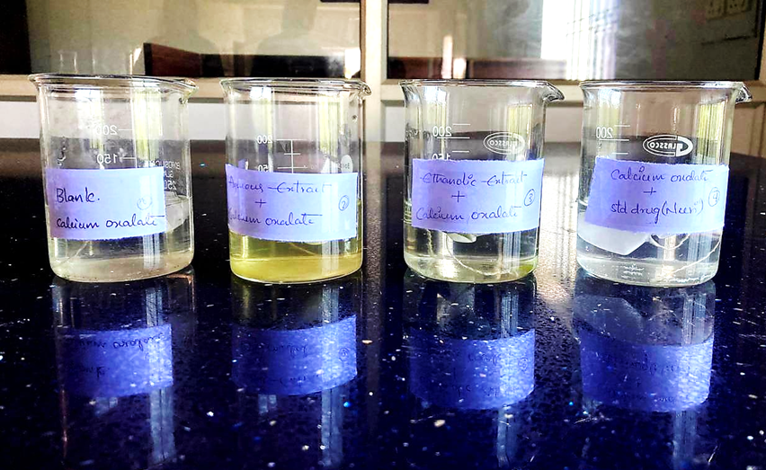
Fig 6: Immersion of egg pouches in Tris buffer solution

3.9. Preparation of 0.9494 N KMnO? Solution:

To prepare a 0.9494 N KMnO? solution, accurately weigh 15 g of potassium permanganate and dissolve it in about 800 mL of distilled water with continuous stirring [62]. The solution is then transferred to a 1000 mL volumetric flask, and the final volume is made up to 1 litre with distilled water. The solution is mixed well and stored in a dark-colored bottle to protect it from light and maintain stability [63].

3.10 Study of In Vitro Anti Urolithiatic Activity Through Titrimetric Method:

After incubation of the egg membrane pouches in Tris buffer, the remaining calcium oxalate content was estimated using the potassium permanganate titrimetric method (permanganometry). This method is commonly used to determine the amount of oxalate present in a sample based on oxidation-reduction reaction [64].

After the incubation period, the contents present inside each egg pouch were carefully transferred into separate conical flasks. Each pouch sample represented different experimental conditions, such as control, plant extracts, and standard drug treatment. To ensure proper acidic conditions for the reaction, 2 mL of 1 N sulphuric acid (H?SO?) was added to each conical flask containing the sample [65].

The titration was then carried out using 0.9494 N potassium permanganate (KMnO?) solution taken in the burette. During the titration process, the potassium permanganate acts as a strong oxidising agent and reacts with oxalate ions present in the sample. As the KMnO? solution was added gradually from the burette, it oxidised the oxalate ions present in the conical flask under acidic conditions [66].

The solution was continuously swirled during titration to ensure uniform mixing and complete reaction. The endpoint of the titration was indicated by the appearance of a light pink colour that persisted for about 30 seconds. This colour change confirmed that the reaction was complete and excess potassium permanganate was present [67, 68].

Each egg pouch sample, including the control pouch containing only calcium oxalate, the pouch with ethanolic extract, the pouch with aqueous extract, and the pouch with the standard drug Neeri, was titrated separately under the same experimental conditions. The volume of potassium permanganate solution consumed during titration was recorded for each sample [69].

The obtained titration values were further used to calculate the amount of calcium oxalate present and to evaluate the anti-urolithiatic activity of the plant extracts by comparing them with the control and standard drug [70].

  1. RESULTS

4.1. Phytochemical Study results For Leaves of Catharanthus Roseus

Table 1: Phytochemical Study results For Leaves of Catharanthus Roseus

Sr. No

Phytochemical Test

Reagents

Procedure

Observation

Result

1

Glycosides (Sodium Nitroprusside Test)

Vinca leaf extract, Sodium nitroprusside solution, Pyridine, Sodium hydroxide solution

About 2 ml of Vinca leaf extract was taken in a test tube. A few drops of sodium nitroprusside solution were added followed by pyridine. The mixture was shaken and made alkaline by adding sodium hydroxide dropwise and allowed to stand for a few minutes.

Reddish colour developed.

Indicates presence of glycosides.


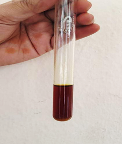
Fig 7: Test for Glycosides.

2

Phenolic Compounds (Ferric Chloride Test)

Vinca leaf extract, 5% Ferric chloride solution, Distilled water

About 2 ml of extract was taken in a test tube and a few drops of freshly prepared 5% ferric chloride solution were added and mixed well.

Deep blue, green, or bluish-black colour appeared.

Confirms presence of phenolic compounds

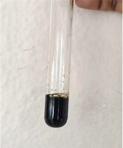
Fig 8: Test for Phenolic compounds.

3

Saponins (Foam Test)

Vinca leaf extract, Distilled water

About 2 ml of extract was diluted with 5 ml distilled water and shaken vigorously for 10–15 seconds, then allowed to stand for 10 minutes.

Formation of stable persistent foam.

Indicates presence of saponins.

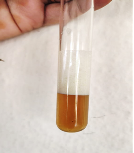
Fig 9: Test for Saponins.

4

Tannins (Ferric Chloride Test)

Vinca leaf extract, 5% Ferric chloride solution, Distilled water

2 ml of extract was taken in a test tube and a few drops of ferric chloride solution were added and mixed gently.

Bluish-black or greenish-black colour formed.

Confirms presence of tannins.

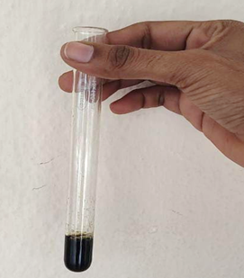
Fig 10: Test for Tannins.

5

Alkaloids (Tannic Acid Test)

Vinca leaf extract, Tannic acid solution

2 ml of extract was taken and a few drops of tannic acid solution were added. The mixture was shaken and allowed to stand for a few minutes.

Buff or pale-yellow precipitate formed.

Indicates presence of alkaloids.

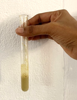
Fig 11: Test for Alkaloids.

6

Terpenoids (Salkowski Test)

Vinca leaf extract, Chloroform, Concentrated sulfuric acid

2 ml of extract was mixed with 2 ml chloroform. Concentrated sulfuric acid was carefully added along the side of the test tube to form a layer.

Reddish-brown colour at the interface of layers.

Indicates presence of terpenoids.

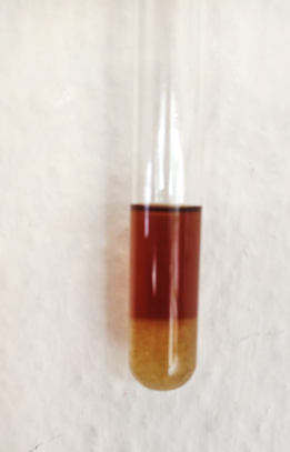
Fig 12:  Test for Terpenoids.

4.2. Titrimetric Method Results:

After the incubation period, the remaining oxalate in the mixture was determined by potassium permanganate (KMnO?) titration, which allowed for an assessment of the ability of the extract to inhibit the crystallization of calcium oxalate. The results of this study were then compared against similar control samples to assess the anti-urolithic effects of the plant extract.

Table 2: Effect of Sample Volumes on Calcium Oxalate Inhibition.

Sr. No

Sample

Volume of KMno4 used (ml)

% Inhibition of calcium oxalate

1.

Blank (Calcium Oxalate)

2.00

0%

2.

Ethanolic Extract + Calcium Oxalate

0.78

61%

3.

Aqueous Extract + Calcium Oxalate

0.71

64.5%

4.

Standard Drug (Neeri) + Calcium Oxalate

0.60

70%

  1. DISCUSSION:

The anti-urolithiatic activity of Catharanthus roseus leaf extracts was evaluated using the egg membrane pouch model, followed by estimation of calcium oxalate through the permanganometric titration method. In this study, both aqueous and ethanolic extracts were tested for their ability to dissolve calcium oxalate crystals, and the results were compared with the standard drug Neeri [71].

The blank sample containing only calcium oxalate showed the highest titration value of 2 mL, indicating the presence of maximum undissolved calcium oxalate crystals. When plant extracts and the standard drug were added, the titration values were significantly reduced, which suggests dissolution or inhibition of calcium oxalate crystals [72].

The ethanolic extract showed a burette reading of 0.78 mL, which corresponds to 61% inhibition of calcium oxalate. The aqueous extract showed a titration value of 0.71 mL, corresponding to 64.5% inhibition. The standard drug Neeri showed the lowest titration value of 0.6 mL, indicating 70% inhibition of calcium oxalate crystals [73].

From these results, it is evident that both extracts of Catharanthus roseus possess considerable anti-urolithiatic activity. The aqueous extract showed slightly better activity than the ethanolic extract, which may be due to the presence of water-soluble phytoconstituents such as flavonoids, alkaloids, and phenolic compounds that help in dissolving or inhibiting calcium oxalate crystal formation [74].

However, the standard drug Neeri demonstrated the highest activity among the tested samples. Even though the plant extracts showed slightly lower activity compared to the standard drug, their significant inhibitory effect indicates that Catharanthus roseus leaves contain bioactive compounds with potential lithotriptic properties [75].

Overall, this in-vitro study suggests that both aqueous and ethanolic extracts of Catharanthus roseus leaves possess promising anti-urolithiatic activity. These findings provide preliminary evidence supporting the traditional use of plant-based medicines in the management of kidney stones. Further in-vivo and clinical studies are required to confirm their therapeutic effectiveness and to identify the active phytochemical constituents responsible for this activity [76].

  1. CONCLUSION:

The present study demonstrated that Vinca leaf extracts possess noticeable anti-urolithiatic activity in an in-vitro model of calcium oxalate crystal formation. Both aqueous and ethanolic extracts showed the ability to reduce oxalate levels when compared with the blank sample, indicating their inhibitory effect on crystal formation. Among the tested samples, the aqueous extract showed relatively higher inhibition, suggesting the presence of water-soluble bioactive constituents responsible for the activity. These findings support the traditional medicinal importance of Vinca leaves and indicate their potential role in the prevention or management of kidney stone formation. Further pharmacological and clinical studies are required to confirm these effects.

  1. AUTHOR CONTRIBUTIONS

Kongi Kavyasudha was responsible for guiding the project and being a primary investigator for the project by being involved in developing the research, designing the study, overseeing the experiments, analyzing the data, and providing a thorough revision to the manuscript. Shaik Afrin, Venumbaka Sravya Sree, Gothala Sushma Chandrika, Cheerla Susmitha, and Vakani Jahnavi were undergraduate pharmacy degree students conducting all experimental aspects of the research as part of their course project and under the direction of the project supervisor. They assisted by processing the plant materials, performing laboratory experiments, collecting data, performing a preliminary analysis of the data, and writing the initial manuscript draft. Dr. Yadala Prapurna Chandra assisted with institutional support for this research by providing laboratory resources, equipment, and administrative approval to conduct the research at the Rathnam Institute of Pharmacy. All authors provided input on the analysis of the data, reviewed the manuscript, and approved the final manuscript for publication.

  1. Funding:

“This research received no external funding”.

  1. Institutional Review Board Statement

The present study involved in-vitro experimental work using plant extracts and did not involve human participants or animal subjects. Therefore, ethical approval from an Institutional Review Board (IRB) was not required for this research.

  1. Informed Consent Statement

Not applicable. The study did not involve human participants or personal data requiring informed consent.

  1. Data Availability Statement

The data supporting the findings of this study are available from the corresponding author upon reasonable request.

  1. Acknowledgments

The authors express their sincere gratitude to the management and Principal of Rathnam Institute of Pharmacy, Pidathapolur, Nellore, for providing laboratory facilities, equipment, and institutional support to carry out this research work. The authors also acknowledge the guidance and supervision provided during the completion of this academic research project.

  1. Conflicts of Interest

The authors declare no conflict of interest regarding the publication of this research work.

  1. Abbreviations
  • KMnO? – Potassium Permanganate
  • CaOx – Calcium Oxalate
  • IRB – Institutional Review Board
  • SD – Standard Deviation
  • UV – Ultraviolet

REFERENCES

  1. Nishikawa, T., Tada, H., Nakagawa-Kamiya, T., Niwa, S., Yoshida, S., Mori, M., Sakata, K., Nohara, A., Higashikata, T., Kato, H., Ino, K., Takemura, H., Takamura, M., & Kawashiri, M. (2020). A case with familial hypercholesterolemia complicated with severe systemic atherosclerosis intensively treated for more than 30 years. Journal of Cardiology Cases, 22(5), 216–220. https://doi.org/10.1016/j.jccase.2020.06.012
  2. Miller, N. L., Gillen, D. L., Williams, J. C., Evan, A. P., Bledsoe, S. B., Coe, F. L., Worcester, E. M., Matlaga, B. R., Munch, L. C., & Lingeman, J. E. (2009). A formal test of the hypothesis that idiopathic calcium oxalate stones grow on Randall’s plaque. BJU International, 103(7), 966–971. https://doi.org/10.1111/j.1464-410X.2008.08193.x
  3. Gore, L., Rothenberg, M. L., O’Bryant, C. L., Schultz, M. K., Sandler, A. B., Coffin, D., McCoy, C., Schott, A., Scholz, C., & Eckhardt, S. G. (2008). A Phase I and Pharmacokinetic Study of the Oral Histone Deacetylase Inhibitor, MS-275, in Patients with Refractory Solid Tumors and Lymphomas. Clinical Cancer Research, 14(14), 4517–4525. https://doi.org/10.1158/1078-0432.CCR-07-1461
  4. Awedew, A. F., Han, H., Berice, B. N., Dodge, M., Schneider, R. D., Abbasi-Kangevari, M., Al-Aly, Z., Almidani, O., Alvand, S., Arabloo, J., Aravkin, A. Y., Ayana, T. M., Bhardwaj, N., Bhardwaj, P., Bhaskar, S., Bikbov, B., Caetano Dos Santos, F. L., Charan, J., Cruz-Martins, N., … Dirac, M. A. (2024). The global, regional, and national burden of urolithiasis in 204 countries and territories, 2000–2021: A systematic analysis for the Global Burden of Disease Study 2021. eClinicalMedicine, 78, 102924. https://doi.org/10.1016/j.eclinm.2024.102924
  5. Rathore, B., Kumar, S., & Ali, M. (2025). A review on herbal drugs in the management of Urolithiasis. Journal of Pharmacognosy and Phytochemistry, 14(1), 283–289. https://doi.org/10.22271/phyto.2025.v14.i1d.15248
  6. Ramadhan, R. D., Wijaya, M. P., & Hidayat, D. R. (2025). Advances in Urology: A Comprehensive Literature Review on Urolithiasis Management and Innovations. Brawijaya Journal of Urology, 6(01), 11–19. https://doi.org/10.11594/bjurology.2025.006.01.3
  7. Pandhare, R. B., Shende, R. R., Avhad, M. S., Deshmukh, V. K., Mohite, P. B., Sangameswaran, B., & Daude, R. B. (2021). Anti-urolithiatic activity of Bryophyllum pinnatum Lam. Hydroalcoholic extract in sodium oxalate-induced urolithiasis in rats. Journal of Traditional and Complementary Medicine, 11(6), 545–551. https://doi.org/10.1016/j.jtcme.2021.06.002
  8. Lim, E. J., Castellani, D., So, W. Z., Fong, K. Y., Li, J. Q., Tiong, H. Y., Gadzhiev, N., Heng, C. T., Teoh, J. Y.-C., Naik, N., Ghani, K., Sarica, K., De La Rosette, J., Somani, B., & Gauhar, V. (2022). Radiomics in Urolithiasis: Systematic Review of Current Applications, Limitations, and Future Directions. Journal of Clinical Medicine, 11(17), 5151. https://doi.org/10.3390/jcm11175151
  9. Rajeshwari, T., Suresh, R., & Sudhakar, M. (2023). Anti-urolithiatic activity of saponin rich fraction from the methanolic extract of Achyranthes aspera against ethylene glycol induced urolithiasis in Wistar rats. Journal of Applied Pharmaceutical Science. https://doi.org/10.7324/JAPS.2023.144912
  10. Pawar, A., & Vyawahare, N. (2015). Anti-urolithiatic activity of standardized extract of Biophytum sensitivum against zinc disc implantation induced urolithiasis in rats. Journal of Advanced Pharmaceutical Technology & Research, 6(4), 176. https://doi.org/10.4103/2231-4040.165017
  11. Das, M., & Malipeddi, H. (2016). Antiurolithiatic activity of ethanol leaf extract of Ipomoea eriocarpa against ethylene glycol-induced urolithiasis in male Wistar rats. Indian Journal of Pharmacology, 48(3), 270. https://doi.org/10.4103/0253-7613.182886
  12. Babita Rana & Umera M Alvi. (2024). Assessment of antiurolithiatic activity of some herbal-fractions using in vitro techniques. International Journal of Biological and Pharmaceutical Sciences Archive, 8(1), 017–024. https://doi.org/10.53771/ijbpsa.2024.8.1.0058
  13. Patel, N. K., Basedia, D. K., & Dubey, B. K. (2022). Assessment of antiurolithiatic potential of gallic acid on calcium oxalate crystallization and ethylene glycol induced lithiasis. Asian Journal of Pharmaceutical Research and Development, 9(6), 104–107. https://doi.org/10.22270/ajprd.v9i6.1020
  14. Keller, J. J., Chen, Y., & Lin, H. (2012). Association between chronic kidney disease and urinary calculus by stone location: A population?based study. BJU International, 110(11c). https://doi.org/10.1111/j.1464-410X.2012.11380.x
  15. Chen, S.-J., Dalanbaatar, S., Chen, H.-Y., Wang, S.-J., Lin, W.-Y., Liu, P.-L., Tsai, M.-Y., Chen, D.-C., Chen, Y.-H., & Chen, W.-C. (2022). Astragalus membranaceus Extract Prevents Calcium Oxalate Crystallization and Extends Lifespan in a Drosophila Urolithiasis Model. Life, 12(8), 1250. https://doi.org/10.3390/life12081250
  16. El Oumari, F. E., Bousta, D., Imtara, H., Lahrichi, A., Elhabbani, R., El Mouhri, G., Al Kamaly, O., Saleh, A., Parvez, M. K., Grafov, A., & Sqalli Houssaini, T. (2022). Chemical Composition and Anti-Urolithiatic Activity of Extracts from Argania spinosa (L.) Skeels Press-Cake and Acacia senegal (L.) Willd. Molecules, 27(13), 3973. https://doi.org/10.3390/molecules27133973
  17. Tinti, F., Umbro, I., D’Alessandro, M., Lai, S., Merli, M., Noce, A., Di Daniele, N., Mazzaferro, S., & Mitterhofer, A. P. (2021). Cholemic Nephropathy as Cause of Acute and Chronic Kidney Disease. Update on an Under-Diagnosed Disease. Life, 11(11), 1200. https://doi.org/10.3390/life11111200
  18. Katekar, V. P. (2026). Cow Urine Distillate and Its Therapeutic Applications: An Experimental Investigation. Journal of Drug Delivery and Therapeutics, 16(2), 129–155. https://doi.org/10.22270/jddt.v16i2.7571
  19. Wróblewski, K., Wróblewska, P., Szukalska, S., Karczewska, M., Lichwala, K., Samborska, A., Balajewicz, B., & Siwek, L. (2026). Current Perspectives on Urolithiasis: Pathogenesis, Clinical Management, and Treatment. Cureus. https://doi.org/10.7759/cureus.101141
  20. Miller, J., Chi, T., Kapahi, P., Kahn, A. J., Kim, M. S., Hirata, T., Romero, M. F., Dow, J. A. T., & Stoller, M. L. (2013). Drosophila melanogaster as an Emerging Translational Model of Human Nephrolithiasis. Journal of Urology, 190(5), 1648–1656. https://doi.org/10.1016/j.juro.2013.03.010
  21. Knauf, F., & Preisig, P. A. (2011). Drosophila: A fruitful model for calcium oxalate nephrolithiasis? Kidney International, 80(4), 327–329. https://doi.org/10.1038/ki.2011.166
  22. Bhardwaj, V. (2025). Eco-Friendly Antibacterial Potential of Catharanthus roseus Leaf Extracts: A Green Approach to Natural Drug Discovery. Journal of Microbes in Health and Disease, 1(1), 100004. https://doi.org/10.53941/jmhd.2025.100004
  23. Doshi, S., & Braganza, V. (2025). Effect of Different Extraction Methods on the Concentration of Phenolics and Flavonoids in Catharanthus roseus and Assessment of Their Efficacy for Lifespan Extension in Model Organism Caenorhabditis elegans. Journal of Pharmaceutical Research International, 37(3), 68–78. https://doi.org/10.9734/jpri/2025/v37i37670
  24. Ho, C., Chen, Y., Wu, P., Chang, C., Chen, H., Man, K., Shen, J., Tsai, F., Lin, W., Lee, Y., & Chen, W. (2013). Effects of commercial citrate?containing juices on urolithiasis in a Drosophila model. The Kaohsiung Journal of Medical Sciences, 29(9), 488–493. https://doi.org/10.1016/j.kjms.2013.01.003
  25. Stamatelou, K., & Goldfarb, D. S. (2023). Epidemiology of Kidney Stones. Healthcare, 11(3), 424. https://doi.org/10.3390/healthcare11030424
  26. Sorokin, I., Mamoulakis, C., Miyazawa, K., Rodgers, A., Talati, J., & Lotan, Y. (2017). Epidemiology of stone disease across the world. World Journal of Urology, 35(9), 1301–1320. https://doi.org/10.1007/s00345-017-2008-6
  27. Tiselius, H. G., & Fornander, A. M. (1981). Evaluation of a routine method for determination of calcium oxalate crystal growth inhibition in diluted urine samples. Clinical Chemistry, 27(4), 565–568. https://doi.org/10.1093/clinchem/27.4.565
  28. Kishore, B., Prasanna Raju, Y., & Chandra Sekhar Kothapalli, B. (2020). Evaluation of Antiurolithiatic Activity of Asparagus Racemosus on In Vitro Calcium Oxalate Crystallization Methods. International Journal of Pharma and Bio Sciences, 10(5). https://doi.org/10.22376/ijpbs/lpr.2020.10.5.P56-62
  29. J, V., & P, N. (2022). EVALUATION OF IN VITRO ANTIUROLITHIATIC ACTIVITY OF ETHANOLIC EXTRACT OF PAVONIA ZEYLANICA (L) CAV. Journal of Pharmaceutical and Scientific Innovation, 11(1), 6–9. https://doi.org/10.7897/2277-4572.111226
  30. Satapathy, T., Pradhan, B., & Sen, K. (2025). Experimental animal models for urolithiasis (kidney stone): A comprehensive review. Animals and Zoonoses, 1(3), 302–311. https://doi.org/10.1016/j.azn.2025.03.001
  31. Gupta, H. (2024). Exploring the Antiurolithiatic and Diuretic Potential of Nausadar and Plant Extracts in Ethylene Glycol-Induced Urolithiasis in Wistar Rats. African Journal OF Biomedical Research, 3464–3472. https://doi.org/10.53555/AJBR.v27i3S.2972
  32. Rani, J., & Kapoor, M. (2019). GAS CHROMATOGRAPHY-MASS SPECTROMETRIC ANALYSIS AND IDENTIFICATION OF BIOACTIVE CONSTITUENTS OF CATHARANTHUS ROSEUS AND ITS ANTIOXIDANT ACTIVITY. Asian Journal of Pharmaceutical and Clinical Research, 461–465. https://doi.org/10.22159/ajpcr.2019.v12i3.30865
  33. Doshi, G. M., Matthews, B. D., & Chaskar, P. K. (2018). GAS CHROMATOGRAPHY-MASS SPECTROSCOPY STUDIES ON ETHANOLIC EXTRACT OF DRIED LEAVES OF CATHARANTHUS ROSEUS. Asian Journal of Pharmaceutical and Clinical Research, 11(6), 336. https://doi.org/10.22159/ajpcr.2018.v11i6.23704
  34. Vera-Ponce, V. J., Sanchez-Tamay, N. M., Ballena-Caicedo, J., Zuzunaga-Montoya, F. E., De Carrillo, C. I. G., & Poemape Mestanza, R. L. (2025). Global prevalence of urolithiasis: A meta-analysis accounting for methodological heterogeneity. Frontiers in Urology, 5, 1705953. https://doi.org/10.3389/fruro.2025.1705953
  35. Department of Physics, Annamalai University, Annamalai Nagar, Tamil Nadu, India, Priadharshini, I. V., & Selvaraju, R. (2025). Growth and Spectral Characterization of Calcium Oxalate Crystals. Indian Journal Of Science And Technology, 18(23), 1862–1872. https://doi.org/10.17485/IJST/v18i23.877
  36. Bhaskar, R., Rani, R., Singh, A. P., & Singh, A. P. (2024). Herbal Drug Use to Treat Urolithiasis: An Updated Review. Journal of Drug Delivery and Therapeutics, 14(11), 146–149. https://doi.org/10.22270/jddt.v14i11.6866
  37. Chmiel, J. A., Stuivenberg, G. A., Alathel, A., Gorla, J., Grohe, B., Razvi, H., Burton, J. P., & Bjazevic, J. (2022). High-Throughput in vitro Gel-Based Plate Assay to Screen for Calcium Oxalate Stone Inhibitors. Urologia Internationalis, 106(6), 616–622. https://doi.org/10.1159/000519842
  38. Jamal, Q. M. S., & Ahmad, V. (2024). Identification of Metabolites from Catharanthus roseus Leaves and Stem Extract, and In Vitro and In Silico Antibacterial Activity against Food Pathogens. Pharmaceuticals, 17(4), 450. https://doi.org/10.3390/ph17040450
  39. Rahamathullah, N., Bagchi, S., Nanda, M., & Dutta, A. (2025). In Vitro Antibacterial Efficacy of Crude Leaf Extracts from Catharanthus roseus: A Natural Therapeutic Prospect. Biomedical and Pharmacology Journal, 18(3), 2249–2257. https://doi.org/10.13005/bpj/3251
  40. Susmita Roy. (2024). In Vitro Antiurolithiatic Activity by Inhibition of Calcium Oxalate Crystallization of Methanol Extract of Imperata Cylindrica L. https://doi.org/10.5281/ZENODO.14742157
  41. Mittal, A., Tandon, S., Singla, S. K., & Tandon, C. (2016). In vitro inhibition of calcium oxalate crystallization and crystal adherence to renal tubular epithelial cells by Terminalia arjuna. Urolithiasis, 44(2), 117–125. https://doi.org/10.1007/s00240-015-0822-0
  42. El Habbani, R., Lahrichi, A., Sqalli Houssaini, T., Kachkoul, R., Mohim, M., Chouhani, B. A., & Chaqroune, A. (2021). In vitro mass reduction of calcium oxalate urinary calculi by some medicinal plants. African Journal of Urology, 27(1), 28. https://doi.org/10.1186/s12301-021-00132-2
  43. Itam, A., Ismail, Z., & Abdul Majid, A. M. S. (2015). IN VITRO STUDIES ON CALCIUM OXALATE CRYSTAL GROWTH INHIBITION OF STROBILANTHES CRISPUS EXTRACTS. Jurnal Riset Kimia, 3(1), 8. https://doi.org/10.25077/jrk.v3i1.30
  44. Hirata, T., Cabrero, P., Berkholz, D. S., Bondeson, D. P., Ritman, E. L., Thompson, J. R., Dow, J. A. T., & Romero, M. F. (2012). In vivo Drosophilia genetic model for calcium oxalate nephrolithiasis. American Journal of Physiology-Renal Physiology, 303(11), F1555–F1562. https://doi.org/10.1152/ajprenal.00074.2012
  45. Ms. Sowmya B A & Dr. Satish Pavuluri. (2023). In-Vitro Analysis of Ethanol Extract of Saponaria Officinalis for Antiurolithiatic Activity. Tropical Journal of Pharmaceutical and Life Sciences, 10(5), 57–62. https://doi.org/10.61280/tjpls.v10i5.147
  46. Ramprasad, R., Nilsha, A., Hameed, S. S., Shifama, J. M. R., & Venkateshan, N. (2019). In-Vitro antiurolithiatic activity of aqueous extract Pavonia lasiopetala. Journal of Drug Delivery and Therapeutics, 9(2), 102–104. https://doi.org/10.22270/jddt.v9i2.2483
  47. Agarwal, K., & Varma, R. (2015). In-vitro Calcium Oxalate Crystallization Inhibition by Achyranthes aspera L. and Bryophyllum pinnatum Lam. British Journal of Pharmaceutical Research, 5(2), 146–152. https://doi.org/10.9734/BJPR/2015/14042
  48. Saha, S., & Verma, R. J. (2013). Inhibition of calcium oxalate crystallisation in vitro by an extract of Bergenia ciliata. Arab Journal of Urology, 11(2), 187–192. https://doi.org/10.1016/j.aju.2013.04.001
  49. Ravindrakumar K, P., & Sandip B, P. (2020). INHIBITION OF CALCIUM OXALATE CRYSTALLIZATION BY AN EXTRACT OF OCIMUM BASILICUM SEEDS: AN IN-VITRO STUDY. Asian Journal of Pharmaceutical and Clinical Research, 166–169. https://doi.org/10.22159/ajpcr.2020.v13i9.37926
  50. Yousefi Ghale-Salimi, M., Eidi, M., Ghaemi, N., & Khavari-Nejad, R. A. (2018). Inhibitory effects of taraxasterol and aqueous extract of Taraxacum officinale on calcium oxalate crystallization: In vitro study. Renal Failure, 40(1), 298–305. https://doi.org/10.1080/0886022X.2018.1455595
  51. Abu Zarin, M., Tan, J. S., Murugan, P., & Ahmad, R. (2020). Investigation of potential anti-urolithiatic activity from different types of Musa pseudo-stem extracts in inhibition of calcium oxalate crystallization. BMC Complementary Medicine and Therapies, 20(1), 317. https://doi.org/10.1186/s12906-020-03113-0
  52. Bargagli, M., Scoglio, M., Howles, S. A., & Fuster, D. G. (2025). Kidney stone disease: Risk factors, pathophysiology and management. Nature Reviews Nephrology, 21(11), 794–808. https://doi.org/10.1038/s41581-025-00990-x
  53. Kant, R., Singh, T. G., & Singh, S. (2020). Mechanistic approach to herbal formulations used for urolithiasis treatment. Obesity Medicine, 19, 100266. https://doi.org/10.1016/j.obmed.2020.100266
  54. Morgan, M. S. C., & Pearle, M. S. (2016). Medical management of renal stones. BMJ, i52. https://doi.org/10.1136/bmj.i52
  55. Skolarikos, A. (2018). Medical treatment of urinary stones. Current Opinion in Urology, 28(5), 403–407. https://doi.org/10.1097/MOU.0000000000000523
  56. Angelopoulos, P., Neofytoy, P., Katsimperis, S., Triantafylloy, P., Manolitsis, I., Lardas, M., Markopoulos, T., Varkarakis, I., Tzelves, L., & Skolarikos, A. (2025). Metabolic evaluation of urolithiasis: A narrative review. Die Urologie, 64(7), 680–685. https://doi.org/10.1007/s00120-025-02617-2
  57. Dai, S., Fang, Q., Li, H.-Y., Sun, R., Zhang, H.-Y., & Wu, W. (2025). Network pharmacology and experimental validation to elucidate the pharmacological mechanisms of OATF against kidney stones. Frontiers in Pharmacology, 16, 1575270. https://doi.org/10.3389/fphar.2025.1575270
  58. Tamborino, F., Cicchetti, R., Mascitti, M., Litterio, G., Orsini, A., Ferretti, S., Basconi, M., De Palma, A., Ferro, M., Marchioni, M., & Schips, L. (2024). Pathophysiology and Main Molecular Mechanisms of Urinary Stone Formation and Recurrence. International Journal of Molecular Sciences, 25(5), 3075. https://doi.org/10.3390/ijms25053075
  59. Ahmad, W., Khan, M. A., Ashraf, K., Ahmad, A., Daud Ali, M., Ansari, M. N., Kamal, Y., Wahab, S., Zaidi, S. A., Mujeeb, Mohd., & Ahmad, S. (2021). Pharmacological Evaluation of Safoof-e-Pathar Phori- A Polyherbal Unani Formulation for Urolithiasis. Frontiers in Pharmacology, 12, 597990. https://doi.org/10.3389/fphar.2021.597990
  60. Mendonce, K. C., Palani, N., Rajadesingu, S., Radhakrishnan, K., Ayyar, M., & Priya, L. S. (2025). Pharmacological potential of bioactive compounds in Catharanthus roseus extract: A comprehensive review. Toxicology Reports, 14, 101998. https://doi.org/10.1016/j.toxrep.2025.101998
  61. Richa Shahwal,  et al. (2025). Phytochemical Characterization and Antimicrobial Potential of Catharanthus roseus Methanol Extract Against Vaginal Microflora. Journal of Chemical Health Risks. https://doi.org/10.52783/jchr.v15.i5.9895
  62. C. T, S., C. K, J., G, J. K. U., K. M, P., & Balachandran, I. (2022). Phytochemical characterization and evaluation of Antiurolithiatic activity of selected source plants of Pashanabheda. Clinical Phytoscience, 8(1), 12. https://doi.org/10.1186/s40816-022-00344-1
  63. Bansal, Y., Mujib, A., Mamgain, J., Dewir, Y. H., & Rihan, H. Z. (2023). Phytochemical Composition and Detection of Novel Bioactives in Anther Callus of Catharanthus roseus L. Plants, 12(11), 2186. https://doi.org/10.3390/plants12112186
  64. Gupta, J., & Gupta, S. (2025). Phytochemical Screening and TLC Profiling of Leaf Extracts of Catharanthus roseus Show Presence of Biopesticidal Secondary Metabolites. Current Journal of Applied Science and Technology, 44(9), 172–179. https://doi.org/10.9734/cjast/2025/v44i94613
  65. Pham, H. N. T., Vuong, Q. V., Bowyer, M. C., & Scarlett, C. J. (2020). Phytochemicals Derived from Catharanthus roseus and Their Health Benefits. Technologies, 8(4), 80. https://doi.org/10.3390/technologies8040080
  66. Mutomba, W. F., Symeonidis, E. N., Mykoniatis, I., Tzelves, L., Tsaturyan, A., Juliebo-Jones, P., Tokas, T., & Sountoulides, P. (2025). Phytotherapy in Urolithiasis: An Updated Overview of Current Knowledge. Journal of Clinical Medicine, 14(9), 2885. https://doi.org/10.3390/jcm14092885
  67. Faridi, M., & Singh, K. (2020). Preliminary study of prevalence of urolithiasis in North-Eastern city of India. Journal of Family Medicine and Primary Care, 9(12), 5939. https://doi.org/10.4103/jfmpc.jfmpc_1522_20
  68. Evan, A. P., Lingeman, J. E., Coe, F. L., Parks, J. H., Bledsoe, S. B., Shao, Y., Sommer, A. J., Paterson, R. F., Kuo, R. L., & Grynpas, M. (2003). Randall’s plaque of patients with nephrolithiasis begins in basement membranes of thin loops of Henle. Journal of Clinical Investigation, 111(5), 607–616. https://doi.org/10.1172/JCI17038
  69. Chen, W.-C., Chou, T.-Y., Chen, H.-Y., Yang, Y.-R., Man, K.-M., Tsai, M.-Y., & Chen, Y.-H. (2019). Salvia miltiorrhiza Bunge (Danshen) for Treatment and Prevention of Urolithiasis: A Drosophila Animal Study. Evidence-Based Complementary and Alternative Medicine, 2019, 1–5. https://doi.org/10.1155/2019/1408979
  70. Wu, T., Liu, Z., Ma, S., Xue, W., Jiang, X., & Ma, J. (2022). Should we support prophylactic intervention for asymptomatic kidney stones? A retrospective cohort study with long-term follow-up. Urolithiasis, 50(4), 431–437. https://doi.org/10.1007/s00240-022-01331-4
  71. Kallidonis, P., & Vagionis, A. (2022). The future of laser technology in kidney stones. Current Opinion in Urology, 32(4), 411–414. https://doi.org/10.1097/MOU.0000000000001006
  72. Awedew, A. F., Han, H., Berice, B. N., Dodge, M., Schneider, R. D., Abbasi-Kangevari, M., Al-Aly, Z., Almidani, O., Alvand, S., Arabloo, J., Aravkin, A. Y., Ayana, T. M., Bhardwaj, N., Bhardwaj, P., Bhaskar, S., Bikbov, B., Caetano Dos Santos, F. L., Charan, J., Cruz-Martins, N., … Dirac, M. A. (2024). The global, regional, and national burden of urolithiasis in 204 countries and territories, 2000–2021: A systematic analysis for the Global Burden of Disease Study 2021. eClinicalMedicine, 78, 102924. https://doi.org/10.1016/j.eclinm.2024.102924
  73. Malhotra, M., & Singh, A. P. (2024). TO EVALUATE THE ANTIUROLITHIATIC ACTIVITY OF AQUEOUS EXTRACT OF CATHARANTHUS ROSEUS IN ETHYLENE-GLYCOL-INDUCED UROLITHIASIS IN RATS. International Journal of Current Pharmaceutical Research, 27–31. https://doi.org/10.22159/ijcpr.2024v16i5.5027
  74. Parihar, S., Sharma, D., Chirania, A., & Telrandhe, U. B. (2022). To Review on the Pharmacology of the Leaf Extract of Catharanthus Roseus. Asian Journal of Pharmaceutical Research and Development, 10(1), 32–37. https://doi.org/10.22270/ajprd.v10i1.1075
  75. Dika, Ž., Mari?, M., & Živko, M. (2025). Treatment of Urolithiasis: A Comprehensive Review. EMJ Urology, 82–97. https://doi.org/10.33590/emjurol/NBZA7146
  76. Sarkar, B. K., Polash, Md. J. I., Islam, Md. J., Hanif, Md. A., Jannat, N., Kundu, S. K., Sarkar, B. K., & Hossain, Md. S. (2025). Unlocking the therapeutic potential of Catharanthus roseus leaves via in-vitro, in-vivo, and in-silico study. Scientific Reports, 15(1), 25909. https://doi.org/10.1038/s41598-025-96643-x

Reference

  1. Nishikawa, T., Tada, H., Nakagawa-Kamiya, T., Niwa, S., Yoshida, S., Mori, M., Sakata, K., Nohara, A., Higashikata, T., Kato, H., Ino, K., Takemura, H., Takamura, M., & Kawashiri, M. (2020). A case with familial hypercholesterolemia complicated with severe systemic atherosclerosis intensively treated for more than 30 years. Journal of Cardiology Cases, 22(5), 216–220. https://doi.org/10.1016/j.jccase.2020.06.012
  2. Miller, N. L., Gillen, D. L., Williams, J. C., Evan, A. P., Bledsoe, S. B., Coe, F. L., Worcester, E. M., Matlaga, B. R., Munch, L. C., & Lingeman, J. E. (2009). A formal test of the hypothesis that idiopathic calcium oxalate stones grow on Randall’s plaque. BJU International, 103(7), 966–971. https://doi.org/10.1111/j.1464-410X.2008.08193.x
  3. Gore, L., Rothenberg, M. L., O’Bryant, C. L., Schultz, M. K., Sandler, A. B., Coffin, D., McCoy, C., Schott, A., Scholz, C., & Eckhardt, S. G. (2008). A Phase I and Pharmacokinetic Study of the Oral Histone Deacetylase Inhibitor, MS-275, in Patients with Refractory Solid Tumors and Lymphomas. Clinical Cancer Research, 14(14), 4517–4525. https://doi.org/10.1158/1078-0432.CCR-07-1461
  4. Awedew, A. F., Han, H., Berice, B. N., Dodge, M., Schneider, R. D., Abbasi-Kangevari, M., Al-Aly, Z., Almidani, O., Alvand, S., Arabloo, J., Aravkin, A. Y., Ayana, T. M., Bhardwaj, N., Bhardwaj, P., Bhaskar, S., Bikbov, B., Caetano Dos Santos, F. L., Charan, J., Cruz-Martins, N., … Dirac, M. A. (2024). The global, regional, and national burden of urolithiasis in 204 countries and territories, 2000–2021: A systematic analysis for the Global Burden of Disease Study 2021. eClinicalMedicine, 78, 102924. https://doi.org/10.1016/j.eclinm.2024.102924
  5. Rathore, B., Kumar, S., & Ali, M. (2025). A review on herbal drugs in the management of Urolithiasis. Journal of Pharmacognosy and Phytochemistry, 14(1), 283–289. https://doi.org/10.22271/phyto.2025.v14.i1d.15248
  6. Ramadhan, R. D., Wijaya, M. P., & Hidayat, D. R. (2025). Advances in Urology: A Comprehensive Literature Review on Urolithiasis Management and Innovations. Brawijaya Journal of Urology, 6(01), 11–19. https://doi.org/10.11594/bjurology.2025.006.01.3
  7. Pandhare, R. B., Shende, R. R., Avhad, M. S., Deshmukh, V. K., Mohite, P. B., Sangameswaran, B., & Daude, R. B. (2021). Anti-urolithiatic activity of Bryophyllum pinnatum Lam. Hydroalcoholic extract in sodium oxalate-induced urolithiasis in rats. Journal of Traditional and Complementary Medicine, 11(6), 545–551. https://doi.org/10.1016/j.jtcme.2021.06.002
  8. Lim, E. J., Castellani, D., So, W. Z., Fong, K. Y., Li, J. Q., Tiong, H. Y., Gadzhiev, N., Heng, C. T., Teoh, J. Y.-C., Naik, N., Ghani, K., Sarica, K., De La Rosette, J., Somani, B., & Gauhar, V. (2022). Radiomics in Urolithiasis: Systematic Review of Current Applications, Limitations, and Future Directions. Journal of Clinical Medicine, 11(17), 5151. https://doi.org/10.3390/jcm11175151
  9. Rajeshwari, T., Suresh, R., & Sudhakar, M. (2023). Anti-urolithiatic activity of saponin rich fraction from the methanolic extract of Achyranthes aspera against ethylene glycol induced urolithiasis in Wistar rats. Journal of Applied Pharmaceutical Science. https://doi.org/10.7324/JAPS.2023.144912
  10. Pawar, A., & Vyawahare, N. (2015). Anti-urolithiatic activity of standardized extract of Biophytum sensitivum against zinc disc implantation induced urolithiasis in rats. Journal of Advanced Pharmaceutical Technology & Research, 6(4), 176. https://doi.org/10.4103/2231-4040.165017
  11. Das, M., & Malipeddi, H. (2016). Antiurolithiatic activity of ethanol leaf extract of Ipomoea eriocarpa against ethylene glycol-induced urolithiasis in male Wistar rats. Indian Journal of Pharmacology, 48(3), 270. https://doi.org/10.4103/0253-7613.182886
  12. Babita Rana & Umera M Alvi. (2024). Assessment of antiurolithiatic activity of some herbal-fractions using in vitro techniques. International Journal of Biological and Pharmaceutical Sciences Archive, 8(1), 017–024. https://doi.org/10.53771/ijbpsa.2024.8.1.0058
  13. Patel, N. K., Basedia, D. K., & Dubey, B. K. (2022). Assessment of antiurolithiatic potential of gallic acid on calcium oxalate crystallization and ethylene glycol induced lithiasis. Asian Journal of Pharmaceutical Research and Development, 9(6), 104–107. https://doi.org/10.22270/ajprd.v9i6.1020
  14. Keller, J. J., Chen, Y., & Lin, H. (2012). Association between chronic kidney disease and urinary calculus by stone location: A population?based study. BJU International, 110(11c). https://doi.org/10.1111/j.1464-410X.2012.11380.x
  15. Chen, S.-J., Dalanbaatar, S., Chen, H.-Y., Wang, S.-J., Lin, W.-Y., Liu, P.-L., Tsai, M.-Y., Chen, D.-C., Chen, Y.-H., & Chen, W.-C. (2022). Astragalus membranaceus Extract Prevents Calcium Oxalate Crystallization and Extends Lifespan in a Drosophila Urolithiasis Model. Life, 12(8), 1250. https://doi.org/10.3390/life12081250
  16. El Oumari, F. E., Bousta, D., Imtara, H., Lahrichi, A., Elhabbani, R., El Mouhri, G., Al Kamaly, O., Saleh, A., Parvez, M. K., Grafov, A., & Sqalli Houssaini, T. (2022). Chemical Composition and Anti-Urolithiatic Activity of Extracts from Argania spinosa (L.) Skeels Press-Cake and Acacia senegal (L.) Willd. Molecules, 27(13), 3973. https://doi.org/10.3390/molecules27133973
  17. Tinti, F., Umbro, I., D’Alessandro, M., Lai, S., Merli, M., Noce, A., Di Daniele, N., Mazzaferro, S., & Mitterhofer, A. P. (2021). Cholemic Nephropathy as Cause of Acute and Chronic Kidney Disease. Update on an Under-Diagnosed Disease. Life, 11(11), 1200. https://doi.org/10.3390/life11111200
  18. Katekar, V. P. (2026). Cow Urine Distillate and Its Therapeutic Applications: An Experimental Investigation. Journal of Drug Delivery and Therapeutics, 16(2), 129–155. https://doi.org/10.22270/jddt.v16i2.7571
  19. Wróblewski, K., Wróblewska, P., Szukalska, S., Karczewska, M., Lichwala, K., Samborska, A., Balajewicz, B., & Siwek, L. (2026). Current Perspectives on Urolithiasis: Pathogenesis, Clinical Management, and Treatment. Cureus. https://doi.org/10.7759/cureus.101141
  20. Miller, J., Chi, T., Kapahi, P., Kahn, A. J., Kim, M. S., Hirata, T., Romero, M. F., Dow, J. A. T., & Stoller, M. L. (2013). Drosophila melanogaster as an Emerging Translational Model of Human Nephrolithiasis. Journal of Urology, 190(5), 1648–1656. https://doi.org/10.1016/j.juro.2013.03.010
  21. Knauf, F., & Preisig, P. A. (2011). Drosophila: A fruitful model for calcium oxalate nephrolithiasis? Kidney International, 80(4), 327–329. https://doi.org/10.1038/ki.2011.166
  22. Bhardwaj, V. (2025). Eco-Friendly Antibacterial Potential of Catharanthus roseus Leaf Extracts: A Green Approach to Natural Drug Discovery. Journal of Microbes in Health and Disease, 1(1), 100004. https://doi.org/10.53941/jmhd.2025.100004
  23. Doshi, S., & Braganza, V. (2025). Effect of Different Extraction Methods on the Concentration of Phenolics and Flavonoids in Catharanthus roseus and Assessment of Their Efficacy for Lifespan Extension in Model Organism Caenorhabditis elegans. Journal of Pharmaceutical Research International, 37(3), 68–78. https://doi.org/10.9734/jpri/2025/v37i37670
  24. Ho, C., Chen, Y., Wu, P., Chang, C., Chen, H., Man, K., Shen, J., Tsai, F., Lin, W., Lee, Y., & Chen, W. (2013). Effects of commercial citrate?containing juices on urolithiasis in a Drosophila model. The Kaohsiung Journal of Medical Sciences, 29(9), 488–493. https://doi.org/10.1016/j.kjms.2013.01.003
  25. Stamatelou, K., & Goldfarb, D. S. (2023). Epidemiology of Kidney Stones. Healthcare, 11(3), 424. https://doi.org/10.3390/healthcare11030424
  26. Sorokin, I., Mamoulakis, C., Miyazawa, K., Rodgers, A., Talati, J., & Lotan, Y. (2017). Epidemiology of stone disease across the world. World Journal of Urology, 35(9), 1301–1320. https://doi.org/10.1007/s00345-017-2008-6
  27. Tiselius, H. G., & Fornander, A. M. (1981). Evaluation of a routine method for determination of calcium oxalate crystal growth inhibition in diluted urine samples. Clinical Chemistry, 27(4), 565–568. https://doi.org/10.1093/clinchem/27.4.565
  28. Kishore, B., Prasanna Raju, Y., & Chandra Sekhar Kothapalli, B. (2020). Evaluation of Antiurolithiatic Activity of Asparagus Racemosus on In Vitro Calcium Oxalate Crystallization Methods. International Journal of Pharma and Bio Sciences, 10(5). https://doi.org/10.22376/ijpbs/lpr.2020.10.5.P56-62
  29. J, V., & P, N. (2022). EVALUATION OF IN VITRO ANTIUROLITHIATIC ACTIVITY OF ETHANOLIC EXTRACT OF PAVONIA ZEYLANICA (L) CAV. Journal of Pharmaceutical and Scientific Innovation, 11(1), 6–9. https://doi.org/10.7897/2277-4572.111226
  30. Satapathy, T., Pradhan, B., & Sen, K. (2025). Experimental animal models for urolithiasis (kidney stone): A comprehensive review. Animals and Zoonoses, 1(3), 302–311. https://doi.org/10.1016/j.azn.2025.03.001
  31. Gupta, H. (2024). Exploring the Antiurolithiatic and Diuretic Potential of Nausadar and Plant Extracts in Ethylene Glycol-Induced Urolithiasis in Wistar Rats. African Journal OF Biomedical Research, 3464–3472. https://doi.org/10.53555/AJBR.v27i3S.2972
  32. Rani, J., & Kapoor, M. (2019). GAS CHROMATOGRAPHY-MASS SPECTROMETRIC ANALYSIS AND IDENTIFICATION OF BIOACTIVE CONSTITUENTS OF CATHARANTHUS ROSEUS AND ITS ANTIOXIDANT ACTIVITY. Asian Journal of Pharmaceutical and Clinical Research, 461–465. https://doi.org/10.22159/ajpcr.2019.v12i3.30865
  33. Doshi, G. M., Matthews, B. D., & Chaskar, P. K. (2018). GAS CHROMATOGRAPHY-MASS SPECTROSCOPY STUDIES ON ETHANOLIC EXTRACT OF DRIED LEAVES OF CATHARANTHUS ROSEUS. Asian Journal of Pharmaceutical and Clinical Research, 11(6), 336. https://doi.org/10.22159/ajpcr.2018.v11i6.23704
  34. Vera-Ponce, V. J., Sanchez-Tamay, N. M., Ballena-Caicedo, J., Zuzunaga-Montoya, F. E., De Carrillo, C. I. G., & Poemape Mestanza, R. L. (2025). Global prevalence of urolithiasis: A meta-analysis accounting for methodological heterogeneity. Frontiers in Urology, 5, 1705953. https://doi.org/10.3389/fruro.2025.1705953
  35. Department of Physics, Annamalai University, Annamalai Nagar, Tamil Nadu, India, Priadharshini, I. V., & Selvaraju, R. (2025). Growth and Spectral Characterization of Calcium Oxalate Crystals. Indian Journal Of Science And Technology, 18(23), 1862–1872. https://doi.org/10.17485/IJST/v18i23.877
  36. Bhaskar, R., Rani, R., Singh, A. P., & Singh, A. P. (2024). Herbal Drug Use to Treat Urolithiasis: An Updated Review. Journal of Drug Delivery and Therapeutics, 14(11), 146–149. https://doi.org/10.22270/jddt.v14i11.6866
  37. Chmiel, J. A., Stuivenberg, G. A., Alathel, A., Gorla, J., Grohe, B., Razvi, H., Burton, J. P., & Bjazevic, J. (2022). High-Throughput in vitro Gel-Based Plate Assay to Screen for Calcium Oxalate Stone Inhibitors. Urologia Internationalis, 106(6), 616–622. https://doi.org/10.1159/000519842
  38. Jamal, Q. M. S., & Ahmad, V. (2024). Identification of Metabolites from Catharanthus roseus Leaves and Stem Extract, and In Vitro and In Silico Antibacterial Activity against Food Pathogens. Pharmaceuticals, 17(4), 450. https://doi.org/10.3390/ph17040450
  39. Rahamathullah, N., Bagchi, S., Nanda, M., & Dutta, A. (2025). In Vitro Antibacterial Efficacy of Crude Leaf Extracts from Catharanthus roseus: A Natural Therapeutic Prospect. Biomedical and Pharmacology Journal, 18(3), 2249–2257. https://doi.org/10.13005/bpj/3251
  40. Susmita Roy. (2024). In Vitro Antiurolithiatic Activity by Inhibition of Calcium Oxalate Crystallization of Methanol Extract of Imperata Cylindrica L. https://doi.org/10.5281/ZENODO.14742157
  41. Mittal, A., Tandon, S., Singla, S. K., & Tandon, C. (2016). In vitro inhibition of calcium oxalate crystallization and crystal adherence to renal tubular epithelial cells by Terminalia arjuna. Urolithiasis, 44(2), 117–125. https://doi.org/10.1007/s00240-015-0822-0
  42. El Habbani, R., Lahrichi, A., Sqalli Houssaini, T., Kachkoul, R., Mohim, M., Chouhani, B. A., & Chaqroune, A. (2021). In vitro mass reduction of calcium oxalate urinary calculi by some medicinal plants. African Journal of Urology, 27(1), 28. https://doi.org/10.1186/s12301-021-00132-2
  43. Itam, A., Ismail, Z., & Abdul Majid, A. M. S. (2015). IN VITRO STUDIES ON CALCIUM OXALATE CRYSTAL GROWTH INHIBITION OF STROBILANTHES CRISPUS EXTRACTS. Jurnal Riset Kimia, 3(1), 8. https://doi.org/10.25077/jrk.v3i1.30
  44. Hirata, T., Cabrero, P., Berkholz, D. S., Bondeson, D. P., Ritman, E. L., Thompson, J. R., Dow, J. A. T., & Romero, M. F. (2012). In vivo Drosophilia genetic model for calcium oxalate nephrolithiasis. American Journal of Physiology-Renal Physiology, 303(11), F1555–F1562. https://doi.org/10.1152/ajprenal.00074.2012
  45. Ms. Sowmya B A & Dr. Satish Pavuluri. (2023). In-Vitro Analysis of Ethanol Extract of Saponaria Officinalis for Antiurolithiatic Activity. Tropical Journal of Pharmaceutical and Life Sciences, 10(5), 57–62. https://doi.org/10.61280/tjpls.v10i5.147
  46. Ramprasad, R., Nilsha, A., Hameed, S. S., Shifama, J. M. R., & Venkateshan, N. (2019). In-Vitro antiurolithiatic activity of aqueous extract Pavonia lasiopetala. Journal of Drug Delivery and Therapeutics, 9(2), 102–104. https://doi.org/10.22270/jddt.v9i2.2483
  47. Agarwal, K., & Varma, R. (2015). In-vitro Calcium Oxalate Crystallization Inhibition by Achyranthes aspera L. and Bryophyllum pinnatum Lam. British Journal of Pharmaceutical Research, 5(2), 146–152. https://doi.org/10.9734/BJPR/2015/14042
  48. Saha, S., & Verma, R. J. (2013). Inhibition of calcium oxalate crystallisation in vitro by an extract of Bergenia ciliata. Arab Journal of Urology, 11(2), 187–192. https://doi.org/10.1016/j.aju.2013.04.001
  49. Ravindrakumar K, P., & Sandip B, P. (2020). INHIBITION OF CALCIUM OXALATE CRYSTALLIZATION BY AN EXTRACT OF OCIMUM BASILICUM SEEDS: AN IN-VITRO STUDY. Asian Journal of Pharmaceutical and Clinical Research, 166–169. https://doi.org/10.22159/ajpcr.2020.v13i9.37926
  50. Yousefi Ghale-Salimi, M., Eidi, M., Ghaemi, N., & Khavari-Nejad, R. A. (2018). Inhibitory effects of taraxasterol and aqueous extract of Taraxacum officinale on calcium oxalate crystallization: In vitro study. Renal Failure, 40(1), 298–305. https://doi.org/10.1080/0886022X.2018.1455595
  51. Abu Zarin, M., Tan, J. S., Murugan, P., & Ahmad, R. (2020). Investigation of potential anti-urolithiatic activity from different types of Musa pseudo-stem extracts in inhibition of calcium oxalate crystallization. BMC Complementary Medicine and Therapies, 20(1), 317. https://doi.org/10.1186/s12906-020-03113-0
  52. Bargagli, M., Scoglio, M., Howles, S. A., & Fuster, D. G. (2025). Kidney stone disease: Risk factors, pathophysiology and management. Nature Reviews Nephrology, 21(11), 794–808. https://doi.org/10.1038/s41581-025-00990-x
  53. Kant, R., Singh, T. G., & Singh, S. (2020). Mechanistic approach to herbal formulations used for urolithiasis treatment. Obesity Medicine, 19, 100266. https://doi.org/10.1016/j.obmed.2020.100266
  54. Morgan, M. S. C., & Pearle, M. S. (2016). Medical management of renal stones. BMJ, i52. https://doi.org/10.1136/bmj.i52
  55. Skolarikos, A. (2018). Medical treatment of urinary stones. Current Opinion in Urology, 28(5), 403–407. https://doi.org/10.1097/MOU.0000000000000523
  56. Angelopoulos, P., Neofytoy, P., Katsimperis, S., Triantafylloy, P., Manolitsis, I., Lardas, M., Markopoulos, T., Varkarakis, I., Tzelves, L., & Skolarikos, A. (2025). Metabolic evaluation of urolithiasis: A narrative review. Die Urologie, 64(7), 680–685. https://doi.org/10.1007/s00120-025-02617-2
  57. Dai, S., Fang, Q., Li, H.-Y., Sun, R., Zhang, H.-Y., & Wu, W. (2025). Network pharmacology and experimental validation to elucidate the pharmacological mechanisms of OATF against kidney stones. Frontiers in Pharmacology, 16, 1575270. https://doi.org/10.3389/fphar.2025.1575270
  58. Tamborino, F., Cicchetti, R., Mascitti, M., Litterio, G., Orsini, A., Ferretti, S., Basconi, M., De Palma, A., Ferro, M., Marchioni, M., & Schips, L. (2024). Pathophysiology and Main Molecular Mechanisms of Urinary Stone Formation and Recurrence. International Journal of Molecular Sciences, 25(5), 3075. https://doi.org/10.3390/ijms25053075
  59. Ahmad, W., Khan, M. A., Ashraf, K., Ahmad, A., Daud Ali, M., Ansari, M. N., Kamal, Y., Wahab, S., Zaidi, S. A., Mujeeb, Mohd., & Ahmad, S. (2021). Pharmacological Evaluation of Safoof-e-Pathar Phori- A Polyherbal Unani Formulation for Urolithiasis. Frontiers in Pharmacology, 12, 597990. https://doi.org/10.3389/fphar.2021.597990
  60. Mendonce, K. C., Palani, N., Rajadesingu, S., Radhakrishnan, K., Ayyar, M., & Priya, L. S. (2025). Pharmacological potential of bioactive compounds in Catharanthus roseus extract: A comprehensive review. Toxicology Reports, 14, 101998. https://doi.org/10.1016/j.toxrep.2025.101998
  61. Richa Shahwal,  et al. (2025). Phytochemical Characterization and Antimicrobial Potential of Catharanthus roseus Methanol Extract Against Vaginal Microflora. Journal of Chemical Health Risks. https://doi.org/10.52783/jchr.v15.i5.9895
  62. C. T, S., C. K, J., G, J. K. U., K. M, P., & Balachandran, I. (2022). Phytochemical characterization and evaluation of Antiurolithiatic activity of selected source plants of Pashanabheda. Clinical Phytoscience, 8(1), 12. https://doi.org/10.1186/s40816-022-00344-1
  63. Bansal, Y., Mujib, A., Mamgain, J., Dewir, Y. H., & Rihan, H. Z. (2023). Phytochemical Composition and Detection of Novel Bioactives in Anther Callus of Catharanthus roseus L. Plants, 12(11), 2186. https://doi.org/10.3390/plants12112186
  64. Gupta, J., & Gupta, S. (2025). Phytochemical Screening and TLC Profiling of Leaf Extracts of Catharanthus roseus Show Presence of Biopesticidal Secondary Metabolites. Current Journal of Applied Science and Technology, 44(9), 172–179. https://doi.org/10.9734/cjast/2025/v44i94613
  65. Pham, H. N. T., Vuong, Q. V., Bowyer, M. C., & Scarlett, C. J. (2020). Phytochemicals Derived from Catharanthus roseus and Their Health Benefits. Technologies, 8(4), 80. https://doi.org/10.3390/technologies8040080
  66. Mutomba, W. F., Symeonidis, E. N., Mykoniatis, I., Tzelves, L., Tsaturyan, A., Juliebo-Jones, P., Tokas, T., & Sountoulides, P. (2025). Phytotherapy in Urolithiasis: An Updated Overview of Current Knowledge. Journal of Clinical Medicine, 14(9), 2885. https://doi.org/10.3390/jcm14092885
  67. Faridi, M., & Singh, K. (2020). Preliminary study of prevalence of urolithiasis in North-Eastern city of India. Journal of Family Medicine and Primary Care, 9(12), 5939. https://doi.org/10.4103/jfmpc.jfmpc_1522_20
  68. Evan, A. P., Lingeman, J. E., Coe, F. L., Parks, J. H., Bledsoe, S. B., Shao, Y., Sommer, A. J., Paterson, R. F., Kuo, R. L., & Grynpas, M. (2003). Randall’s plaque of patients with nephrolithiasis begins in basement membranes of thin loops of Henle. Journal of Clinical Investigation, 111(5), 607–616. https://doi.org/10.1172/JCI17038
  69. Chen, W.-C., Chou, T.-Y., Chen, H.-Y., Yang, Y.-R., Man, K.-M., Tsai, M.-Y., & Chen, Y.-H. (2019). Salvia miltiorrhiza Bunge (Danshen) for Treatment and Prevention of Urolithiasis: A Drosophila Animal Study. Evidence-Based Complementary and Alternative Medicine, 2019, 1–5. https://doi.org/10.1155/2019/1408979
  70. Wu, T., Liu, Z., Ma, S., Xue, W., Jiang, X., & Ma, J. (2022). Should we support prophylactic intervention for asymptomatic kidney stones? A retrospective cohort study with long-term follow-up. Urolithiasis, 50(4), 431–437. https://doi.org/10.1007/s00240-022-01331-4
  71. Kallidonis, P., & Vagionis, A. (2022). The future of laser technology in kidney stones. Current Opinion in Urology, 32(4), 411–414. https://doi.org/10.1097/MOU.0000000000001006
  72. Awedew, A. F., Han, H., Berice, B. N., Dodge, M., Schneider, R. D., Abbasi-Kangevari, M., Al-Aly, Z., Almidani, O., Alvand, S., Arabloo, J., Aravkin, A. Y., Ayana, T. M., Bhardwaj, N., Bhardwaj, P., Bhaskar, S., Bikbov, B., Caetano Dos Santos, F. L., Charan, J., Cruz-Martins, N., … Dirac, M. A. (2024). The global, regional, and national burden of urolithiasis in 204 countries and territories, 2000–2021: A systematic analysis for the Global Burden of Disease Study 2021. eClinicalMedicine, 78, 102924. https://doi.org/10.1016/j.eclinm.2024.102924
  73. Malhotra, M., & Singh, A. P. (2024). TO EVALUATE THE ANTIUROLITHIATIC ACTIVITY OF AQUEOUS EXTRACT OF CATHARANTHUS ROSEUS IN ETHYLENE-GLYCOL-INDUCED UROLITHIASIS IN RATS. International Journal of Current Pharmaceutical Research, 27–31. https://doi.org/10.22159/ijcpr.2024v16i5.5027
  74. Parihar, S., Sharma, D., Chirania, A., & Telrandhe, U. B. (2022). To Review on the Pharmacology of the Leaf Extract of Catharanthus Roseus. Asian Journal of Pharmaceutical Research and Development, 10(1), 32–37. https://doi.org/10.22270/ajprd.v10i1.1075
  75. Dika, Ž., Mari?, M., & Živko, M. (2025). Treatment of Urolithiasis: A Comprehensive Review. EMJ Urology, 82–97. https://doi.org/10.33590/emjurol/NBZA7146
  76. Sarkar, B. K., Polash, Md. J. I., Islam, Md. J., Hanif, Md. A., Jannat, N., Kundu, S. K., Sarkar, B. K., & Hossain, Md. S. (2025). Unlocking the therapeutic potential of Catharanthus roseus leaves via in-vitro, in-vivo, and in-silico study. Scientific Reports, 15(1), 25909. https://doi.org/10.1038/s41598-025-96643-x

Photo
Kongi Kavyasudha
Corresponding author

Associate Professor, Rathnam Institute of Pharmacy, Pidathapolur, Nellore.

Photo
Shaik Afrin
Co-author

Rathnam Institute of Pharmacy, Pidathapolur, Nellore.

Photo
Venumbaka Sravya Sree
Co-author

Rathnam Institute of Pharmacy, Pidathapolur, Nellore.

Photo
Gothala Sushma Chandrika
Co-author

Rathnam Institute of Pharmacy, Pidathapolur, Nellore.

Photo
Cheerla Susmitha
Co-author

Rathnam Institute of Pharmacy, Pidathapolur, Nellore.

Photo
Vakani Jahnavi
Co-author

Rathnam Institute of Pharmacy, Pidathapolur, Nellore.

Photo
Dr. Yadala Prapurna Chandra
Co-author

Principal, Rathnam Institute of Pharmacy, Pidathapolur, Nellore.

Kongi Kavyasudha, Shaik Afrin, Venumbaka Sravya Sree, Gothala Sushma Chandrika, Cheerla Susmitha, Vakani Jahnavi, Dr. Yadala Prapurna Chandra, Anti-Urolithiatic Potential of Catharanthus Roseus Aqueous and Ethanolic Leaf Extractions: An Invitro Study, Int. J. of Pharm. Sci., 2026, Vol 4, Issue 3, 3219-3236. https://doi.org/10.5281/zenodo.19229079

More related articles
A Review Article of Clinical Research Studies and ...
Sonawane Diksha, Kandalkar Nilam, Gangawane Khushi, Bhandalkar Ga...
Formulation and Evaluation of Herbal Conditioning ...
Sonali Malik , Dr. Mrudula Bele, ...
Azadirachtin Indica: A Potential Substance with An...
Samiksha Thorat , Ashok jagdale, ...
Preparation And Evaluation Of Polyherbal Antacid Powder...
Mr. Akash D. Surve , Pooja K. Khanzode, Prathamesh S. Shinde, Samiksha M. Pakhare, Shyam G. Rekhe,...
Continuous Manufacturing and Process Analytical Technology in the Pharmaceutical...
Nishita Nagpure , Deepak Askar , Harshal Raut , Dr. Tirupati Rasala , ...
Related Articles
Extraction of Natural Dyes from Agricultural Waste for Textile Applications...
Jayendar Dansena, Dr. Gyanesh Kumar, Vanshika Yadav, Devendra Tandan, Anushka Sahu, Nutan Sahu, Dali...
Recent Advancements In Pharmaceutical Industry : Emulgel...
Saleha Firoz Tamboli , Suhana Afjal Tamboli , Shital B Gondkar , ...
Microneedle-Based Transdermal Delivery of Lovastatin for Enhanced Wound Healing:...
Rajni dubey, Rani Namdev, Dr. Bhaskar Kumar Gupta, Mariya Beg, ...
A Review On Electrospinning Techniques And Its Applications...
V. V. Pongade, Iraa Gupta, Prathamesh C. Rumde, Sridhar Satyabhushanam Vannam, Tushar Mahadev Patil...
A Review Article of Clinical Research Studies and Pharmacovigilance for Healthca...
Sonawane Diksha, Kandalkar Nilam, Gangawane Khushi, Bhandalkar Gauri, Sonawane Kajol, ...
More related articles
A Review Article of Clinical Research Studies and Pharmacovigilance for Healthca...
Sonawane Diksha, Kandalkar Nilam, Gangawane Khushi, Bhandalkar Gauri, Sonawane Kajol, ...