1,2,3,4Latur College of Pharmacy Hasegaon, Latur, Maharashtra 413512.
5Bharat Shikshan Sansthas Tatyaraoji More College of Pharmacy, Umerga, Dist. Dharashiv, 413606.
6Dr. Rajendra Gode College of Pharmacy, Mardi Road, Amravati, Maharashtra-444602.
7Usha Dwarkadas Pathrikar Institute of Pharmacy, Dongargaon Kawad, Phulambri Chh. Sambhajinagar, Maharashtra- 431111.
8Shri Sai Institute of Pharmacy and Research Chhatrapati Sambhajinagar, Maharashtra–431006.
Liposomes, spherical vesicles composed of lipid bilayers, have gained immense attention as versatile carriers in pharmaceutical and biomedical fields. Their unique ability to encapsulate both hydrophilic and lipophilic drugs makes them ideal for targeted drug delivery, controlled release, and reduced systemic toxicity. This review provides a comprehensive overview of liposome characterization techniques including particle size, zeta potential, encapsulation efficiency, morphology, drug release, and stability. Furthermore, it highlights the wide spectrum of pharmaceutical applications such as cancer therapy (e.g., Doxil®), antifungal treatments (e.g., AmBisome®), mRNA vaccine delivery, gene therapy, and antimicrobial agents. Biomedical applications are also discussed, focusing on liposomes as imaging agents for MRI and PET, in theranostic systems, tissue engineering, immunotherapy, and targeted delivery to organs such as the brain and liver. Despite their advantages, challenges like stability and scale-up persist, necessitating further research. The integration of liposomes with nanotechnology, ligand targeting, and theranostic approaches continues to expand their clinical potential across therapeutic and diagnostic domains.
Drug delivery systems (DDSs) hold promise in optimizing drug therapy by augmenting drug concentration, prolonging residence time in target cells, and minimizing side effects. This approach involves transporting potentially active drugs to their site of action using nano-vehicles to enhance pharmacological properties and mitigate undesirable features. By improving drug pharmacokinetics and biodistribution, as well as serving as drug reservoirs, DDSs aim to enhance therapeutic efficacy.1-3 These nano-vehicles, typically nanoparticles (NPs), vary in size from a few nanometers to several hundred nanometers depending on their intended application. They are crafted from a range of materials, including ceramics, polymers, metals, and lipids, giving rise to nanostructures like micelles and liposomes.4-7 Therapeutic drugs are incorporated into NPs primarily through physical interactions such as entrapment, surface attachment, or encapsulation.8 leveraging the diverse properties of different NPs can improve the characteristics of conventional therapeutics, opening avenues for innovative drug design and delivery in the nanoscale range.8Nanomedicine offers a platform for designing novel therapeutic options that can deliver various active biomedical ingredients for treating, preventing, and diagnosing a myriad of diseases.1,9 However, despite significant advancements, many nanoparticle-based drug delivery systems suffer from inadequate loading capacity and lack specificity towards their targets.10 Therefore, further progress in this field necessitates the development of high-capacity nanocarriers equipped with recognition ligands tailored to specifically target unique or overexpressed biomarkers.11 The fusion of the Greek words 'Lipos,' meaning fat, and 'Soma,' meaning body, gave rise to the creation of spherical concentric vesicles known as liposomes.12 These structures consist of sac-like formations composed of phospholipid molecules, encapsulating a water droplet, especially when formed artificially for drug delivery into tissue membranes. Liposomes, with a typical size of around 100nm, were first described by Bangham in 1961.13 This discovery was accidental, occurring when he dispersed phosphatidylcholine molecules in water, observing the formation of closed bilayer structures enclosing an aqueous segment trapped within a lipid bilayer.14 Liposomes serve as carriers for various drugs, offering potential therapeutic benefits. They are part of a range of carriers, including nanoparticles, microparticles, polysaccharides, lectins, and liposomes themselves, utilized to target drugs to specific sites. Liposomal drug delivery is garnering attention for its applications in drug delivery, cosmetics, and understanding biological membrane structure. A liposome comprises a tiny bubble or vesicle, featuring a membrane composed of a phospholipid bilayer. These membranes typically consist of phospholipids such as phosphatidylethanolamine and phosphatidylcholine. Phospholipids exhibit amphiphilic properties, with polar heads being hydrophilic and hydrocarbon tails being hydrophobic.15 The simplest method for creating drug-loaded liposomes begins by drying a lipid solution in an organic solvent onto the surface of a flask or tube. Subsequently, the lipid is hydrated and dispersed by adding a buffer and vortexing. To incorporate the drug into the liposomes, it can be added to the buffer if it is water-soluble or to the organic solvent if it is hydrophobic.
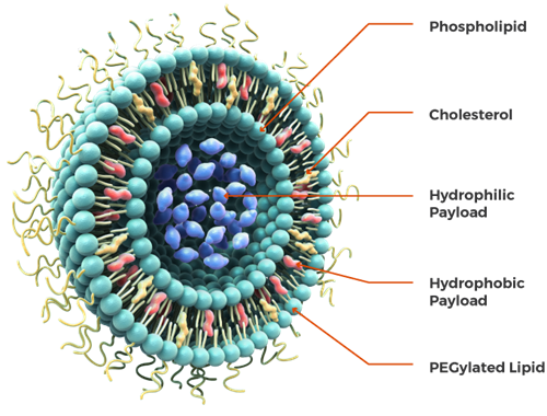
Figure 1: Structure of liposome34
History of Liposomes
In the mid-1960s, British haematologist Dr. Alec D. Bangham serendipitously discovered liposomes while investigating cell membrane structure using an electron microscope.13 These tiny spherical structures, composed of lipid bilayers akin to cell membranes, opened new avenues in biomedical research. Dr. Bangham's pioneering work laid the groundwork for comprehending and harnessing liposomes for diverse applications.16 Over subsequent decades, liposomes gained prominence, particularly in drug delivery. Their unique ability to encapsulate drugs and transport them to specific bodily sites transformed pharmacology.17 Liposomal drug delivery systems facilitated controlled release and mitigated side effects associated with many medications.18 Since then, liposomes have found utility not only in drug delivery but also in cosmetics, food technology, and gene therapy. Researchers have developed myriad liposome variations, varying in size, composition, and surface modifications, to optimize performance for specific applications.19-21. Today, liposomes remain indispensable in medicine and biotechnology, with ongoing research focused on enhancing their efficacy and versatility in delivering therapeutic agents and other bioactive compounds.22-24
Features of Liposomes:
Classification of Liposomes25-33
The Liposomes classification are based on-
Oligolamellar vesicles consist of 2 to 10 lipid bilayers surrounding a relatively large internal volume. This structure provides OLVs with increased stability and drug encapsulation capacity compared to unilamellar vesicles. They are particularly useful for sustained drug release and targeted delivery due to their multi-layered composition.
Multilamellar vesicles are characterized by the presence of several lipid bilayers, which can compartmentalize the aqueous volume in numerous ways. These vesicles vary in structure depending on the method of preparation, with possible arrangements including onion-like concentric layers of large unilamellar vesicles (LUVs) or multilamellar vesicles enclosing smaller SUVs. MLVs offer versatility in drug delivery and are commonly used for encapsulating hydrophobic and hydrophilic drugs simultaneously, allowing for controlled release and enhanced therapeutic efficacy.
Based on the method of liposome preparation, various types of liposomes with unique properties and applications can be obtained:
REV (Reverse-Phase Evaporation):
REV liposomes are typically single or oligolamellar vesicles created using the Reverse-Phase Evaporation Method. This method involves the formation of liposomes by dispersing lipids in an organic solvent, followed by evaporation to form a lipid film. Subsequent hydration with an aqueous phase result in the formation of liposomes. REV liposomes offer advantages such as high encapsulation efficiency and stability, making them suitable for drug delivery applications requiring sustained release.
MLV-REV (Multilamellar Vesicles - Reverse-Phase Evaporation):
MLV-REV liposomes are multilamellar vesicles prepared using the Reverse-Phase Evaporation Method. This technique involves the formation of multilamellar vesicles by evaporating a lipid mixture containing multiple lipid layers, followed by hydration to form vesicles. MLV-REV liposomes are characterized by their multilayered structure, offering increased drug loading capacity and potential for controlled release.
SPLV (Stable Plurilamellar Vesicles):
SPLV liposomes are stable plurilamellar vesicles characterized by their multilayered structure and enhanced stability. These liposomes are prepared using specialized methods that promote the formation of multiple lipid layers, resulting in vesicles with prolonged shelf life and improved storage stability.
FATMLV (Frozen and Thawed MLV):
FATMLV liposomes are multilamellar vesicles prepared using the Frozen and Thawed MLV method. This technique involves freezing MLV liposome suspensions followed by thawing, which induces vesicle fusion and results in larger, more stable liposomes. FATMLV liposomes are commonly used in research applications requiring vesicles with specific size and stability properties.
VET (Vesicles prepared by Extrusion Technique):
VET liposomes are vesicles prepared using the extrusion technique, which involves forcing liposome suspensions through porous membranes to control vesicle size and homogeneity. VET liposomes exhibit uniform size distribution and enhanced stability, making them ideal for drug delivery applications requiring precise control over vesicle size and drug release kinetics.
DRV (Dehydration-Rehydration Method):
DRV liposomes are prepared using the Dehydration-Rehydration Method, which involves dehydrating lipid films followed by rehydration with an aqueous phase to form vesicles. DRV liposomes are characterized by their ability to encapsulate hydrophilic and hydrophobic drugs efficiently, making them versatile carriers for various therapeutic agents.
Based on composition and application, liposomes exhibit diverse characteristics and functionalities tailored for specific purposes:
Conventional Liposomes (CL):
CLs consist of neutral or negatively charged phospholipids along with cholesterol. These liposomes serve as versatile carriers for various drugs due to their biocompatibility and stability.
Fusogenic Liposomes (RSVE):
RSVE liposomes are composed of reconstituted Sendai virus envelopes, enabling them to fuse with cell membranes and facilitate efficient drug delivery. These liposomes are particularly useful for delivering therapeutic agents to target cells with high precision.
pH-Sensitive Liposomes:
pH-sensitive liposomes are formulated with phospholipids such as PE or DOPE, along with either CHEMS or OA. These liposomes exhibit pH-dependent membrane destabilization, releasing encapsulated drugs in response to acidic conditions found in specific cellular compartments or tumor microenvironments.
Cationic Liposomes:
Cationic liposomes comprise cationic lipids in combination with DOPE. These liposomes possess positively charged surfaces, enabling efficient interaction with negatively charged cell membranes and enhancing cellular uptake of encapsulated drugs or nucleic acids.
Long Circulatory (Stealth) Liposomes (LCL):
LCLs are equipped with polyethylene glycol (PEG) derivatives attached to their surface, a process known as pegylation. This modification decreases their detection by the reticuloendothelial system (RES), prolonging their circulation time in the bloodstream. LCLs are ideal for delivering drugs requiring sustained release and extended systemic exposure.
Immuno-Liposomes (IL):
Immuno-liposomes are formulated by conjugating conventional (CL) or long-circulating (LCL) liposomes with monoclonal antibodies or recognition sequences. These liposomes selectively target specific cell types or biomarkers, enhancing drug delivery efficiency and reducing off-target effects. Immuno-liposomes hold promise for precision medicine and targeted therapy in various diseases, including cancer and inflammatory disorders.
Based on Conventional Liposomes, various subtypes are distinguished by their composition and properties:
Normalized Mixtures of Natural Lecithin (PC):
These liposomes are formulated using normalized mixtures of natural lecithin, primarily phosphatidylcholine (PC). Lecithin, a naturally occurring phospholipid found in egg yolk and soybeans, serves as the main component of these liposomes. These liposomes exhibit biocompatibility and stability, making them suitable for a wide range of pharmaceutical and biomedical applications. Their composition closely mimics natural cell membranes, enhancing their compatibility with biological systems.
Glycolipid-loaded Liposomes:
Glycolipid-loaded liposomes are formulated by incorporating glycolipids into the lipid bilayers of conventional liposomes. Glycolipids are amphiphilic molecules containing both lipid and carbohydrate moieties. By integrating glycolipids into the liposomal membranes, these liposomes gain additional functionalities such as enhanced stability, cellular targeting, and immunomodulation. Glycolipid-loaded liposomes are utilized in drug delivery systems, vaccine formulations, and biomaterial engineering due to their unique properties.
Synthetic Phospholipids with the Same Chain as Natural Phospholipids:
This subtype of conventional liposomes employs synthetic phospholipids with lipid chains identical to those found in natural phospholipids. By using synthetic phospholipids, liposome formulations can be precisely tailored to achieve desired characteristics such as membrane fluidity, stability, and drug release kinetics. These liposomes offer advantages in terms of reproducibility, scalability, and versatility in drug delivery applications. Moreover, they allow for the incorporation of specific functional groups or modifications to optimize drug encapsulation and targeting. Synthetic phospholipid-based liposomes hold promise for the development of advanced drug delivery systems with enhanced efficacy and safety profiles.
Carbohydrate Coated:
Carbohydrate-coated liposomes feature a surface layer of carbohydrates, which provide additional functionality and targeting capabilities. These liposomes can selectively interact with carbohydrate-binding proteins or receptors on cell surfaces, facilitating targeted drug delivery and enhancing cellular uptake. Carbohydrate-coated liposomes hold promise for applications in cancer therapy, immunotherapy, and vaccine delivery, where specific cell targeting is desired.
Bipolar Fatty Acid:
Bipolar fatty acid liposomes contain fatty acids with both hydrophilic and hydrophobic regions, allowing them to incorporate into lipid bilayers with ease. These liposomes exhibit enhanced membrane stability and drug loading capacity compared to conventional liposomes. Bipolar fatty acid liposomes are particularly useful for delivering hydrophobic drugs or bioactive compounds, offering improved solubility and bioavailability.
Lipoprotein Coated:
Lipoprotein-coated liposomes are enveloped by natural or synthetic lipoproteins, mimicking the structure and function of lipoprotein particles found in the body. These liposomes benefit from the targeting and transport properties of lipoproteins, enabling efficient delivery of drugs or therapeutic agents to specific tissues or cells. Lipoprotein-coated liposomes hold potential for applications in cardiovascular disease treatment, lipid metabolism modulation, and cancer therapy.
Methyl/Methylene X-linked:
Methyl or methylene X-linked liposomes are modified liposome formulations containing methyl or methylene cross-linkers between lipid molecules. These cross-linkers enhance liposome stability and structural integrity, preventing premature drug release and improving circulation time in vivo. Methyl/methylene X-linked liposomes are employed in drug delivery systems requiring sustained release profiles and prolonged therapeutic effects.
Multiple Encapsulated:
Multiple encapsulated liposomes are designed to encapsulate multiple drug molecules or therapeutic agents within a single liposomal carrier. These liposomes offer advantages such as synergistic drug combinations, controlled release kinetics, and reduced toxicity compared to conventional drug delivery systems. Multiple encapsulated liposomes are utilized in combination therapy, targeted drug delivery, and personalized medicine approaches to maximize therapeutic efficacy while minimizing side effects.
Table 1: Classification of Liposomes Based on Structural and Functional Features
|
Type of Liposome |
Description |
Size Range |
Applications |
|
Small Unilamellar Vesicles (SUVs) |
Single lipid bilayer |
20–100 nm |
Drug delivery, imaging |
|
Large Unilamellar Vesicles (LUVs) |
Single lipid bilayer with larger core |
100–1000 nm |
Gene delivery, cancer therapy |
|
Multilamellar Vesicles (MLVs) |
Multiple concentric lipid bilayers |
500–5000 nm |
Vaccine delivery, sustained release |
|
Stealth Liposomes |
PEGylated for extended circulation time |
80–200 nm |
Cancer, antifungal therapies |
|
Cationic Liposomes |
Positively charged surface |
Variable |
Gene and siRNA delivery |
|
Targeted Liposomes |
Ligand-functionalized for specific cell targeting |
Variable |
Brain/liver-specific drug delivery |
Structural Components of Liposomes35,36
Phospholipids
Phospholipids, predominantly glycerol-containing, constitute the primary component of liposome formulations, accounting for over 50% of the lipid weight in biological membranes. These phospholipids are derived from Phosphatidic acid and form the backbone of liposomal molecules. The molecular structure consists of a glycerol moiety, with the C3 hydroxyl group esterified to phosphoric acid, while the hydroxyl groups at C1 and C2 are esterified with long-chain fatty acids, imparting lipidic properties. One of the remaining hydroxyl groups of phosphoric acid can further esterify with various organic alcohols such as glycerol, choline, ethanolamine, serine, and inositol. Thus, the parent compound of this series is the phosphoric ester of glycerol.
Examples of phospholipids include:
For stable liposomes, saturated fatty acids are typically used, while unsaturated fatty acids are generally avoided due to their potential to destabilize liposome structures.
2) Sphingolipids:
Sphingolipids are essential components found in both plant and animal cells, featuring a backbone of sphingosine or a related base. They consist of three characteristic building blocks: a molecule of fatty acid, a molecule of sphingosine, and a head group that can range from simple alcohols like choline to complex carbohydrates. Among the most common sphingolipids are sphingomyelin and glycosphingolipids. Gangliosides, which contain complex saccharides with sialic acid residues in their polar head groups, are also utilized in liposome formulations to provide a layer of surface-charged groups, aiding in targeting and stability.
3) Cholesterol:
Cholesterol and its derivatives are frequently incorporated into liposomes for various purposes, including decreasing the fluidity or micro-viscosity of the bilayer, reducing membrane permeability to water-soluble molecules, and stabilizing the membrane in biological fluids like plasma. Liposomes containing cholesterol exhibit reduced interactions with plasma proteins, thus enhancing their stability and minimizing phospholipid depletion. Cholesterol acts as a "mortar" in bilayers, filling empty spaces among phospholipid molecules and anchoring them more securely. Its molecular shape and solubility properties enable it to intercalate within the bilayers, contributing to membrane stability. Typically, liposomes comprised solely of phospholipids often lack sufficient rigidity due to their low phase transition temperature and the presence of unsaturated fatty alkyl chains, which can lead to defects in the cell membrane structure during packaging. This can result in leakage of encapsulated drugs from the liposomes. To address this issue, one or two bilayer stabilizers are commonly included in liposome formulations. Among the most widely used stabilizers are cholesterol and alpha-tocopherol. The quality of liposome encapsulation varies depending on the composition of the phospholipid bilayer. Cholesterol plays a crucial role in enhancing the characteristics of liposomes. While it does not form bilayer complexes on its own, it can integrate into high concentrations of phospholipid membranes. This integration promotes rigidity through compact stacking of the bilayers and reduces the permeability of water-soluble molecules. By enhancing bilayer durability, cholesterol contributes to the stability of hydrophilic drugs encapsulated within liposomes. Moreover, cholesterol reduces fluidity above the phase transition temperature (Tc), resulting in a more ordered bilayer structure. The tricyclic ring structure of cholesterol is wedged among the first few carbons of the fatty acyl chains, with the hydroxyl group exposed to the liquid phase. This molecular arrangement allows cholesterol to fit in with the phospholipid molecules and orient itself among them. In cellular membranes, cholesterol can be incorporated at significant concentrations, with ratios of up to 1:1 or even 2:1 compared to phospholipids. The absence of cholesterol in liposomes can lead to increased interactions with blood proteins such as albumin, macroglobulin, and transferrin. These interactions make cholesterol-free liposomes more prone to instability, leading to a decline in their usage as therapeutic delivery vehicles.
4) Synthetic Phospholipids:
Synthetic phospholipids play a crucial role in liposome formulation, offering tailored properties for specific applications. Saturated phospholipids such as dipalmitoyl phosphatidylcholine (DPPC) and distearoyl phosphatidylcholine (DSPC) are commonly used for their stability and compatibility. Unsaturated phospholipids like dioleoyl phosphatidylcholine (DOPC) provide flexibility and fluidity to liposomal membranes, influencing drug release kinetics and membrane permeability.
5) Polymeric Materials:
Polymeric materials, including synthetic phospholipids with diacetylenic groups in the hydrocarbon chain, can polymerize under UV exposure, forming polymerized liposomes with enhanced permeability barriers. Lipids containing conjugated dienes or methacrylate groups are also utilized for polymerizable liposomes. Additionally, polymerizable surfactants are synthesized for liposome formulation, offering diverse properties and applications.
6) Polymer-Bearing Lipids:
Polymer-bearing lipids are designed to stabilize liposome surfaces through repulsive electrostatic forces, achieved by coating liposomes with charged polymers. Non-ionic and water-compatible polymers like polyethylene oxide and polyvinyl alcohol enhance solubility but may lead to liposome leakage. Covalently attaching polymers to phospholipids, such as diacyl phosphatidylethanolamine with PEG polymer linked via a carbon or succinate bond, yields optimal stability and functionality for liposome formulations.
7) Cationic Lipids:
Cationic lipids play a significant role in liposome formulation, offering unique properties that enhance their applications in drug delivery and gene therapy. For example, Dioctadecyl dimethyl ammonium bromide or chloride (DODAB/C) and Dioleoyl propyl trimethyl ammonium chloride (DOTAP) are commonly used cationic lipids. DOTAP, an analogue of DODAB, is frequently employed due to its favourable properties for gene delivery. Additionally, various analogues of DOTMA and cationic derivatives of cholesterol are utilized to tailor liposome characteristics for specific applications. Cationic lipids facilitate the formation of lipoplexes, which are complexes formed between cationic lipids and nucleic acids, enhancing cellular uptake and gene transfection efficiency.
8) Other Substances:
In addition to phospholipids and cationic lipids, a variety of other substances are used in liposome formulations to modulate their properties and functions. These substances include various lipids and surfactants that contribute to the formation and stability of liposomes. Many single-chain surfactants can form liposomes when mixed with cholesterol, providing versatility in liposome design. Non-ionic lipids are also utilized to enhance liposome stability and biocompatibility. Polyglycerol and polyethoxylated mono and dialkyl amphiphiles are commonly incorporated into liposomes, particularly in cosmetic preparations, due to their emulsifying and moisturizing properties. Lipids with fluoro carbon chains, whether single or double-chain, can form exceptionally stable liposomes, making them suitable for specialized applications where prolonged stability is required. Substances like sterylamine and dicetyl phosphate are incorporated into liposomes to impart either a negative or positive surface charge, influencing interactions with biological membranes and cellular uptake. Overall, the inclusion of these diverse substances allows for the customization of liposome properties to meet specific requirements in various biomedical and cosmetic applications.
Advantages of Liposomes:12,37,38,39,40
Disadvantages of Liposomes:12,37,38,40,41.
Handling of Liposomes:
Method of Preparation47,48
Liposomes can be created using various formulation methods, with the manufacturing process and the type of phospholipids utilized playing crucial roles in determining the characteristics of the final liposomes. The fabrication procedures for liposomes can generally be categorized into:
Thin film hydration method (Bangham method)
In this approach, all lipids and the hydrophobic drug are dissolved in an appropriate organic solvent within a round-bottom flask. Subsequently, the organic solvent is gently evaporated under reduced pressure to form a thin film layer.44 This thin film is then hydrated with an aqueous buffer solution at a temperature above the transition temperature (Tm) of the lipid used. The hydration solution may contain hydrophilic drugs intended for loading into the aqueous core of the liposomes. The rate at which hydration occurs directly influences the efficiency of drug encapsulation,45 with slower hydration rates resulting in higher encapsulation efficiencies.46 Control over liposome resizing, lamellarity types, and particle distributions can be achieved through extrusion using polycarbonate membranes of specific pore sizes or through the use of bath or probe sonicators. The extrusion method ensures the production of stable liposomes with enhanced encapsulation efficiency compared to sonication. Sonication typically yields small unilamellar vesicles (SUVs) but may also lead to the degradation or hydrolysis of encapsulated drugs and/or lipids. Furthermore, probe sonication exposes liposome suspensions to potential metal contamination.
Reverse-phase evaporation method48
This technique is commonly employed for encapsulating RNA and various enzymes. It involves injecting an aqueous solution of the drug into an organic solvent containing lipid, followed by sonication of the biphasic mixture. This process results in the formation of a water-in-oil type of emulsion. Subsequently, the emulsion is dried using a rotary evaporator to yield a semi-solid gel. Mechanical agitation of the gel induces phase inversion, converting the emulsion from water-in-oil to oil-in-water type. During this agitation process, some water droplets collapse to form the external phase, while the remaining portion forms the entrapped aqueous volume. This method is well-suited for encapsulating high molecular weight molecules, although therapeutic peptides may undergo denaturation due to the presence of organic solvents and the conditions during sonication.
Solvent Injection Methods47
Injection methods have been categorized based on the type of organic solvent utilized.49 In these methods, an organic solvent, containing lipids and hydrophobic active agents, is rapidly injected into an aqueous phase. For instance, diethyl ether facilitates direct solvent evaporation during the mixing process, achieved at a temperature above the boiling point of the solvent used.50 Ethanol, another solvent option, requires a 10-to-20-fold aqueous solution, and the ethanol can be subsequently evaporated under vacuum using a rotary evaporator, dialysis, or filtering. However, formulations prepared with this method often exhibit higher polydispersity indexes (PDI).51 Moreover, continuous exposure to high temperatures and organic solvents may lead to reduced stability of both the drug and lipids.52
Detergent removal method
In this method, lipids and a surfactant with a high critical micelle concentration (CMC) are dissolved in an appropriate organic solvent within a round bottom flask. Gentle evaporation of the solvent yields a thin film at the bottom of the flask.53 The lipid film is then hydrated in an aqueous solution containing the drug molecules, resulting in the formation of a mixed micelle solution.54 Subsequently, the surfactant is removed through techniques such as dialysis, size-exclusion chromatography, adsorption onto hydrophobic beads, or dilution.55-58. Finally, liposomes with large unilamellar vesicles (LUVs) are formulated after concentrating the solution.59 A notable drawback of this method is that most hydrophilic drugs tend to separate from the liposomes during the detergent removal step.60
Dehydration-rehydration method
This technique offers an organic solvent-free approach to produce Large Unilamellar Vesicles (LUVs) through sonication. The method involves directly dispersing lipids at low concentrations into an aqueous solution containing drug molecules, followed by sonication.61 Initially, a dehydration step is undertaken to evaporate water under nitrogen, creating a multilayered film that entraps the drug molecules. Subsequently, a hydration step ensues to form large vesicles encapsulating the drug molecules. While this method is straightforward, it often results in high heterogeneity in liposome sizes.47,62
Heating Method
This technique is also solvent-free. Here, lipids are directly hydrated with an aqueous solution and heated for at least one hour above the transition temperature (Tm) of the phospholipids used, in the presence of a 3–5% hydrating agent such as glycerin or propylene glycol. When cholesterol is added to the formulation, the suspension can be heated up to 100°C.63 These hydrating agents serve as stabilizers and isotonic additives, preventing coagulation and sedimentation of nanoparticles. Additionally, they offer a cryoprotective effect,64 making the heating method an efficient approach for formulating powder inhalable liposomes.47
Microfluidic channel method
The microfluidic channel method has emerged as a recent innovative approach for liposome preparation. Microfluidics technology enables precise manipulation of liquids within microscopic channels.65 In this method, lipids are dissolved in ethanol or isopropanol, and the resulting solution is injected either vertically or in the opposite direction to the aqueous medium within the micro-channels. Continuous axial mixing of the organic and aqueous solutions facilitates the formation of liposomes. To prevent coagulation and separation, surfactants are used to stabilize the liposomes.66. The microfluidic channel method controls the mixing process of organic and aqueous phases, ensuring reproducible liposomes with appropriate average size, polydispersity, morphology, and lamellarity.67
Supercritical Fluid Method
The injection method developed by Castor and Chu bears resemblance to the supercritical liposome approach introduced in 1994 by the Supercritical Fluid Method. This method generates small unilamellar vesicles (SUVs) with particle sizes ranging from 20 to 50 nm. During the procedure, lipids and cholesterol are dissolved in supercritical carbon dioxide. The hydrophilic molecule intended for trapping is then enclosed within an aqueous phase formed by rapidly expanding the solution. However, the encapsulation efficiency achieved with this method tends to be less effective compared to conventional liposome production methods.68
Sonication
Sonication stands out as one of the most widely utilized methods for Small Unilamellar Vesicle (SUV) preparation. In this process, Multilamellar Vesicles (MLVs) are subjected to sonication either with a bath type sonicator or a probe sonicator, typically under passive atmospheric conditions. However, this method has several drawbacks, including low internal volume/encapsulation efficacy, potential degradation of phospholipids and encapsulated compounds, elimination of large molecules, metal contamination from the probe tip, and the coexistence of MLVs alongside SUVs.68
There are two primary sonication techniques:
Lyophilisation
Natural extracts often undergo degradation due to oxidation and other chemical reactions before reaching their intended destination. Freeze-drying, a common technique in the production of many pharmaceutical products, has become a standard practice. The majority of these products are lyophilized from basic aqueous solutions, where water is the sole solvent that needs to be removed through the freeze-drying process.68
Purification of Liposomes:
Liposomes are typically purified using methods such as gel filtration chromatography, dialysis, and centrifugation. Sephadex-50 is commonly employed in gel filtration chromatography for this purpose. Hollow fiber dialysis cartridges may be utilized in the dialysis method. For centrifugation, small unilamellar vesicles (SUVs) in normal saline can be separated by centrifuging at 200,000 g for 10-20 hours, while multilamellar vesicles (MLVs) are separated by centrifuging at 100,000 g for less than one hour.68
Mechanism of Liposome Transport:
The effectiveness of liposome drug carriers hinges on their interaction with cells and their fate in vivo post-administration. Studies, both in vivo and in vitro, have revealed that liposomes primarily interact with cells through either simple adsorption (involving specific interactions with cell-surface components, electrostatic forces, or non-specific weak hydrophobic forces) or through endocytosis (engagement with phagocyte cells of the reticuloendothelial system, such as macrophages and neutrophils). Fusion with the plasma cell membrane, where the liposome's lipid bilayer integrates into the plasma membrane, leading to the simultaneous release of liposomal contents into the cytoplasm, is less common. Another possible interaction involves the exchange of bilayer components, such as cholesterol, lipids, and membrane-bound molecules, with components of cell membranes.68
Evaluation Or Characterization of Liposomes69-72
1. Particle Size and Size Distribution (DLS, NTA)
The particle size and size distribution are fundamental parameters that significantly influence the biological behaviour, drug release profile, and cellular uptake of liposomes. Dynamic Light Scattering (DLS) is one of the most commonly used techniques to determine the average size and polydispersity index (PDI) of liposomal formulations. A lower PDI (typically < 0.3) indicates a uniform size distribution, which is essential for consistent pharmacokinetics and bioavailability. Nanoparticle Tracking Analysis (NTA) is another powerful method that tracks the Brownian motion of individual particles in a suspension to provide more detailed insights into size distribution. The optimal size range for liposomes intended for systemic drug delivery generally falls between 50–200 nm, ensuring prolonged circulation time and enhanced tissue penetration, especially in tumor-targeting applications through the enhanced permeability and retention (EPR) effect.
2. Zeta Potential
Zeta potential is a measure of the surface charge of liposomes and is a crucial indicator of colloidal stability. It reflects the degree of electrostatic repulsion or attraction between adjacent particles in a dispersion. Liposomes with high absolute zeta potential values (either strongly positive or negative, typically > ±30 mV) are more likely to remain stable in suspension due to reduced aggregation. This parameter also influences the interaction of liposomes with biological membranes, proteins, and cells. Positively charged liposomes can enhance cellular uptake, particularly in gene and protein delivery systems, while negatively charged or neutral liposomes may offer better systemic circulation properties. Measurement of zeta potential is typically done using laser Doppler electrophoresis.
3. Encapsulation Efficiency (EE %)
Encapsulation efficiency refers to the percentage of drug or bioactive molecule successfully entrapped within the liposomal vesicles relative to the total amount used during formulation. It is a critical parameter affecting the therapeutic efficacy and dosage requirements of the liposomal drug delivery system. The EE% is influenced by factors such as the physicochemical properties of the drug (hydrophilic or lipophilic), lipid composition, preparation method, and vesicle size. High encapsulation efficiency ensures optimal drug loading and sustained release characteristics. Quantification of EE% is usually performed by separating unencapsulated drug (via ultracentrifugation, dialysis, or gel filtration) followed by spectroscopic or chromatographic analysis.
4. Morphology (TEM, SEM, AFM)
Morphological analysis provides insights into the shape, surface structure, and lamellarity (number of bilayers) of liposomes. Transmission Electron Microscopy (TEM) is widely used to observe the internal structure and confirm the spherical nature and uniformity of liposomal vesicles. Scanning Electron Microscopy (SEM) provides detailed surface images, allowing for the observation of surface texture and vesicle integrity. Atomic Force Microscopy (AFM) offers topographical mapping at nanometer resolution and allows characterization under near-physiological conditions. These microscopic techniques are essential for validating the quality and consistency of liposome formulations, particularly in research and development settings.
5. Drug Release Studies
Drug release studies are conducted to assess the rate and extent of active pharmaceutical ingredient (API) release from liposomes under simulated physiological conditions. These studies help predict the in vivo performance and therapeutic efficiency of the formulation. Typically, in vitro release studies are performed using dialysis membrane methods, Franz diffusion cells, or USP dissolution apparatus. Factors such as lipid composition, vesicle size, surface modifications, and encapsulated drug properties influence the release kinetics. Drug release profiles are often analyzed using mathematical models (e.g., Higuchi, Korsmeyer–Peppas) to understand the mechanism of release—whether it is diffusion-controlled, erosion-controlled, or a combination of both.
6. Stability Studies
Stability studies are crucial to evaluate the shelf-life and storage conditions of liposomal formulations. Liposomes are inherently prone to degradation through processes like aggregation, fusion, leakage of the encapsulated drug, or oxidation of lipids. Stability testing includes monitoring changes in particle size, zeta potential, pH, drug content, and encapsulation efficiency over time under various storage conditions (e.g., 4°C, 25°C, and 40°C/75% RH). These studies may also include stress testing, freeze-thaw cycling, and photostability testing. A stable liposome formulation ensures consistent therapeutic outcomes, safety, and regulatory compliance during commercialization.
Table 2: Characterization Techniques for Liposomes
|
Parameter |
Analytical Method |
Purpose |
|
Particle Size |
Dynamic Light Scattering (DLS), NTA |
Determines average diameter and PDI |
|
Zeta Potential |
Laser Doppler Electrophoresis |
Evaluates surface charge and colloidal stability |
|
Encapsulation Efficiency |
UV-Vis Spectroscopy, HPLC |
Measures drug loading efficiency |
|
Morphology |
TEM, SEM, AFM |
Visualizes shape, size, lamellarity |
|
Drug Release |
Dialysis, Franz Diffusion Cell |
Studies release kinetics and profiles |
|
Stability |
Storage at various conditions |
Monitors size, charge, leakage over time |
Advancements In Liposomes
Liposomes in biomedical research applications
An Experiment The use of liposomes in healthcare has the potential to provide innovative and effective therapies for a variety of pathological diseases. At the trial in vitro and in vivo stages there appears to be a significant increase in lipid-based therapeutic carrier research. Liposomes are being used to transport a broad variety of therapeutic and diagnostic components including therapeutic molecules, bioactive agents and gene therapy.19 Alterations in lipid content, charge and the inclusion of surface coatings and ligands are all being looked into to increase effectiveness, reduce RES clearance, and limit toxicity.20,21 For a number of biological applications, active targeting methods involving the conjugation of targeting ligands to the surface of liposomes have been widely explored at the early stage of the research especially following parenteral injection.22-24 Targeting ligands are utilised to enhance the selectivity of encapsulated cargo delivery to and retention in disordered cellular components with even less non-target deposition. Following the accumulation of nano carriers in affected tissues, it's likely that adding targeting moieties boosts receptor-mediated absorption of drug-encapsulated liposomes into target cells ultimately improving therapy efficacy.25,26 Despite the fact that ligand-targeted liposomes have enhanced bio distribution and therapeutic results in a majority of preclinical investigations, the positive impacts have been minimal in clinical trials thus far.27 The optimal density of targeting ligands on the surface of each liposome is yet unclear, and will most likely be determined by the molecular target's properties. We are learning more about the more relevant clinical indications for ligand-targeted liposomal formulations as a result of our comprehensive testing. Furthermore, charged lipids have been used to modify the lipid bilayer, which has gotten a lot of interest.28 The incorporation of charged lipids into the liposomal bilayer has aided in the development of bioadhesive, mucoadhesive, and nucleic acid-based delivery systems. Using triggering mechanisms for site-specific release of medicines from liposomes is another way to increase therapeutic effectiveness of liposomal formulations. Knowing the accomplishments in liposomal innovation thus far as well as the obstacles that remain, will enable further research to enhance on legacy systems and solve present translational and regulatory restrictions.29,30
Applications Of Liposome:73-78
Liposome research has seen significant growth in the past three decades, enabling the design of various types of liposomes tailored to specific needs. These liposomes can vary in size, phospholipid and cholesterol composition, and surface morphology, offering versatility for different applications. Utilizing liposome carriers, targeting organs like the liver and spleen becomes feasible, while tomography can distinguish between benign and malignant tissues effectively. In transdermal drug delivery systems, liposomes prove highly useful. Moreover, in plant cell therapy, liposomal drug delivery mechanisms play a crucial role in reducing toxicity and enhancing drug efficacy. By binding to specific cell receptors through a series of amino acids, liposomes effectively target their functional areas.
Liposomes In Pharmaceutical Applications
1. Cance Therapy
Liposomes have emerged as a powerful platform in cancer therapy due to their ability to encapsulate chemotherapeutic agents and deliver them selectively to tumor sites while minimizing systemic toxicity. One of the most well-known liposomal formulations is Doxil®, a PEGylated liposomal formulation of doxorubicin. PEGylation (attachment of polyethylene glycol chains) enhances circulation time by evading recognition by the mononuclear phagocyte system (MPS), thereby increasing drug accumulation in tumor tissues through the enhanced permeability and retention (EPR) effect. Liposomes in cancer therapy also reduce adverse effects like cardiotoxicity commonly associated with free doxorubicin. Beyond Doxil®, various liposomal formulations are under clinical investigation for delivering other anticancer agents such as paclitaxel, irinotecan, and vincristine. Overall, liposomal systems represent a promising strategy to improve the safety and efficacy of anticancer drugs.
2. Antifungal Agents
Liposomes are extensively used in the delivery of antifungal agents, especially for drugs that are poorly soluble or highly toxic. AmBisome®, a liposomal formulation of amphotericin B, is a prime example used in the treatment of systemic fungal infections like cryptococcosis, candidiasis, and aspergillosis. Traditional amphotericin B is highly nephrotoxic, but liposomal encapsulation significantly reduces renal toxicity while preserving its potent antifungal action. The lipid bilayer of liposomes sequesters amphotericin B, allowing its gradual release and targeted action at infection sites. This improved therapeutic index makes liposomal formulations a superior alternative, especially for immunocompromised patients, such as those undergoing chemotherapy or organ transplantation.
3. Vaccines
Liposomes, and more specifically lipid nanoparticles (LNPs), have revolutionized vaccine technology, particularly for mRNA-based vaccines. In the context of the COVID-19 pandemic, liposomal formulations played a critical role in the successful development and delivery of vaccines such as those by Pfizer-BioNTech and Moderna. These formulations protect the fragile mRNA from degradation by RNases and facilitate efficient uptake into host cells, where the mRNA is translated into viral proteins that stimulate an immune response. The liposomal carriers also act as adjuvants, enhancing the immunogenicity of the vaccine. The success of mRNA COVID-19 vaccines has opened the door for liposomal applications in vaccines against other infectious diseases, as well as in therapeutic cancer vaccines.
4. Gene Delivery
Liposomes serve as excellent vectors for gene therapy, enabling the delivery of genetic material such as small interfering RNA (siRNA) and plasmid DNA into target cells. Cationic liposomes, which possess a positive surface charge, can complex with negatively charged nucleic acids to form lipoplexes. These complexes protect the genetic material from enzymatic degradation and facilitate cellular uptake through endocytosis. Once inside the cell, the liposomes can escape the endosomal compartment and release their genetic payload into the cytoplasm or nucleus. This approach is widely studied for treating genetic disorders, cancers, and viral infections. Liposomal gene delivery systems are also being optimized for CRISPR/Cas9-based gene editing, offering exciting opportunities for precision medicine.
5. Antiviral and Antibacterial Therapies
Liposomes enhance the therapeutic efficacy of antiviral and antibacterial agents by improving their bioavailability, reducing toxicity, and enabling targeted delivery. For antiviral applications, liposomes have been used to deliver drugs such as acyclovir, zidovudine, and remdesivir for diseases including herpes, HIV, and COVID-19. They facilitate intracellular drug accumulation, which is essential for viral inhibition. In the case of bacterial infections, liposomes can deliver antibiotics like ciprofloxacin, vancomycin, or gentamicin directly to infected tissues or biofilms, enhancing antimicrobial activity while minimizing resistance. Liposomal systems also show promise in targeting intracellular pathogens such as Mycobacterium tuberculosis, offering potential solutions for difficult-to-treat infections.
6. Transdermal and Ophthalmic Drug Delivery
Liposomes are increasingly employed in transdermal and ophthalmic drug delivery systems due to their biocompatibility and ability to enhance drug penetration through biological barriers. In transdermal applications, liposomes can deliver drugs like anti-inflammatory agents or analgesics through the skin by merging with skin lipids and improving permeation. Elastic or deformable liposomes, such as transfersomes and ethosomes, are specially designed to penetrate the stratum corneum more effectively. In ophthalmology, liposomal formulations are used to prolong the residence time of drugs on the ocular surface, improving bioavailability and therapeutic outcomes. Liposomal eye drops have been developed for drugs like cyclosporine A and timolol for the treatment of glaucoma, dry eye, and other ocular diseases.
Table 3: Pharmaceutical Applications of Liposomes
|
Application Area |
Examples |
Advantages |
|
Cancer Therapy |
Doxil®, Marqibo® |
Reduced toxicity, EPR effect targeting |
|
Antifungal Therapy |
AmBisome® |
Decreased nephrotoxicity, improved efficacy |
|
Vaccines |
mRNA COVID-19 vaccines |
Protection of RNA, enhanced immune response |
|
Gene Delivery |
Lipoplexes with siRNA/DNA |
Efficient transfection, nuclease protection |
|
Antiviral/Antibacterial |
Liposomal acyclovir, gentamicin |
Enhanced cellular uptake, reduced resistance |
|
Transdermal Delivery |
Liposomal NSAIDs |
Improved skin penetration, sustained release |
|
Ophthalmic Delivery |
Liposomal cyclosporine |
Prolonged contact time, better bioavailability |
Liposomes In Biomedical Applications
1. Imaging Agents (MRI, CT, PET Contrast Agents)
Liposomes have gained substantial interest as carriers for diagnostic imaging agents due to their biocompatibility, structural versatility, and ability to encapsulate both hydrophilic and lipophilic contrast materials. In Magnetic Resonance Imaging (MRI), liposomes can be loaded with paramagnetic agents such as gadolinium (Gd³?) to enhance image contrast at targeted sites. For Computed Tomography (CT), iodine-containing compounds can be encapsulated in liposomes to improve the visualization of vascular structures or tumors. Similarly, for Positron Emission Tomography (PET), liposomes can carry radiotracers like fluorine-18 (¹?F) or copper-64 (??Cu) to facilitate functional imaging. Liposomal encapsulation allows for longer circulation times, targeted delivery, and reduced toxicity, thereby improving imaging precision and minimizing background noise. The integration of targeting ligands such as antibodies or peptides further enables site-specific imaging, especially in cancer diagnostics and cardiovascular imaging.
2. Theranostics (Combined Diagnostic and Therapeutic Applications)
Theranostic liposomes are multifunctional nanocarriers that combine both diagnostic and therapeutic capabilities within a single system. These formulations allow real-time monitoring of drug distribution and therapeutic response, which is particularly valuable in personalized medicine. Typically, theranostic liposomes co-encapsulate therapeutic agents (like chemotherapeutics or genes) along with imaging agents (e.g., fluorophores, MRI contrast agents, or radionuclides). For instance, a liposome loaded with doxorubicin and a near-infrared dye can be used to treat and simultaneously monitor tumor regression using optical imaging. These platforms enable controlled drug release, site-specific targeting, and non-invasive visualization, significantly improving treatment accuracy and efficacy. Theranostic liposomes are being widely researched for applications in oncology, cardiovascular disease, and neurodegenerative disorders, marking a major advancement in nanomedicine.
Table 4: Biomedical Applications of Liposomes
|
Application Area |
Liposome Role |
Specific Utility |
|
Imaging (MRI/PET/CT) |
Carrier of contrast agents |
Enhanced imaging contrast, prolonged circulation |
|
Theranostics |
Dual imaging and drug delivery |
Real-time monitoring of therapy |
|
Tissue Engineering |
Delivery of growth factors/genes |
Promotes regeneration and cell proliferation |
|
Immunotherapy |
Antigen and cytokine delivery |
Enhances immune activation or tolerance |
|
Targeted Delivery |
Functionalized liposomes |
Brain, liver, and tumor-specific delivery |
3. Tissue Engineering
In the field of tissue engineering, liposomes serve as delivery vehicles for growth factors, cytokines, and genes that promote tissue regeneration and repair. They can be incorporated into scaffolds or hydrogels to provide sustained and localized release of bioactive molecules, creating a favorable microenvironment for cell proliferation, differentiation, and extracellular matrix deposition. Liposomes can also encapsulate anti-inflammatory or angiogenic agents to enhance wound healing and vascularization in engineered tissues. Their biocompatibility and ability to merge with cellular membranes make them ideal for supporting cell–material interactions. Research has demonstrated the use of liposomal formulations in regenerating bone, cartilage, skin, and nerve tissues, establishing them as a promising adjunct in regenerative medicine strategies.
4. Immunotherapy and Immunomodulation
Liposomes are extensively utilized in immunotherapy and immunomodulation due to their ability to deliver antigens, adjuvants, and immune modulators directly to immune cells. By encapsulating immunologically active substances, liposomes can enhance antigen presentation and stimulate robust immune responses, making them ideal for vaccine development and cancer immunotherapy. Cationic or mannosylated liposomes can target antigen-presenting cells (APCs) such as dendritic cells and macrophages, improving immune recognition. In cancer immunotherapy, liposomes can deliver tumor-associated antigens, immune checkpoint inhibitors, or cytokines to modulate the tumor microenvironment and stimulate T-cell activation. Moreover, liposomes are also being studied for treating autoimmune diseases by delivering tolerogenic agents that suppress overactive immune responses. This dual role in both stimulating and suppressing immunity makes liposomes a versatile tool in immunological research and therapy.
5. Targeted Delivery to Specific Organs/Tissues
One of the most remarkable features of liposomes is their ability to be engineered for targeted delivery to specific organs or tissues, including the brain, liver, and tumors. Passive targeting is achieved through the enhanced permeability and retention (EPR) effect, particularly in tumor tissues where leaky vasculature allows nanoparticles to accumulate. For active targeting, liposomes can be functionalized with ligands such as antibodies, peptides, or aptamers that bind to overexpressed receptors on target cells—for example, transferrin or folate receptors in cancer cells, or lactobionic acid for hepatocyte targeting. In brain delivery, liposomes are modified to cross the blood–brain barrier (BBB) using ligands like transferrin or glutathione. For liver targeting, glycosylated liposomes can be directed to hepatocytes or Kupffer cells. These approaches enhance the therapeutic index of drugs, reduce off-target effects, and enable precision medicine strategies in treating complex diseases such as cancer, Alzheimer's disease, and hepatic fibrosis.
Table 5: FDA-Approved Liposomal Drug Products
|
Application Area |
Examples |
Advantages |
|
Cancer Therapy |
Doxil®, Marqibo® |
Reduced toxicity, EPR effect targeting |
|
Antifungal Therapy |
AmBisome® |
Decreased nephrotoxicity, improved efficacy |
|
Vaccines |
mRNA COVID-19 vaccines |
Protection of RNA, enhanced immune response |
|
Gene Delivery |
Lipoplexes with siRNA/DNA |
Efficient transfection, nuclease protection |
|
Antiviral/Antibacterial |
Liposomal acyclovir, gentamicin |
Enhanced cellular uptake, reduced resistance |
|
Transdermal Delivery |
Liposomal NSAIDs |
Improved skin penetration, sustained release |
|
Ophthalmic Delivery |
Liposomal cyclosporine |
Prolonged contact time, better bioavailability |
CONCLUSION:
Liposomes have revolutionized the landscape of modern drug delivery and biomedical engineering due to their biocompatibility, flexibility in drug encapsulation, and targeted delivery capabilities. Their application spans a wide range—from FDA-approved anticancer and antifungal therapies to cutting-edge mRNA vaccines and gene delivery systems. In biomedical domains, liposomes serve as powerful tools for non-invasive imaging, combined diagnostic-therapeutic systems (theranostics), and tissue regeneration. While they offer numerous benefits such as reduced toxicity and enhanced efficacy, challenges like physicochemical instability, scalability, and regulatory hurdles still need to be addressed. Future advancements in lipid nanotechnology, surface modification, and personalized medicine will likely unlock the full therapeutic and diagnostic potential of liposomal systems, solidifying their place as pivotal components in the next generation of healthcare solutions.
REFERENCES


Deepak A. Joshi*, Ganesh V. Bansode, Sneha S. Vairagkar, Madhuri M. Landge, Dasrao A. Patil, Akshay Bhashkarrao Ghanmode, Sandhya Bhagwat Puri, Madhuri Pratap Shrawane, A Comprehensive Review of Liposomes in Pharmaceutical and Biomedical Applications, Int. J. of Pharm. Sci., 2025, Vol 3, Issue 7, 3958-3983. https://doi.org/10.5281/zenodo.16573346
 10.5281/zenodo.16573346
