RJS College of Pharmacy, Kokamthan, Kopargaon.
The development of patient-specific constructions, functioning implants, and customized drug delivery systems made possible by three-dimensional (3D) printing technologies has drastically changed biomedical research.[1] By utilizing bio-inks, scaffolds, and computational design, bioprinting techniques enable the accurate creation of tissues and organs, thereby bridging the gap between naturally occurring and designed structures. Advances in computer-aided modelling, biomaterials, and stem cell biology have increased the potential for creating structures that closely mimic natural anatomy and function. Aside from tissue engineering, additive manufacturing has been used to create ex-vivo disease models, customized dosage forms, and drug-eluting implants.[3] Natural architecture-inspired biomimetic techniques have improved the creation of materials with better biological performance that are multi-scale and multifunctional. To get beyond restrictions in print resolution, mechanical strength, and biocompatibility, hybrid approaches that combine additive and traditional manufacturing are being investigated more and more. Notwithstanding these developments, there are still difficulties, such as the requirement for standardized regulatory pathways, scalability for clinical usage, and restricted vascularization in thick tissues. Future studies will focus on developing bio-ink formulations, adding stimuli-responsive smart materials, and developing 4D printing for dynamic, adaptive systems. When combined, 3D and 4D printing have enormous potential for orthopedic implants, personalized therapies, and regenerative medicine, opening the door for next-generation medical advancements.[6]
Overview Regenerative medicine and medication delivery technology have advanced significantly during the last few decades.[2] Although traditional dose forms including pills, capsules, and injectables have been used extensively to treat illnesses, these methods frequently have drawbacks such systemic toxicity, inconsistent drug levels, and inadequate site-specific action. Tissue engineering has emerged as a viable method for replacing or repairing damaged organs and tissues at the same time. The creation of bio-printed implants that combine tissue regeneration and therapeutic drug delivery is the result of these two fields coming together, made possible by 3D bioprinting.[4]
Historical Background
When 3D printing methods like stereolithography and fused deposition modeling were first developed for industrial manufacture in the 1980s, it is when 3D bioprinting got its start.[15] These methods were modified for use in biomedicine by the early 2000s, which resulted in the creation of hydrogel-based formulations of bio-inks that could encapsulate living cells and macromolecules. After printing basic tissue constructs like skin patches and cartilage scaffolds, researchers progressively progressed to more intricate structures like organ models and vascularized tissues.[6] At the same time, drug delivery methods were changing from straightforward immediate-release formulations to targeted and regulated systems that made use of implanted devices, polymers, and nanocarriers. As the scaffold promotes regeneration, the concept of embedding medications into 3D-printed scaffolds became apparent as a natural next step, enabling therapeutic molecules to be given straight to the damaged tissue. Bio-printed implants are now being researched in fields like wound healing, dentistry, orthopedics, and oncology.[11]
Advantages of 3D Bio printed Implants
1. Personalization: Using imaging data (CT/MRI scans), patient-specific implants can be created, guaranteeing anatomical precision and customized drug delivery.[7]
2. Localized therapy: Reduces systemic exposure and adverse effects by administering medications directly to the site of harm.[13]
3. Sustained release: Drugs can be released gradually over a period of days, weeks, or months using bio printed hydrogels and polymers.[17]
4. Dual functionality: Implants serve as both therapeutic agent reservoirs and structural scaffolds for tissue healing.[10]
5. Fewer surgical interventions: Recurrent operations or systemic treatments can be avoided by integrating regeneration and therapy into a single implant.[3]
6. Increased bioactivity: To promote healing and enhance results, growth hormones, stem cells, and bioactive compounds can be added.[16]
Disadvantages and Challenges
This technique has significant drawbacks despite its potential:
1. Manufacturing complexity: Bioprinting calls very sophisticated machinery, sterile conditions, and exact process control.[5]
2. Material limitations: Not all biomaterials can be printed, and it's still challenging to guarantee both mechanical strength and biocompatibility.[8]
3. Regulatory obstacles: Comprehensive safety and effectiveness assessments are necessary before bio printed implants can be approved for clinical use.[12]
4. Expensive: The procedure is costly due to the need for equipment, raw materials, and trained labor.[15]
5. Problems with cell viability: It might be difficult to keep living cells viable and functional both before and after printing.[9]
6. Drug stability: Adding medications to bio-inks may change how they work or result in an early release.[4]
Ideal Characteristics of Bio printed Implants
The following characteristics are necessary for a 3D bio printed implant used for drug delivery to be successful: biocompatibility: non-toxic, non-immunogenic materials that integrate with host tissue; mechanical stability: Sufficient strength to tolerate physiological stress while promoting tissue growth; controlled degradability: The scaffold should break down at a rate that corresponds with tissue regeneration; sustained drug release: The capacity to deliver consistent, long-term release of incorporated drugs; customizability: Easily adapted to the anatomy of each patient and therapeutic needs; sterilizability: Must withstand sterilization without compromising integrity or functionality; Vascularization potential: Should permit nutrient and oxygen transport to support cell survival.[13]
Current Applications
1. Critical-Sized Bone Defect Repair: Large bone defects that cannot mend on their own, like those caused by trauma, tumor resections, or congenital deformities, can be repaired using 3D bio printed scaffolds. The implants encourage the formation of new bone while offering structural support.[2]
2. Craniofacial Reconstruction: After surgery, customized bone grafts that are printed to fit intricate cranial and facial geometries improve both the functional and aesthetic results.[8]
3. Orthopedic Implants: Unlike conventional implants, customized implants for long bones, such the femur or tibia, are made to meet the anatomical needs of each patient. This improves integration and lowers problems.[6]
4. Drug Delivery and Growth Factor Incorporation: Bio printed scaffolds can be loaded with pharmaceuticals that promote bone regeneration, osteogenic factors, or stem cells, providing a dual purpose of biological enhancement and structural repair.[14]
5. Preclinical Disease Modeling: Before being employed in clinical settings, 3D bio printed bone constructions are used in lab settings to research bone illnesses, test medications, and model the healing process of bones.[12]
6. Spinal and Vertebral Reconstruction: By offering mechanical support and encouraging bone regeneration, patient-specific bio printed implants are being investigated for the repair of injured vertebrae or intervertebral bone structures.
7. Integration with Joint Implants: To promote osseointegration, lessen implant loosening, and increase long-term stability, 3D bio printed bone scaffolds are utilized in conjunction with joint replacements (such as hip or knee prosthesis).[7]
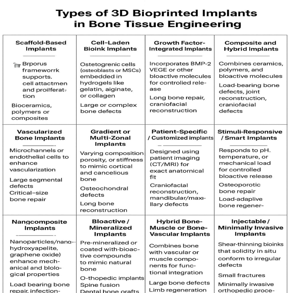
Applications of 3D Bioprinted Implants in Bone Tissue Engineering
1. Customized Repair for Bone: Defects Scaffolds customized for each patient that fit the dimensions, form, and geometry of bone deformities can be made using 3D bioprinted implants. Especially in intricate craniofacial, maxillofacial, and long bone restorations, this personalization improves implant integration and lowers surgical difficulties.[11]
2. Encouragement of Vascularization and Osteogenesis: To promote bone formation and vascularization, bioprinted constructions can be loaded with angiogenic and osteoinductive growth factors, including bone morphogenetic proteins (BMPs). The scaffold's precise cell and bioactive molecule placement maximizes the local microenvironment, facilitating quicker and more effective bone regeneration.[8]
3. Delivery of Biomaterials and Stem Cells: Mesenchymal stem cells (MSCs) and other stem cells can be added to bioprinted scaffolds to help the creation of bone tissue by differentiating into osteoblasts. The gradual integration with native bone tissue is facilitated and cell viability is guaranteed by the combination of biocompatible hydrogels and bioinks.[6]
4. Mechanical and load-bearing assistance: Implants with mechanical characteristics that can be adjusted to resemble natural bone can be created using sophisticated 3D bioprinting processes. In some situations, these implants can replace metal implants by temporarily supporting physiological stresses and encouraging tissue regeneration.[13]
5. Drug-Eluting Bone Implants: Bioprinted scaffolds can serve as local drug delivery systems, releasing antibiotics, anti-inflammatory agents, or osteogenic drugs directly at the defect site. This dual function supports bone healing while minimizing systemic side effects.[17]
6. Disease Modeling and Preclinical Testing: 3D bioprinted bone constructs provide physiologically relevant in vitro models for studying bone diseases, testing drug efficacy, and optimizing scaffold designs before clinical application. This reduces reliance on animal models and accelerates translational research.[9]
7. Complex Bone Tissue Reconstruction: Multi-material and multi-cell bioprinting allow the creation of heterogeneous structures that mimic the composition of native bone, including cortical and cancellous regions. This capability is particularly useful for reconstructing complex anatomical structures, such as the jaw or vertebrae.[14]
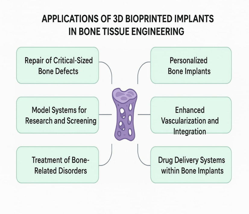

Fig – Applications of 3d bioprinting implants bone tissue.
Drug selection criteria for Dexamethasone:
|
Parameter |
Dexamethasone profile |
Relevance for bone tissue implant |
|
Drug class |
Synthetic Glucocorticoid |
Reduce local inflammation and provide controlled localised action |
|
Molecular weight |
392.46 g/mol |
Easy diffusion through polymeric or hydrogel matrices in scaffolds, enable controlled and sustain release |
|
Log p (lipophilicity) |
1.83 (moderately lipophilic) |
Moderate lipophilicity allows good cell membrane penetration |
|
Aqueous solubility |
~0.089 mg/ml (poorly water soluble) |
Limitations: can be improved with nanoparticles, microspheres and encapsulation |
|
Half-life (t1/2) |
36- 54 hrs (long acting) |
Support sustained / controlled release |
|
Therapeutic plasma level |
~5-20 ng/ml |
Plasma level guides safe systemic exposure, while local scaffold concentration is optimized independently |
|
Daily dose requirement |
0.75 - 9mg/day |
Small fraction of systemic dose to be safely deliver |
|
Current dosage forms |
Oral tablet, parentral injection, eye drop, cream etc. |
Bone tissue implant offers most relevant and promotes osteogenic differentiation |
|
Mechanism of action |
Bind glucocorticoid receptor, alters gene expression, reduce inflammation. |
Implant site: prevent immune mediated damage and improve scaffold integration |
|
Stability for bone tissue implant |
Chemically stable in dry state, neutral pH, room temperature. |
Preserve bioactivity allow controlled local release and ensure safe effective bone regeneration |
|
Suitability for bone tissue implant |
High potency, long half-life, stable dosage form |
Strong candidate for localized, sustained and osteogenic stimulation is desired |
The long-acting synthetic glucocorticoid dexamethasone has drawn interest in bone tissue engineering because of its dual osteogenic and anti-inflammatory qualities.[13] Its weak water solubility (~0.089 mg/mL) and moderate lipophilicity (LogP ~1.83) enable regulated, localized release when added to biodegradable scaffolds such hydroxyapatite composites, PCL, GelMA, or PLGA. Dexamethasone decreases inflammation and encourages mesenchymal stem cell development into osteoblasts by modifying gene expression via glucocorticoid receptors. If the dosage is carefully managed to avoid cytotoxicity or systemic side effects, its chemical stability within scaffolds, lengthy half-life (36–54 hours), and potential for sustained release make it an excellent choice for 3D-printed bone implants.[8]
Composition and Excipient Used in 3D bioprinting bone tissue implants:
|
Components |
Examples |
Function in implants |
Relevance to bone regeneration |
|
Dexamethasone (DEX) |
DEX powder, polycaprolactone microsphere, nanoparticles |
API that drives MSCs to form bone and reduces inflammation.” |
Guides stem cells to form bone, reduces inflammation, and provides controlled drug release. |
|
Biomaterial scaffold (bioink) |
Polylactic acid,gelatin, alginate, bioactive glass |
A 3D scaffold that supports cell attachment, growth, and differentiation, mimicking natural bone’s extracellular matrix. |
Porous scaffold mimics bone, supports cells and nutrients, provides mechanical strength, and guides bone regeneration. |
|
Cellular materials |
Human mesenchymal stem cells (hMSCs) derived from sources like bone marrow,adipose tissue, or dental pulp |
Serves as the implant’s living component, supplying biological elements for direct tissue regeneration. |
Patient-derived cells form new bone and improve implant integration, lowering immune rejection risk. |
|
Growth factors and bioactive molecules |
Bone morphogenetic protein 2 (BMP-2), copper ions |
Bioactive molecules that, along with DEX, initiate key processes for bone repair. |
VEGF and copper stimulate blood vessel growth, while BMP-2 boosts DEX-driven bone formation. |
|
Cross-linking agents |
Calcium chloride for alginate glutaraldehyde for gelatin |
Trigger hydrogel solidification post-printing to preserve the implant’s 3D shape. |
Cross-linking preserves hydrogel structure, maintaining shape and functionality. |
|
Plasticizers |
Polyethylene glycol (PEG), sucrose acetate iso butyrate (SAIB) |
Enhances flow and printability of polymer-based bioinks in 3D printing. |
Ensures smooth bioink extrusion for precise, high-resolution 3D structures |
|
Solvents |
Dichloromethane (DCM) for polymer solutions, phosphate-buffered saline (PBS) for aqueous inks |
Help dissolve or disperse bioink components, later removed during processing. |
Ensures uniform mixing of components before printing, with non-toxic residues. |
Methods for Preparation of 3D Bioprinted Bone Tissue Implants
1. Bioprinting using inkjet: This method involves carefully applying bioinks that include growth stimulants and osteogenic cells as tiny droplets onto a substrate. Although the variety of printable biomaterials is limited by viscosity restrictions, it enables high printing speeds and the incorporation of multiple cell types.[9]
2. Based on Extrusion: The process of bioprinting Extrusion bioprinting, the most popular technique for creating bone tissue constructions, extrudes continuous filaments of bioink using mechanical or pneumatic force. In order to simulate the mineral phase of bone, it promotes the use of hydrogels mixed with ceramic nanoparticles (such as hydroxyapatite and β-tricalcium phosphate). The technique makes it possible to build bigger scaffolds that are mechanically stable.[16]
3. Bioprinting with Laser Assistance (LAB): LAB transfers cell-rich bioinks onto a collector surface using concentrated laser pulses. This method avoids direct nozzle contact and provides good resolution and cell survival. It is especially useful for precisely positioning mesenchymal stem cells or osteoblasts within intricate bone morphologies.[11]
4. Digital Light Processing (DLP) and Stereolithography (SLA): Both techniques use UV or visible light to photo-crosslink light-sensitive bioresins. High-resolution bone scaffolds with complex internal channels that assist vascularization can be produced using SLA and DLP. Osteoconductivity is increased by adding growth factors or nanoceramics to photo-curable resins.[12]
5. Bioprinting that is hybrid: To take use of their complementing advantages, this tactic blends two or more printing methods. For example, the mechanical framework can be formed by extrusion, and cells or growth factors can be precisely seeded using inkjet or laser techniques. These combinations show promise for replicating native bone's biological milieu and load-bearing capacity.[8]
6. Maturation and Post-Processing: Constructs usually go through crosslinking, mineralization, and culture in bioreactors after printing. Functional bone regeneration requires osteogenic differentiation and the development of vascular networks, both of which are facilitated by dynamic culture conditions.[17]
Evaluation Methods of 3D Bioprinted Bone Tissue Implants
1. Analysis of Structure and Morphology: Microscopy methods including confocal microscopy and scanning electron microscopy (SEM) are used to analyze the printed constructions' layer-by-layer fidelity, pore size, and surface shape. In order to evaluate the internal structure, porosity, and interconnectivity of scaffolds—all of which are essential for vascular ingrowth and nutrient diffusion—micro-computed tomography, or micro-CT, offers high-resolution three-dimensional imaging.[10]
2. Mechanical Description: Compression testing, tensile testing, and nanoindentation are used to determine the strength, stiffness, and elastic modulus of bone implants because they must endure physiological loads. Viscoelastic characteristics are assessed using dynamic mechanical analysis (DMA) in settings that mimic those found in vivo.[14]
3. Assessment by Physicochemical FTIR and XRD: are examples of spectroscopic techniques that verify the mineral distribution, crystallinity, and chemical composition of structures. Studies on water absorption, swelling ratio, and degradation rate aid in forecasting implant stability and resorption patterns inside the body.[2]
4. Biological Evaluation in Vitro Assays: like MTT, Alamar Blue, or Live/Dead staining are used to assess cell viability and proliferation in order to ascertain the bioink's cytocompatibility. Alkaline phosphatase activity, calcium deposition tests, and gene expression indicators (RUNX2, osteocalcin, and osteopontin) are used to assess osteogenic differentiation. Co-culture research involving osteogenic and endothelial cells sheds light on their angiogenic potential.[11]
5. Preclinical Assessment in Vivo: The ability to regenerate bone, vascularization, and immune response are evaluated using animal models (e.g., rabbit femur defect, rat calvarial defect). Micro-CT measures the volume and density of bone, whereas histological staining (H&E, Masson's trichrome) verifies the creation of new bone. Biodegradation rate and functional integration with host bone are demonstrated by long-term implantation investigations.[15]
6. Testing for Functional Performance: After implantation, the load-bearing capability and resistance to mechanical stress are evaluated. To assess implant viability over time, vascular network development and nutrient delivery are evaluated.[7]
RESULTS AND DISCUSSION
The creation of bone tissue implants with exact geometry, repeatable architecture, and regulated medication distribution has shown great promise thanks to 3D bioprinting. Research continuously shows that interconnected-pore bioprinted scaffolds promote osteogenic differentiation, cell adhesion, and proliferation.[8] Incorporating bioactive substances like growth hormones, BMP-2, and dexamethasone (Dex) improves integration into host tissue by promoting osteogenesis and angiogenesis. Successful bone regeneration depends on sustained drug release from hydrogels or microspheres inside the scaffold, which has been demonstrated to lower local inflammation and sustain therapeutic levels for prolonged periods of time.[13]
Optimized polymer blends, such as PLGA, PCL, and gelatin, can produce enough compressive strength to temporarily support physiological loads while gradually deteriorating to allow for natural bone regeneration, according to mechanical
tests of printed structures. Furthermore, by demonstrating higher cell proliferation and upregulation of bone-specific markers like osteocalcin, osteonectin, and collagen type I, in vitro tests like Alamar Blue and RT-PCR validate the bioactivity of printed scaffolds.[15]
There are still difficulties in spite of these developments. Ongoing research priorities include vascularization throughout big implants, managing the spatial distribution of various cell types, and guaranteeing mechanical stability over the long term. In order to facilitate clinical translation, future research will concentrate on multi-material printing, the integration of smart biomaterials that are sensitive to the local microenvironment, and scalable manufacturing methodologies.[14]
Marketed Examples
|
Brand name |
Company |
Strength |
|
CMFlex |
Dimension Inx |
Hyperelastic Bone, a flexible composite of hydroxyapatite and PLG, is osteoconductive and promotes rapid tissue and vessel ingrowth for bone regeneration. |
|
My bone |
Cehrum |
Cerhum uses patient-specific hydroxyapatite scaffolds with a patented porous design that enhances vascularization and accelerates bone regeneration. |
|
P3D |
Ossiform |
β-TCP scaffolds with tailored porosity mimic cancellous/cortical bone, offering strength and supporting rapid, vascularized bone regeneration. |
|
OsteoFab |
Oxford Performance materials (OPM) |
PEKK-based implants offer bone-like mechanics, radiolucency, and a micro-textured surface that supports osteoconduction. |
|
RESOMER polymer implants |
Bellaseno/ Evonik |
RESOMER® bioresorbable polymers allow customizable mechanics and degradation rates to match patient-specific requirements. |
Recent research/ Advances on Dexamethasone
Delivery strategies developed recently:
1. Microspheres filled with drugs and embedded in bioinks
It is currently standard practice to encapsulate DEX in biodegradable microspheres (usually PLGA) and then combine those particles with printable hydrogels or composite inks. Microspheres shield the medication during printing and offer adjustable release kinetics (burst vs. sustained); many studies demonstrate that PLGA-DEX microspheres in printable bioinks promote better matrix mineralization and prolonged osteoinduction.[13]
2. Direct integration into composites and polymer matrices (PCL, PCL/nHAp)
DEX has been placed into solvent-cast or melt-extruded polymer blends (polycaprolactone with nano-hydroxyapatite, for instance) for gradual release. These mechanically sturdy structures can be coupled with a cell-supporting hydrogel phase and are appropriate for load-bearing locations. In order to achieve both mechanical support and local osteoinduction, recent hybrid designs combine rigid PCL-based frames with soft phases that release DEX.[16]
3. Self-healing injectable hydrogels and hydrogel microparticles
Minimally invasive injection or extrusion printing is made possible by hydrogels that are self-healing or shear-thinning that contain DEX, either free or encapsulated. They can allow cell penetration while providing a local osteogenic environment and conforming to irregular flaws. UPy-modified gelatin or cellulose derivatives are also being investigated in recent studies as matrix for extended DEX release.[6]
4. Hierarchical and sequential release systems
Some groups strive for time-staged delivery, which involves releasing DEX gradually to promote osteogenesis after an early angiogenic trigger (growth factors or ions). In animal models, this phased method has demonstrated enhanced vascular ingrowth and subsequent mineralization, reflecting physiological healing (vascularization first, followed by bone deposition).[10]
Beyond osteogenesis, biological impacts include immunological regulation and cell-to-cell communication. Significant new research demonstrates that scaffold-released DEX actively regulates local immunological responses in addition to promoting MSC development. Prolonged local DEX can improve MSC-macrophage interaction that supports bone regeneration by changing macrophage polarization toward a reparative (M2-like) phenotype.[8] It is becoming more widely acknowledged that this immunomodulatory function is a purposeful treatment mechanism as opposed to merely a side effect. Advances in printing and translating Compatibility with hybrid printing and extrusion: Printability and cell viability are maintained during extrusion by encasing DEX in particles or embedding it in distinct hydrogel phases; multiple papers from 2023–2025 show printable formulations that maintain DEX activity after printing.[13]
Composite scaffolds for load-bearing repair:
From benchtop experiments to bigger animal models, this technique combines stiff biopolymers/ bioceramics for mechanical support with a soft phase that releases DEX. Compared to non-drug scaffolds, early preclinical studies show improved bone quality and defect filling.[7]
CONCLUSION:
In addition to improving osteogenesis and enabling localized medication delivery, 3D bioprinted bone tissue implants provide a patient-specific method of bone healing.[13] Improved integration and healing are demonstrated by current research, suggesting that they could be a secure and efficient substitute for traditional grafts.[17]
REFERENCES


Pooja Lohakane, Disha Kandalkar, Bhagyashri Murtadak, Janvi Joshi, Sonali Kote, 3D Bio Printed Implants: Combining Dosage Form with Bone Tissue Engineering, Int. J. of Pharm. Sci., 2025, Vol 3, Issue 12, 3047-3057. https://doi.org/10.5281/zenodo.18000137
 10.5281/zenodo.18000137
