Department of pharmaceutics, Government College of pharmacy karad
The advantages of transdermal drug delivery over traditional methods make it seem like the most important drug delivery method. There are several ways to boost transdermal delivery, including as iontophoresis, electrophoresis, sonophoresis, microneedles, vesicular systems (liposomes, niosomes, and elastic liposomes like ethosomes and transfersomes), and chemical permeation enhancers. The transfersomal system outperformed all of these tactics in terms of efficiency. An edge activator and a lipid bilayer containing phospholipids make up the ultradeformable vesicles known as transferosomes. Transferosomes are more effective than liposomes at delivering higher concentrations of active substances to deeper layers of the skin following topical therapy. In general, molecules that weigh more than 500 Daltons are unable to pass through skin. Consequently, only a few drugs can be administered by this method. Therefore, the medication can be encapsulated in a transferosome to resolve this problem. This paper attempts to clarify the idea of transfersomes, the process, several ways to prepare and manufacture materials, characteristics that affect transfersomes, and their most recent uses in the delivery of pharmaceuticals.
In addition to intravenous and oral methods, transdermal administration is another practical and appropriate way to provide medications. Patients are spared the agony of intravenous injections, and it is especially necessary for diseases involving motion sickness, nausea, and vomiting. [1,2] The transdermal route offers a number of benefits over other conventional routes, including preventing liver metabolism, reducing undesirable side effects, delivering drugs efficiently with a short half-life, improving pharmacological and physiological response, reducing blood level fluctuations, and most importantly—improving patient compliance. [3,4] One of the main issues with dermal and transdermal drug delivery methods is the permeability of the stratum corneum. Made up of flattened, keratinized epidermal cells, the stratum corneum is the outermost layer of the epidermis. Chemical transfer is hampered by watertight cells with a strong, flexible membrane, making this mode of administration inadequate for therapeutic use. A new method for getting beyond the stratum corneum barrier without damaging it is to encapsulate the medication in a vesicular system. Vesicles are composed of an interior hydrophilic core enclosed in a bilayer wall of phospholipids or surfactants, according to structural analysis. Many researchers have introduced different types of alterations to make a novel vesicles which have significantly improved their penetration across the entire skin. A few examples of such modified vesicles include transferosomes, invasomes, flexosomes, ethosomes, menthosomes & niosomes.[5] Using a specific type of composite body called "transferosomes" and rational membrane design, a novel vesicular drug carrier system is created. Transfersomes are the most promising way to build an unique dosage form that delivers the medicine slowly and safely with minimal side effects. Transferosome is a trademark of IDEA AG and refers to patented drug delivery. The name is a mix of transfero and soma. Transferosome means to carry across in Latin, and soma means body in Greek, therefore it is a “carrying body”. Transferosomes are artificial vesicles meant to mimic a cell vesicle or a cell in exocytosis, making them ideal for controlled and potentially targeted medication delivery[6].
TRANSFEROSOMES:
The term "Transfersome" refers to an artificial vesicle that is intended to mimic the properties of a cell vesicle or a cell that is exocytosing, making it appropriate for controlled and possibly targeted drug administration. An extremely flexible and stress-responsive complex aggregation, a transfersome is a highly deformable vesicle with an aqueous core encased in a complex lipid bilayer. Because the structure of the bilayer and the local composition are interdependent, the vesicle is self-regulating and self-optimizing. This makes the transfersome more capable of successfully navigating a range of transit obstacles. [7]
Transdermal delivery makes drug distribution simple, which makes it a desirable approach. In cutaneous and transdermal drug delivery systems, the stratum corneum permeability is a major issue. The stratum corneum, which is formed of flattened, keratinized epidermal cells, is the outermost layer of the epidermis. The robust, flexible membrane of watertight cells prevents the transmission of chemicals, making this mode of administration inadequate for therapeutic use.[8] In order to get across the barrier of skin penetration, transfersomes squeeze themselves along the stratum corneum's intracellular sealing lipid. This is allowed for by the high vesicle deformability, which allows entry owing to the surrounding mechanical stress in a self-adapting way.[9]
HISTORICAL BACKGROUND:
Cevc[10]chased the term" transferosome," which refers to the first generation of ultra deformable vesicles and has been the focus of multitudinous patents and publications since the 1990s Transfersomes, a trademark of IDEA AG, Munich, Germany). The skin saturation and penetration of these elastic vesicles affect from a synergic medium among the carrier parcels and the access enrichment capability. Transfersomes are supramolecular, ultradeformable lipid packets of summations made up of at least one interior waterless member girdled by a lipid bilayer with acclimated characteristics that are suitable when surfactants( edge activator( EA)) are present in the vesicular membrane. Although liposomes are generally allowed to only access the remotest subcaste of the stratum corneum, giving them a localizing effect for medicines or cosmetics within the skin, transfersomes are said to inoculate as complete vesicles through the layers of the skin to the entire rotation. On the base of confirmation, thenon-steroidalanti-inflammatory drug( NSAID) ketoprofen was successful and veritably well- liked by consumers. Swiss Croaker, a nonsupervisory body in Switzerland, approved ketoprofen in 2007. It was retailed under the brand name" Ketoprofen transdermal" and produced by" IDEA AG" medicinals Pvt. Ltd.[11- 15]
COMPOSITION OF TRANSFEROSOMES. [16]
Despite TFS's enhanced stability profile, this deformable nanosystem is mostly made up of a number of components.Nevertheless, some are used in the synthesis of TFS, while others, including phospholipid and edge activator, are essential for maintaining its structural integrity and are covered in depth below.Table I is a list of all the frequently used ingredients for TFS production.
Table 1: Composition Of Transferosome
|
Examples |
Class |
Uses |
|
|
|
|
|
Egg phosphotidylcholine,soya phosphotidylcholine,dipalamitoyl phosphotidylcholine. |
Phospholipid |
Vesicle forming agent. |
|
Sod.cholate,sod.deoxycholate,Tween 80,span 80,Tween 20 |
Surfactant |
Vesicle forming componenent (edge activator) |
|
Ethanol,Methanol,Choloroform,Isopropyl Alcohol |
Solvent |
As a solvent |
|
Saline phosphate buffer(pH 6.4),Phosphate Buffer (pH 7.4) |
Buffering agent |
As a medium |
ROLE OF EACH INGREDIENT:[17,18,19,20,21,22,23]
1.Phospholipid:
2.Edge Activator:
STRUCTURE OF TRANSFEROSOME.
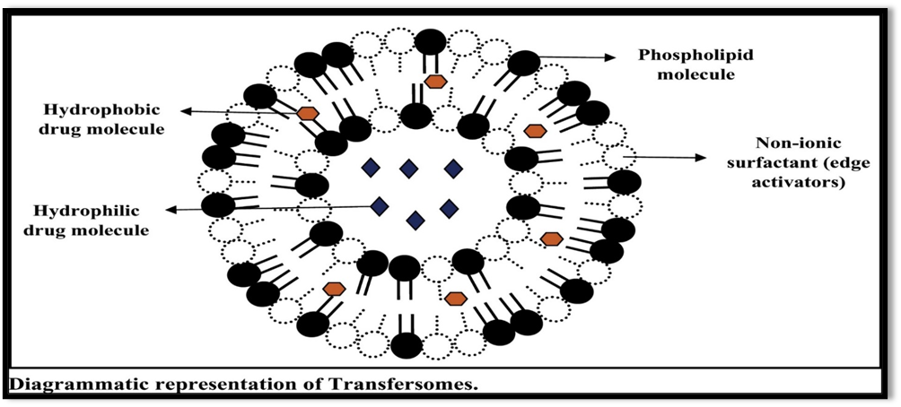
Figure 1: Diagrammatic representation of Transferosome
ADVANTAGES OF TRANSFEROSOME:[24,25,26,27]
DISADVANTAGES OF TRANSFEROSOME;[28]
LIMITATION OF TRANSFEROSOME;[29,30,31]
STRUCTURE OF SKIN;
About 15% of an adult's body weight is made up of the skin, making it the biggest organ in the body. Along with preventing excessive water loss from the body and playing a part in thermoregulation, it also protects the body from external physical, chemical, and biological threats. The mucous membranes that cover the body's surface are part of the continuous skin.[32]An average adult's skin has a surface area of around 2 m2, absorbs almost one-third of the blood that circulates throughout the body, and acts as a permeability barrier to prevent the transdermal absorption of different biological and chemical agents. The underlying blood circulation network is isolated from the external environment by the skin. It acts as a defense against assaults from microbes, chemicals, and physical forces. It maintains body temperature by acting as a thermostat. In addition to protecting the human body from UV radiation, it is crucial for controlling blood pressure. The primary determinant of medication penetration and absorption via the dermis is the skin. [10]
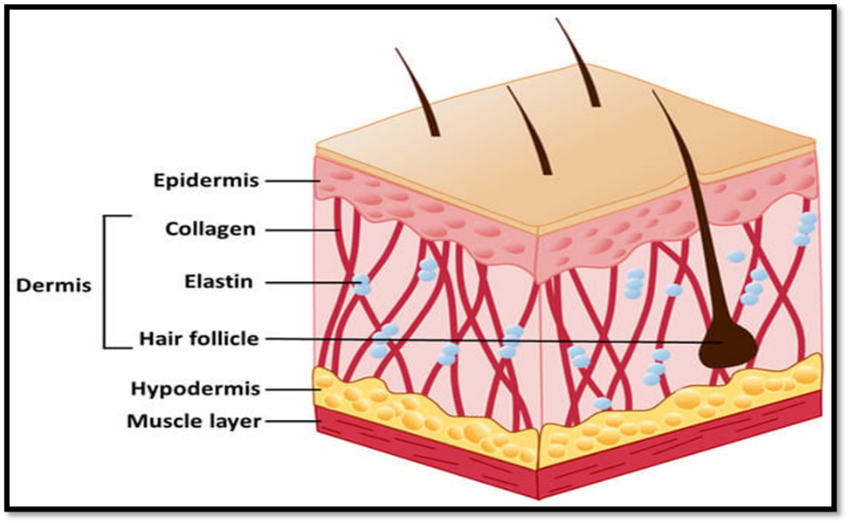
Figure 2: Structure of skin
Subcutaneous fat layer:
The layer that connects the dermis to the body's underlying tissues is called the hypodermis, or subcutaneous fat layer. This layer covers most parts of the body in a thick layer several centimeters thick. Insulation and mechanical protection against physical stress are the main functions of this layer of adipose tissue. Major blood vessels and nerves are delivered to the skin by the subcutaneous adipose layer, which may eventually offer a quick supply of high-energy molecules. [35]
Dermis:
The dermis includes sweat glands like eccrine and apocrine, pilosebaceous units like hair follicles and sebaceous glands, nerve endings, and blood and lymphatic arteries. It comprises the majority of human skin and gives the epidermis physiological support. This layer, which is typically described as simply gelled water, provides a slight barrier to the majority of polar drugs when it comes to transdermal drug delivery; nevertheless, when highly lipophilic compounds are being administered, the dermal barrier should be addressed.
Epidermis:
The epidermis consists of 10–20 cell layers. Additionally, this pluristratified epithelium contains Langerhans' cells, which are involved in immunological responses and antigen presentation, and melanocytes, which contribute to skin pigmentation. Like all epithelium, the dermal vascular network provides nourishment to the epidermis. Numerous layers make up the epidermis. Stratum germinativum is the most basic layer of the epidermis. Above the base layer are the stratum corneum, stratum lucidum, stratum granulosum, and stratum spinosum.
Stratum Corneum:
Multilayer corneocytes that are flat, polyhedral, nonnucleated, and 2-3 μm thick make up the stratum corneum, which is 10-20 μm thick. Mostly composed of insoluble bundled keratins, corneocytes are held together by covalently bound lipids and cross-linked proteins within a cell envelope. The membrane junctions known as corneodesmosomes serve to stabilize the stratum corneum by joining corneocytes. Lipids that comprise the intercellular gap between corneocytes are mostly derived by exocytosis of lamellar structures during keratinocyte terminal development.
To maintain the integrity of the epidermal barrier, these lipids are necessary. The stratum corneum serves as the skin's main defense against permeability and penetration. With a penetrating molecule that penetrates the watery environment of the underlying living epidermis and upper dermis as well as the lipophilic stratum corneum to reach the dermal vasculature, the skin can be conceptualized as a bilaminated membrane in the most basic sense.
HOW TRANSFEROSOME WORK? [9,36,37]
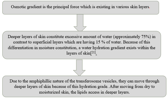
The presence of edge activators / surfactants plays a pivotal role in the enhancement of permeation of transferosomes topically & transdermally. Several researchers have reported the mechanism by which the edge activators/ surface active agents enhance the penetration of these vesicles.[38,39,40]It has been reported that existence of particular molecular inclination of surfactants, supports the vesicles to pervade the SC and in due course upsurge the drug’s transdermal permeation.[40]
TRANSFEROSOMES VS OTHER CARRIER SYSTEM:
Liposome:
Research on liposomes for many decades has been conducted to provide the medications via topical and transdermal methods. However, because of their stiff shape, liposomes cannot enter the deeper layer of skin and instead remain atop the stratum corneum.Unlike liposomes, TFS can pass through the SC barrier and deliver medications to the skin's inner layers.The edge activator makes TFS more flexible than liposomes [41].According to a study comparing liposomes and TFS, the former had a lower entrapment efficiency (49.76 ± 2.71%) than the latter (81.97 ± 1.5%) [42]. In contrast to liposomes, TFS demonstrated greater entrapment efficiency and deformability, according to another study comparing the two [43].
Ethosome:
Ethosomes can readily pass through the epidermal barrier and are also flexible.Ethosomes and TFS vary mostly in their vesicle makeup.Phospholipids and edge activators make up TFS, whereas phospholipids and ethanol make up ethosomes.Furthermore, ethosomes' increased ethanolic concentration may result in cutaneous irritation [44].
Micelle:
Furthermore, there are two ways in which TFS differ from mixed micelles.The body of a TFS vesicle is filled with water, whereas a micelle is just a fatty droplet.Thus, while mixed micelles can only entrap lipophilic medicines, TFS can transport both hydrophilic and hydrophobic medicines.In terms of size, TFS are typically one to two orders of magnitude bigger than typical lipid micelles [45].
Microneedle:
Although they both have the ability to deliver drug molecules into deep dermal tissue, TFS and microneedles have distinct methods of penetration. As the name suggests, the microneedle device consists of 10–50 µm wide and 10–2000 µm high micron-sized needles that are aligned on a patch [46]. TFS convey drug molecules across the stratum corneum, whereas microneedles send them directly into the tissues of the dermis, avoiding the stratum corneum barrier.There are further limitations related to the microneedle delivery system, including the possibility of localized inflammation at the application site, the danger of microneedle tip breaking, and skin irritation from an allergic reaction [47].
Hydrogel:
Due to physical or chemical cross-linking of the individual polymer chain, hydrogels are a three-dimensional (3D) system of hydrophilic polymers that can expand and retain a significant quantity of water while maintaining structure [48].Both hydrophobic and hydrophilic parts make into the structure of TFS, which allows them to carry both kinds of medicinal molecules.Since the hydrogels' structure is mostly hydrophilic in nature, they are incompatible with lipophilic medicinal compounds [49]. One similarity between TFS and hydrogels is that they both exhibit a high level of biocompatibility and biodegradability.
Cubosome:
Cubosomes are sustainable lipid-based nano-delivery systems made up of a stabilizing agent and phospholipid (such phytantriol and glyceryl monooleate) [50].However, TFS are made up of phospholipids (such as egg phosphatidylcholine and soy phosphatidylcholine) and edge activator.The findings of a published study comparing TFS with cubosomes revealed that TFS had a high entrapment efficiency of 84.20%, but cubosome vesicles only shown 59.82% entrapment [51]. Similar to TFS, cubosomes function as drug repositories and offer focused, regulated delivery [52].
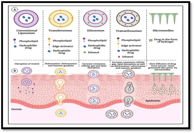
Figure 3: Transferosome Vs Other Carrier System
TRANSFEROSOME UNDER CLINICAL TRIAL:
A number of transfersome-based formulations are now being assessed at different stages of clinical studies. To treat osteoarthritis in the knees, for example, a phase III clinical trial was carried out to investigate the safety and effectiveness of ketoprofen integrated in transfersomes (Diractin®). It has been shown that the drug encapsulated in transfersomal carriers exhibits comparable fewer adverse effects and greater therapeutic efficacy in treating knee osteoarthritis pain over a six-week treatment period when compared to a placebo [53]. Similarly, topical application of insulin-loaded transfersomes (Transfersulin®) for hypoglycemic effects is being investigated under early-stage clinical trials. Transfersulin® was found to lower blood glucose levels in rabbits with alloxan-induced diabetes during the preclinical study [54]. A randomized controlled trial was conducted to evaluate the risk-benefit ratio of topical triamcinolone acetonide in transfersomes versus commercially available triamcinolone acetonidecontaining cream and ointment. It has been determined that the risk-benefit ratio of topical triamcinolone acetonide may be considerably enhanced by transfersomes [55]. As a result, transfersomes are recognized as the most exceptional and revolutionary transdermal medication carrier available today [56].
Table 2: Recent research activities on transferosome
|
Drug |
Category |
Therapeutic activity |
|
Dexamethasone |
Corticosteroid |
Anti-edema activity |
|
Diclofenac |
NSAID |
Formulation optimization |
|
Tacrolimus |
Immunosupressive |
Atopic dermatitis |
|
Pentoxifylin |
Xanthinese derivative |
Chronic occulusive arterial disease. |
|
Eprosartan mesylate |
Angiotensin receptor blocker |
Management of Hypertension |
|
Ciprofloxacin |
Quinoline antibiotic |
Otitis media |
|
Timolol maleate |
Non selective B adrenergic blocker |
Management of hypertension |
|
Ketoconazole |
Azole antifungal |
Antimicrobial activity |
|
Diclofenac diethylamine,curcumin |
NSAID and natural phenol curcuminiod |
Analgesic and antiinflammatory |
|
Itraconazole |
Antifungal triazole |
Formulation optimization |
|
Parmomycin sulfate |
Antibiotic |
Cutaneous leishmanisis |
|
Risperidone |
Antipsychotic |
Formulation optimization |
|
Minoxidil and caffeine |
Antihypertensive Vasodilator |
Alopecia |
|
Raloxifene hydrochloride |
Selective oestrogen receptor modulator |
Treatment of oesteoporosis |
|
Sinomenine hydrochloride |
Alkaloid |
Rheumatism |
|
Embelin |
Benzoquinone derivative |
Treatment of cancer |
|
Stavudine |
Reverse transcriotase inhibitor |
Prevention and tratment of HIV/AIDS |
PATENT ON TRANSFEROSOME:[58]
Table 3: Some US Patent on transferosome
|
Application number of patent |
Applicant |
Result |
|
US20020048596A1(2002) |
Gregor cevc |
The patent claim the use of nsaid in transferosome for transport through natural barrier and constriction of skin. |
|
US7175850B2(2007) |
IDEA,AG Munich(DE) |
Described the administaration of corticosteroids via transferosome on mice skin for edema suppression activity. They were tested against commercial reference cream. |
|
US20070042030A1(2007) |
IDEA,AG Munich(DE) |
It is non invasive and painless therapy,resulted in >90% of the applied drug dose reaching the desitend organ of the body. |
|
US7591949 B2 (2009) |
IDEA,AG,Munich (DE) |
Claimed the penetrant capability of transferosome because these deformable complex droplets adapt the pore of the skin along the natural moisture gradient rather than coalescing locally. |
METHOD OF PREPARATION OF TRANSFEROSOME [59,60,61,62,63,64]
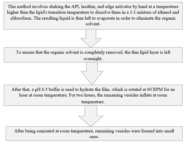
1.Rotary Film Evaporation method:
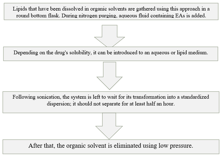
2.Reverse Phase Evaporation Method
The design will now change to a viscous gel, and then the vesicles will be arranged. Dialysis or centrifugation can be used to make the nonencapsulated material and leftover solvents indifferentiable.
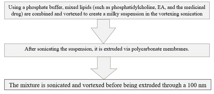
3.Vortexing sonication method:
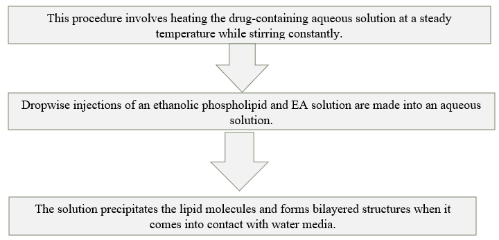
4.Ethanol Injection Method
EVALUATION OF TRANSFEROSOME:[65]
Transferosomes are referred to as liposomes, niosomes, or micelles. It is necessary to address the following transferosome characterisation requirements.
1.Vesicle size distribution and zeta potential; Vesicle size, size distribution, and zeta potential were evaluated using the Malvern Zetasizer Dynamic Light Scattering instrument.
|
(Total number of Transferosomes counted × dilution factor × 4000) / Total number of squares counted is the total number of Transferosomes per cubic mm. |

3. Entrapment efficiency. The proportion of medication entrapment is used to compute entrapment efficiency. A mini-column centrifugation technique was used to separate the unentrapped medication in order to ascertain the entrapment efficiency. 0.1 percent Triton X-100 or 50 percent n-propanol were used to rupture the vesicles during centrifugation.
The entrapment efficiency is expressed as:

4. Drug content : According to the pharmacopoeia drug's analytical method, one of the instrumental analytical techniques, such as the modified high-performance liquid chromatography method (HPLC) with a UV detector, column oven, auto sample, pump, and computerized analysis software, may be used to determine the drug content.
5. Degree of deformability or permeability measurement : Permeability analysis is one of the most crucial and unique features for characterizing transferosomes. The deformability test uses pure water as a control. The process of creating transferosomes involves passing them through a huge number of different-sized pores. Particle size and size distributions are recorded using dynamic light scattering (DLS) measurements following each pass.
6.Penetration ability : Fluorescence microscopy can be used to evaluate the penetration capacity of transferosomes.
7.Surface charge and charge density :. Zetasizer can be used to measure the surface charge and charge density of transferosomes.
8. In vitro drug release: The penetration rate is determined using an in vitro drug release research. Prior to more costly in vivo study, the formulation is optimized using data from in vitro trials, the time required to reach steadystate permeation, and the permeation flow at a steady-state. Transferosome suspension is incubated at 37°C for several hours in order to measure drug release. Samples are obtained at various points in time, and the free drug is separated by microcolumn centrifugation. The initial amount of drug entrapped is used as the starting point for an indirect calculation of the amount of drug released.
9.In vitro Skin permeation Studies :This study made use of a modified Franz diffusion cell, which has an effective diffusion area of 2.50 cm2 and a receiver compartment capacity of 50 ml. Goat skin was used in an in vitro drug research in a phosphate buffer solution (pH 7.4). The skin from the abdomen of a fresh goat that was acquired from the slaughterhouse was used for the penetration tests. A typical saline solution was used to moisturize the skin following the removal of the abdominal hairs.
The adipose tissue layer was removed by rubbing the skin’s adipose tissue layer with a cotton swab. The skin was maintained at 0-40°C in an isopropyl alcohol solution. The treated skin was placed horizontally atop the receptor compartment of the Franz diffusion cell, with the stratum corneum side looking upwards toward the donor compartment. The effective penetration area from the donor compartment to the receptor compartment was 2.50cm2, and the receptor compartment had a capacity of 50 ml. A magnetic bar was used to swirl 50 ml of phosphate-buffered saline (pH 7.4) into the receptor compartment at 100RPM.
The formulation was applied to the skin (equivalent to 10 mg of medicine), and the diffusion cell’s top was covered. 1 ml aliquots of the receptor medium were taken at regular intervals and replaced with an equal quantity of fresh phosphate buffers to maintain sink conditions (pH 7.4). Correction factors were used to compute the release profile for each aliquot. The materials were examined using any instrumental analytical approach.
10.Physical stability :The original quantity of medication entrapped in the formulation was determined, and it was preserved in sealed glass ampoules. The ampoules were stored at 4 ± 2°C, 25 ± 2°C, and 37 ± 2°C for at least three months. Samples from each ampoule were analyzed after 30 days to check whether there was any pharmaceutical leaking. By keeping the original drug entrapment at 100%, the percent drug loss was calculated.
When it comes to topical drug administration, transferosomes—lipid-based nanosystems—offer benefits like improved solubility and permeability for medications with low bioavailability . In numerous trials, they have been used to administer a variety of medications, including as ferulic acid and antifungal agents. Here are a few noteworthy uses of transferosomes:
Transferosomes are a successful non-invasive therapeutic delivery system for these big molecular weight medications. Insulin is often supplied through an uncomfortable subcutaneous method. Transfersulin, which is insulin encapsulated in transferosomes, solves all of these issues. The initial indications of systemic hypoglycemia appear 90 to 180 minutes after applying transfersulin to intact skin, depending on the particular carrier composition.
A Box-Behnken design was used in a study to encapsulate carvedilol, a medication with a limited bioavailability (25–35%), in transferosomes loaded with nanostructured lipid carriers (NLC). Comparing the modified formulation to a traditional formulation, better dermato pharmacokinetic and pharmacodynamic characteristics were observed.
The Rotary Flask Evaporation-Sonication method was used to create transferosomes with an antifungal drug. The Plackett-Burman design was used to determine important formulation and process factors, such as the volume of ethanol and hydration medium, the amount of lipid and surfactant, and the hydration time, that have an impact on vesicle size .
The transdermal delivery of anti-cancer medications such as methotrexate was investigated using transferosome technology, with encouraging outcomes and a viable therapeutic approach, especially for the treatment of skin cancer. In addition to various therapeutic uses, research has demonstrated the potential of transferosomes in delivering phytoconstituents with anticancer effects. Transferosomes may be useful for the targeted delivery of anti-cancer drugs, according to these findings, and they merit more research in the area of cancer treatment. Transferosome-embedded paclitaxel hydrogels were used by Jiang et al. (2018) to exhibit successful topical chemotherapy for melanoma, demonstrating effective tumor tissue penetration with components of sodium deoxycholate, tween 80, and phosphatidylcholine .
There are serious gastrointestinal adverse effects from several NSAIDs. Transferosome-based transdermal distribution provides an answer to these problems. Research on diclofenac and ketoprofen shows encouraging outcomes. Notably, in 2007, Swissmedic, a regulatory body in Switzerland, approved a transferosome formulation of ketoprofen, which was to be sold under the name "Diractin." IDEAAG states that other therapeutic solutions using transferosome technology are still in the clinical development stage. Conversely, non-steroidal anti-inflammatory medications, or NSAIDs, are used to treat fever, pain, and inflammation. Their prospective application in the pharmacotherapy of cancer, diabetes, cardiovascular, and neurological illnesses has been assessed .
CONCLUSION:
The transdermal route has been the most popular way to administer drugs due to its special and adaptable qualities. But the main issue with transdermal distribution is that the stratum corneum is impenetrable, which makes it impossible for medications to enter at all. Therefore, the transferosomal system focuses on successfully delivering amphiphilic substances as well as hydrophilic and hydrophobic medications. Transferosomes are a good and appropriate method because of their increased topical applications, greater loading capacity, decreased dosing frequency, and improved stability features. Site-specific active drug delivery is a promising application for transferosomes, which are also used in a variety of cosmetic procedures. There are still a number of issues that need to be addressed with regard to purity, retention properties, and oxidative degradation. Therefore, additional considerations and technology developments are necessary for every prospective process improvement. Additionally, to enable future prospects of these brilliant nanocarriers, improvement in synergistic potential of components and active compounds also has to be researched across the globe. It is also emphasized that in order to obtain the data necessary to determine the safety aspect of difficult medications prior to industrial scale-up, advanced research based on convincing preclinical and clinical investigations is needed. Advances in scientific perspectives are still required for the creation of novel transferosomes, which are likely to concentrate on better treatment plans employing more sophisticated, promising, and well-structured new techniques. To reduce the current disadvantages of transferosomes, it is also critical to investigate novel medicinal excipients with extra properties. Industrial pharmaceutical firms may investigate novel prospects for important developmental properties of transferosomes with suitably customized features in the future.
REFERENCES


Rutuja Kadam*, A. H. Hosmanii, S. V. Potdar, R. M. Savakhande, S. G. Patil, Transfersomes: Pioneering Nanocarriers for Enhanced Drug Delivery, Int. J. of Pharm. Sci., 2025, Vol 3, Issue 3, 1-17. https://doi.org/10.5281/zenodo.14950131
 10.5281/zenodo.14950131
