Dr. Rajendra Gode College of Pharmacy, Amravati.
Cancer remains the second leading cause of death in the United States, as reported by the American Cancer Society. Over the past five years, the field of theranostic nanomedicine— which integrates both treatment and imaging—has emerged as a promising approach for delivering precise, timely, and personalized cancer therapies. This review highlights recent advancements in this area, primarily from the authors' research, and explores its potential medical applications. It covers fundamental principles, key characteristics, and future directions while addressing the challenges that lie ahead in theranostic nanomedicine research. Theranostic nanomedicine, an emerging field integrating diagnostics and therapy, offers a promising approach for personalized cancer treatment. By leveraging nanotechnology, theranostic agents enable real-time monitoring of treatment efficacy while delivering precise therapeutic payloads. This review discusses the latest advancements, design principles, clinical applications, and future prospects of theranostic nanomedicine in oncology.
Cancer is a major health issue, with about 1.66 million new cases expected to be diagnosed in 2013 alone, according to the American Cancer Society. Sadly, cancer is also projected to cause nearly 580,350 deaths that year, making it responsible for almost 1 in every 4 deaths. Early detection and effective treatment are crucial for saving lives, and a major contributor to cancer-related deaths is the spread of cancer cells, known as metastasis. This happens when cancer cells, called circulating tumor cells (CTCs), enter the bloodstream. Because of this, scientists and doctors are focused on developing new ways to detect CTCs at earlier stages, which can help doctors treat patients more effectively and predict the progression of cancer. Although CTCs have been known since 1869, they are generally only detectable in patients with advanced cancer. Detecting them early is particularly difficult because they appear in extremely low numbers—just a few CTCs per million healthy blood cells. To address this challenge, improving methods to separate and enrich these cells is essential. One promising approach involves using nanomaterials as agents for both detecting and treating cancer. While this technology is still in its early stages, it holds a lot of potential for improving cancer detection and treatment in the future. Nanoparticles, with their unique properties, could play a key role in this new era of cancer care. Nanomaterials have unique photothermal properties, making them useful for both detecting cancer and treating it noninvasively through photothermal therapy. Over the past five years, researchers have shown that it's possible to combine diagnosis and treatment into a single, multifunctional nanomaterial known as "theranostic nanoparticles." The ideal theranostic material should have a few key qualities: (1) the ability to selectively accumulate in diseased tissue, (2) the ability to deliver targeted therapeutic actions, and (3) safety, meaning it can be broken down by the body into harmless substances. We believe that creating effective theranostic materials is crucial for the early detection and treatment of cancer in the modern era. In this review, we’ll discuss recent breakthroughs in the development of "theranostic nanomedicine" based on our own research. We’ll also explore where the future of this exciting field could take us in terms of research and real-world applications for treating cancer.
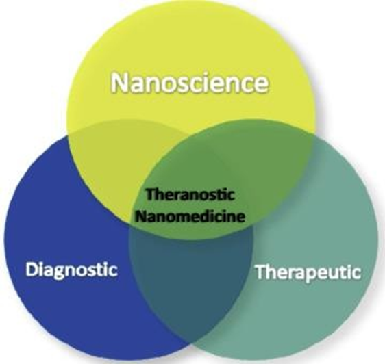
Fig1: Shows A Diagram That Combines Both Therapeutic and Diagnostic Functions into A Single Theranostic Nanomedicine
2.Theranostic nanomedicine Over the past decade, researchers have shown that nanoparticles ranging from 4 to 100 nanometers—around 1,000 to 10,000 times smaller than regular human cells—interact strongly with biomolecules like enzymes,
receptors, and antibodies, both on the cell surface and inside cells. These nanoparticles can be chemically modified on their surface, allowing them to be coated and functionalized with various bioconjugates for specific detection and treatment purposes. This ability has led to exciting progress in nanomedicine, particularly in cancer detection, diagnosis, and treatment. Thanks to advances in nanoscience, we're now able to integrate three critical functions—targeting, diagnosis, and therapy—into a single nanomedicine. This innovation, called theranostic nanomedicine, combines both therapeutic and diagnostic roles into one nanoparticle, creating a powerful tool for personalized medicine and cancer care.
4.Theranostic nanomedicine for cancer Therapy Cancer nanotechnology therapeutics go beyond simply acting as carriers for drugs or biomolecules, like proteins or siRNA. Theranostic nanomedicine, in particular, offers the potential to address several of the challenges previously mentioned. One major advantage is that nanoparticles can deliver drugs directly to tumor sites, ensuring more precise treatment. They also allow for controlled release of specific drugs at different locations, which improves the effectiveness of the therapy. Additionally, by altering the pharmacokinetic profile of the drugs, nanoparticles can increase the half-life of the drugs at disease sites, meaning they remain active for a longer time. To tackle these challenges, researchers are engineering nanomaterials that can actively bind to specific cells after they leave the blood vessels (extravasation). This selective binding is accomplished by conjugating biomolecules, such as antibodies, aptamers, or peptides, with the nanomaterials through chemical bonds. These functionalized nanoparticles will then recognize and attach to target cells, ensuring the drug reaches the precise location where it’s needed most.
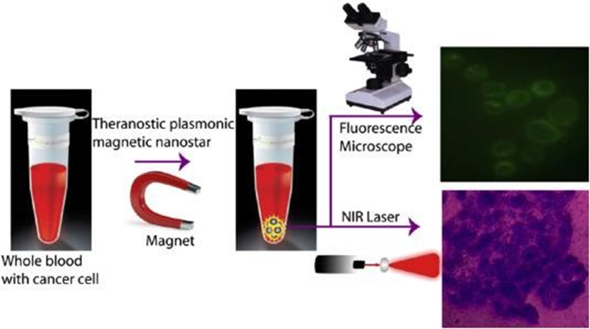
Fig 2 A Process Where Specific Cancer Cells Are Separated Using S6 Aptamer-Conjugated Plasmonic/Magnetic Nanoparticles.
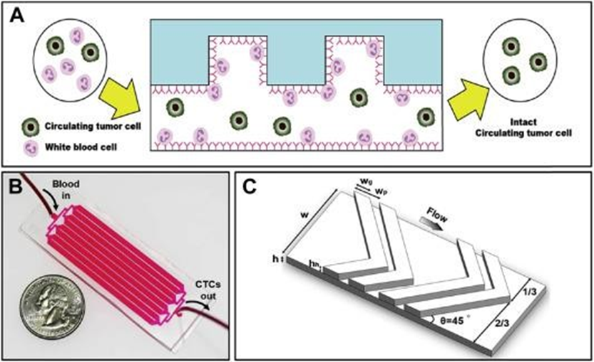
Fig 3. (A) Pictorial presentation of negative enrichment (side view). When the blood sample enters the inlet, the nontarget cells (i.e., leukocytes) are specifically captured on the CD45 antibody-coated channels, whereas the target cells (i.e., CTCs) are eluted through the outlet. The ridges on the top of the main straight channel create a helical flow that creates considerable interaction between the cells and the antibody-coated channel surface. (B) A photograph of the fabricated GASI chip. The GASI chip consists of an inlet, eight channels, and an outlet. Each channel is 2.1 mm in width and 50 mm in length. To prevent breakdown of the PDMS channel due to their flexibility, a wall 400 mm in width bears the pressure. (C) herringbone structure and key parameters used in this study
3.Types of Theranostic Nanoparticles
Various nanoparticles have been developed for theranostic applications, including: Lipid-based nanoparticles (liposomes, micelles): Used for effective drug transport and imaging.
Polymeric nanoparticles: Serve as biodegradable
carriers for controlled drug release.
Metallic nanoparticles (gold, silver, iron oxide): Applied in photothermal therapy and MRI contrast enhancement.
Quantum dots: Aid in fluorescent imaging and drug tracking.
Carbon-based nanoparticles (graphene, carbon nanotubes): Employed for drug delivery and
thermal-based cancer treatment.
4.Applications in Cancer Management
a. Precision Drug Delivery
Nanoparticles (NPs): Various carriers such as liposomes, dendrimers, and polymer-based nanoparticles can transport chemotherapy drugs
directly to cancerous tissues, minimizing harm to healthy cells. Enhanced Permeability and Retention (EPR) Effect: Due to the leaky blood vessels in tumors, nanoparticles accumulate at the tumor site, increasing drug concentration where needed.
Controlled Drug Release: Smart nanocarriers can
be programmed to release medication in response to specific stimuli, including temperature, pH
changes, or enzymatic activity.
b. Early Cancer Detection and Diagnosis
Quantum Dots & Gold Nanoparticles: These nanomaterials improve imaging precision,
allowing for the early identification of tumors with exceptional sensitivity.
Magnetic Nanoparticles: Used in MRI scans to enhance contrast, leading to more accurate tumor visualization.
c. Photothermal & Photodynamic Therapy (PTT & PDT)
Gold & Carbon-Based Nanoparticles: These particles absorb light and generate localized heat, selectively destroying cancer cells.
Photosensitizers: Encapsulated in nanocarriers, these light-activated drugs achieve better
solubility and targeting for photodynamic therapy.
d. Gene Therapy & Immunotherapy
Nanovectors for Gene Editing: Deliver gene-modifying tools like CRISPR/Cas9, siRNA, or miRNA to alter cancer-related genes.
Nanoparticles in Immunotherapy: Enhance the transport of immune-stimulating agents such as
cancer vaccines, cytokines, or immune checkpoint inhibitors, boosting the body's defence against
tumors.
e. Theragnostic (Combination of Therapy & Diagnostics)
Multifunctional Nanoparticles: These allow
simultaneous imaging and treatment, enabling real-time monitoring of therapeutic progress.
f. Combating Drug Resistance:
Multidrug-Loaded Nanoparticles: Deliver multiple drugs together, attacking cancer through different mechanisms and reducing the likelihood of resistance.
Nanocarriers for Efflux Pump Inhibition: Prevent cancer cells from expelling chemotherapy drugs, increasing treatment effectiveness.
5.Challenges and Future Directions
While promising, theranostic nanomedicine encounters several hurdles, including:
Biocompatibility and Toxicity: The long-term effects of nanoparticles require extensive
research.
Manufacturing and Scalability: Consistent large- scale production remains a challenge.
Regulatory Approvals: Clinical acceptance demands rigorous safety and efficacy evaluations.
Looking ahead, advancements in AI-driven nanoparticle design, intelligent biosensors, and patient-specific treatment plans will further enhance the effectiveness of theranostic nanomedicine, making cancer therapy more precise and individualized.
REFRENCES


Narendra Umale*, Akash Malthankar, Nurvi Deshmukh, Sakshi Thakur, Dr. H. Sawarkar, Theranostic Nanomedicine for Cancer Diagnosis and Treatment, Int. J. of Pharm. Sci., 2025, Vol 3, Issue 3, 1379-1384. https://doi.org/10.5281/zenodo.15030636
 10.5281/zenodo.15030636
