1Department of pharmaceutics R. R. College of pharmacy chikkabanavara, Bangaluru
2Department of pharmaceutical Chemistry Vutkoor laxhmiah College of Pharmacy Raichur
3TVM College of Pharmacy, Bellary, Karnataka
A straightforward and efficient UV-Spectrophotometric technique was created and approved for the measurement of diclofenac sodium in tablet form [1]. Diclofenac standard solution was scanned in a 1 cm quartz cell using a twin beam UV spectrophotometer in the UV range (400-200 nm) [2]. With a %RSD of less than 2%, the approach proved reliable and exact. Up to four hours, the solution was found to be stable. In accordance with ICH criteria, the suggested technique was precise and targeted (Q2) [3].
Chemically, Diclofenac Sodium (DS) is the sodium salt of acetic acid 2-[{2,6-dichlorophenyl} amino] benzene [4]. It has antipyretic, analgesic, and anti-inflammatory properties in both humans and animals. It is frequently employed to treat cancer-related persistent discomfort [5]. Diclofenac sodium is a member of the cyclo-oxygenase (COX) inhibitor or non-steroidal anti-inflammatory medication (NSAID) family [6]. Numerous clinical illnesses, including degenerative joint diseases, acute and chronic musculoskeletal disorders, sports injuries, and post-surgical analgesia in people and animals, are associated with pain, fever, and inflammation that are commonly managed with it. It has anticancer properties as well [7]. Diclofenac sodium works as an analgesic by inhibiting COX-2, which lowers the amount of inflammatory prostaglandins that are made from arachidonic acid [8]. Many pharmaceutical companies are currently producing diclofenac under their own brands in Bangladesh due to high demand. One of Bangladesh's industries with the quickest rate of growth is pharmaceuticals, which exports medications to Europe and other international markets. Adequate dosage of medications is crucial for alleviating symptoms and minimising adverse effects; without it, accurate illness management is unattainable. Diclofenac is a necessary medication that is produced locally by businesses so that everyone can access it. It is a non-prescription medication that is accessible in developed nations as well [9]. One area of research that examines how electromagnetic radiation interacts with matter is called spectroscopy. The matter absorbs or emits energy in definite amounts known as quanta, which is the most significant effect of such interaction. One of the most effective tools for studying atomic and molecular structure is spectroscopy, which is applied to the investigation of a variety of samples. The area of the electromagnetic spectrum between 100 Å and 400μm is included in optical spectroscopy [10]. According to various studies (Adeyeye& Li, 1990; Reynold, 1993; Willis et al., 1979; Degen et al., 1988; Said &Sharaf, 1981; and Landesdorfer, 1990), the half-life of diclofenac sodium in plasma ranges from one to three hours. The mean peak plasma levels of approximately 0.5 µg/ml and 1.0 µg/ml are observed approximately two hours after single doses of 25 mg and 50 mg of enteric-coated tablets, respectively (Riess) [11]. One of the most helpful NSAIDs is diclofenac sodium, which dissolves in water and intestinal fluid but is nearly insoluble in acidic solutions (pKa=4.0). Several swellable controlled-release pharmacological dosage forms have been developed in an attempt to eliminate the gastrointestinal side effects of this medicine. Velasco et al. assessed the effects of the drug, the polymer, the ratio of Diclofenac Sodium to HPMC, and compression force on drug release from HPMC matrices. Their findings indicated that the drug primarily controls the rate and mechanism of drug release [12]. There are several ways to administer DIC, including orally, rectally, or intramuscularly. DIC is a cyclooxygenase inhibitor with a potency that is significantly higher than that of naproxen, indomethacin, and a number of other medications (2–5). Although DIC has been used extensively in clinical settings for more than 20 years, little is known about its pharmacokinetics in animals, particularly rats [13]. The International Conference on Harmonisation (ICH) standards for specificity, linearity, limit of detection (LOD), limit of quantification (LOQ), accuracy, precision, and robustness were all met by the method's validation. With a lower limit of detection of 12.5 ng/ml, the calibration curve was linear over the concentration range of 10 to 200 µg/ml. The method's precision and accuracy fell between the permissible ranges of ±15% at other concentrations and ±20% at the lower quantitation limit. Diclofenac was unstable at room temperature; after 24 hours, there was a loss of more than 25% [14]. Currently, government laboratories, the pharmaceutical industry, and other businesses place a premium on the development of appropriate techniques for the routine analysis of pharmaceuticals in pharmaceutical preparations. Pharmaceutical development, production, and quality control programs depend heavily on the effectiveness of analytical quality. International pharmacopoeias have tightened their standards in recent years for the quality control of pharmaceuticals, requiring advanced pharmaceutical analysis together with quick, dependable, and affordable outcomes [15]. Its main mode of action is the inhibition of prostaglandin synthesis through inhibition of cyclooxygenase (COX), which also has antipyretic and analgesic properties. By preventing the synthesis of bacterial DNA, it also seems to have bacteriostatic effects (Dutta et al., 2007). Additionally, inhibition of COX reduces prostaglandin in the stomach epithelium, increasing the sensitivity of the epithelium to gastric acid corrosion. Additionally, this is the primary side effect of diclofenac. A number of techniques have been put up for diclofenac analysis. An exact and reliable spectrophotometric method for determining the presence of diclofenac sodium in bulk materials and pharmaceutical preparations is described by Sastry et al. (1989) [16]. The method's precision and recovery were determined to be within acceptable bounds. The suggested approach was verified in accordance with ICH Guidelines and tested for accuracy, linearity, precision, limit of quantification (LOQ), and limit of detection (LOD). Beer-Lambert's law is followed by the drug in concentration. The suggested techniques were discovered to be easy to use, highly accurate, repeatable, economical, and suitable for practical quality control analysis. [17]. The Beer-Lambert law is the underlying principle that underpins quantitative spectrophotometric analysis.
Beer's Law
It says that as the number of absorbing molecules increases, the intensity of a parallel monochromatic radiation beam drops exponentially. Stated differently, absorbance is directly related to concentration.
Lambert's legislation
It says that as a beam of parallel monochromatic radiation travels through a medium with a uniform thickness, its intensity drops exponentially. The result of combining these two laws is the Beer-Lambert law [18]. Before and after formulation, drug and excipient quality controls, as well as quantitative analysis, are commonly carried out using UV-vis spectrophotometry. Even for the most basic single-component drug systems, spectral overlap and non-specific irrelevant absorption can cause varying intercepts on the absorbance axis and systematic mistakes in the graphs of absorbance vs concentration, making it difficult to interpret the data [19]. Neural networks are known to be superior to other chemometrics techniques like principal component regression (PCR) and partial least squares (PLS) in the modelling of systems with non-linear signal-answer dependencies. As a result, there is a steady increase in the use of neural networks for spectral data analysis. However, networking techniques are mostly employed as tools for categoriation, and it is uncommon to use this approach to solve quantification problems [20]. The most common way that drug compounds are given orally is through solid dosage forms, such as tablets and capsules. Tablets are solid unit dosage forms with flat or convex surfaces that are compressed or moulded into solid cylindrical shapes holding a medication and excipients. Tablet shapes include oblong, triangular, cylindrical, and spherical. A coating is put on a tablet to change how the active substance released from it. Nowadays, many tablets are coated because this can improve the look, prevent the components from breaking down, and mask or lessen the disagreeable taste of some medications [21]. Remove DCF from water resources through degradation by ultraviolet (UV) irradiation in the presence of different catalysts. This study assessed the best way to degrade DCF under UV light irradiation using biogenic selenium nanoparticles (Se NPs) in the presence of H2O2 (with the help of central composite design) in a photoreactor, and then identified the metabolites that were produced (using the GC-MS (EI) technique). Light intensity (W/m2), Se NPs concentration (μg/mL), pH, and H2O2 concentration (mM) were the four key factors that were selected in order to evaluate the impact of parameters on the UV/Se NPs/H2O2-assisted degrading efficiency of DCF [22]. Many NSAIDs' efficacy in diverse clinical contexts has been well assessed. They are frequently used as monotherapy in rheumatologic practice to treat osteoarthritis. The most prevalent type of arthritis is osteoarthritis (OA), sometimes referred to as osteoarthrosis or degenerative joint disease. One prevalent cause of long-term impairment in adulthood is osteoarthritis (OA). Although prevalence varies by population, it always rises with age. A certain percentage of these will be symptomatic and eventually develop into disabilities. The distal and proximal interphalangeal joints, as well as the first carpometacarpal joint, are frequently impacted by hand OA. Osteoarthritis-related X-ray abnormalities have been detected in 22.1% of hand joints in males and 32.7% in women over 70. An aging population means that OA will always have a socioeconomic impact on North America [23]. Pain is a result of joint deterioration and inflammation associated with OA, which leads to functional restrictions, increased use of healthcare resources, and a lower standard of living among those who suffer from this illness [5, 14, 15]. Nonsteroidal anti-inflammatory medicines (NSAIDs) are widely recommended for the management of pain in patients with osteoarthritis (OA) according to national and international English-language guidelines (explained and cited in Supplementary Table 1). Nineteen of these guidelines support the application of topical NSAIDs as a means of treating osteoarthritis (OA) pain. The American College of Rheumatology and Arthritis Foundation's 2019 recommendation for managing osteoarthritis (OA) of the hand, hip, and knee is the most recent to suggest topical NSAIDs for treating knee OA and conditionally recommends them for treating hand OA [24]. Since diclofenac is widely accessible over-the-counter and the most widely used NSAID in low-, middle-, and high-income countries,5 The cardiovascular risk profile is crucial for both public health and therapeutic practice. Consequently, the European Medicines Agency has once more demanded that diclofenac undergo a safety evaluation. Six In response, we carried out a number of cohort studies that emulated the stringent clinical trial design requirements (also known as a "emulated trial design"). The purpose of these studies was to compare the rates of major adverse cardiovascular events among diclofenac initiators with those among non-initiators or among non-initiators or initiators of active comparator drugs [25]. Furthermore, research results suggesting the potential for repurposing diclofenac therapy for illnesses like neurodegeneration and cancer are also covered. Repurposing or repositioning pharmaceuticals is becoming into a more successful tactic for re-examining well-known, outdated medications for novel therapeutic uses, cutting down on the expenses and time associated with de novo drug development. A few recent instances include the successful results of minoxidil, which was once created to treat hypertension, in preventing hair loss. Once intended to treat Parkinson's disease, bromocriptine is now used to treat diabetes mellitus. Similar to this, dexamethasone-assisted thalidomide treatment for multiple myeloma was approved. Thalidomide was first created to relieve nausea in pregnant women [26]. Additionally, the FDA has approved the use of diclofenac in ophthalmology to treat cataract extraction, ocular discomfort, and photophobia. It is a non-steroidal anti-inflammatory medicine (NSAID), and while it can help control pain during inflammatory processes, it is unable to stop or reverse the long-term joint damage caused by rheumatoid arthritis and osteoarthritis. Diclofenac is the most often prescribed NSAID globally, having been developed in 1973. Off-label uses of diclofenac include the treatment of biliary colic, corneal abrasions, fever, gout, migraines, myalgia, and pain following episiotomy. In February 2020, the over-the-counter distribution of Diclofenac 1% gel was approved for the treatment of arthritic pain. Other than that, diclofenac can only be obtained with a prescription [27]. Diclofenac can cause renal failure in wildlife, according to environmental studies (Oaks et al., 2004); at doses found in the environment, it can have toxic effects (Fent et al., 2006). One of the main processes leading to the breakdown of diclofenac has received particular attention: photolysis. Numerous research works have suggested possible diclofenac degradation routes when exposed to various radiant radiation sources. UV light (wavelengths greater than 315 nm) was employed by Moore et al. (1990), Buser et al. (1998), Agüera et al. (2005), and Eriksson et al. (2010) to study solar degradation in natural light [28]. Thus, the UV technique was created for the same purpose. The suggested analytical technique has been verified in terms of linearity, accuracy, precision, and LOQ. The development and validation of a spectroscopic approach for the measurement of Diclofenac sodium swab samples is the main focus of this work [29]. The novel approach has the clear benefits of simplicity, speed, sensitivity, and less expensive equipment. It may be used as a quick analytical technique for diclofenac sodium tablets [30].
Combination Of Diclofenac Sodium:
1. Diclofenac sodium + Paracetamol
2. Diclofenac sodium + Rabeprazole
3. Diclofenac sodium + Tramadol Hydrochloride
4. Diclofenac sodium + Tolperisone Hydrochloride
5. Diclofenac sodium + Pantaprazole
6. Diclofenac sodium + Paracetamol + Chlorozoxazone
7. Diclofenac sodium + Oxytetracycline
8. Diclofenac sodium + Omeprazol
9. Diclofenac sodium + Diflunisal
10. Diclofenac sodium + Serratiopeptidase
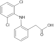
Figure 1: Diclofenac sodium
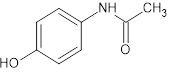
Figure 2: Paracetamol
Figure 3: Rabeprazole
Figure 4: Tramadol
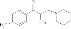
Figure 5: Tolperisone
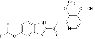
Figure 6: Pantaprazole
Figure 7: Chlorozoxazone
Figure 8: Oxytetracycline
Figure 9: Omeprazole
Figure 10: Diflunisal
Figure 11: Serratiopeptidase
Diclofenac and Paracetamol: UV Method
Introduction:
The hydrotropic solubilising agent used in the current study to solubilise the poorly water-soluble medication paracetamol for spectrophotometric analysis was 1.0 M urea solution. The suggested method for the simultaneous estimate of paracetamol and diclofenac sodium in bulk medication and tablet dosage form is novel, straightforward, environmentally friendly, accurate, repeatable, exact, and statistically validated. The estimation of both medications has been done using the dual wavelength method, absorption ratio method, and simultaneous equation approach. All three approaches exhibit good precision and accuracy, which has been statistically confirmed.
Materials And Methods:
UV-visible double beam spectrophotometer, Shimadzu model-1700, with wavelength precision and a spectral bandwidth of 3 nm ±1 nm, using quartz cells that measured 1 cm. Zenith Pharma Ltd., Indore (MP), provided a gift sample of pure PA and DS. Diclogesic, a tablet dosage form that contains 500 mg of PA and 50 mg of DS, was purchased from the local market in Indore, India. The choice of hydrotropic solubilising agent was 1.0 M urea. The other materials utilized were all of the grade of analytical reagent.
Conclusion:
In conclusion, it is clear that there is a good match between the estimated quantities and those that the makers assert. The percent label claims have low coefficients of variation, standard error, and standard deviation, all of which are very close to 100. Therefore, it can be said that the suggested approach is novel, easy to use, environmentally benign, and could be very significant for pharmaceutical analysis [31].
Diclofenac + Papaverine HCL: HPLC method
Introduction:
Diclofenac sodium and papaverine hydrochloride in tablets were simultaneously determined using an HPLC method that was devised and verified. A Zorbax SB ñ C18 column was used for the determination; the mobile phase consisted of methanol and water (60:40, v/v), with a flow rate of 1 mL Σ min-1 and UV detection at 278 nm.
Materials And Methods:
Papaverine hydrochloride (P) was bought from Caesar and Loretz, GmbH, Hilden, Germany, which produced diclofenac sodium (D). Polyvinylpyrrolidone (PVP), mannitol (M), potato starch (PS), hydroxypropyl methylcellulose (HPMC), microcrystaline cellulose (MC), magnesium stearate (MS), trisodium citrate dihydrate, citric acid monohydrate, methanol, and water were among the products produced by Merck, Germany and distributed to Galfarm PPH, CefarmLublin, Poland. The remaining reagents were all pure for analysis and of analytical grade.
Conclusion:
For the simultaneous measurement of diclofenac sodium and papaverine hydrochloride in tablets, the HPLC approach can be suggested since it is easy to use, quick, and precise [32].
HPTLC method: Diclofenac and Ibuprofen
Introduction:
A high-performance thin-layer chromatographic (HPTLC) approach that is precise, sensitive, and accurate was developed and validated for the detection of methocarbamol (ME) and related substances. (guaifenesin (GU)) in two ternary combinations containing potassium diclofenac and ibuprofen (IB). Using ethyl acetate–acetone–triethylamine–formic acid (62:35:6:0.3, by volume) as the developing system, the method separates and quantifies the drugs under study on TLC silica gel 60 F254 plates. Densitometric measurement of the bands at 222 nm for the first mixture containing methocarbmol, IB, and GU and at 278 nm for the second mixture containing methocarbmol, diclofenac potassium, and GU is then performed. The suggested techniques were effectively used to measure diclofenac potassium, ME, and IB when ME-related material (GU) was present, either in bulk powder.
Reagents:
The following analytic grade chemicals and reagents were employed without further purification: methanol HPLC grade (SDS, France). Tri ethylamine, acetone, formic acid, and ethyl acetate (E. MERCK, Germany).
Materials:
October Pharma S.A.E. (6th of October city, Egypt) graciously provided standard ME and DI with confirmed purities of 99.80% and 99.97%, respectively. Standard IB was graciously provided has a confirmed purity of 100.06%, by El Kahira Co. for Pharmaceutical and Chemical Industries (Cairo, Egypt). With a confirmed purity of 99.95%, Standard GU was graciously provided by RAMEDA Co. for Pharmaceutical Industries and Diagnostic Reagents (6th of October city, Egypt).
Conclusion:
The benefits of the HPTLC spectro densitometric approach are its ability to give excellent sensitivity and several sample runs with a modest amount of mobile phase selectivity. The advantages of the proposed method over the current HPLC methods are better sensitivity and the ability to identify both degradation products. The methods developed for the first mixture are the first to report ME and IB in the presence of GU, and the method developed for the second mixture differs from the reported one in that it is thought to be more practical and simpler than gradient elution for determining ME and DI in the presence of GU. Both methods yielded high selectivity and sensitivity [33].
RP-HPLC method: Diclofenac + Tramadol + chlorozone
Introduction:
The current study's goal was to create and validate an RP-HPLC method for simultaneously estimating the amounts of diclofenac sodium, chlorzoxazone, and tramadol hydrochloride from combination tablet dosage forms.
Methods:
The present technique outlines the RP-HPLC method for estimating the combination tablet dose form of diclofenac sodium, chlorzoxazone, and tramadol hydrochloride. The 0.05M Disodium Hydrogen Phosphate buffer pH 3.5 was adjusted with 10% v/v Ortho Phosphoric acid (50:50 v/v) and Hypersil ODS C18. Acetonitrile was the mobile phase utilized.
Conclusion:
The estimate of Tramadol hydrochloride, Chlorzoxazone, and Diclofenac sodium from their combination tablet dose form was found to be easy, accurate, exact, and suitable for using this method [34].
Mechanism of action of diclofenac sodium:
Diclofenac works by preventing the production of prostanoids, which are vital elements of the inflammatory and nociceptive response, such as prostaglandin-E2 (PGE2), prostacyclin, and thromboxane. This inhibits the activities of cyclooxygenase-1 (COX-1) and cyclooxygenase-2 (COX-2). It prevents arachidonic acid from binding to COX-1 and COX-2 in a competitive manner. Although data suggest that diclofenac has selective COX-2 inhibition, almost four times that of COX-1 inhibition during in vitro research, it inhibits COX-1 and COX-2 relatively similarly. Diclofenac's efficacy can be more precisely compared to celecoxib, although it is still far from the claimed 20-fold selectivity of COX-2 inhibition of the more selective COX-2 inhibitors, such as rofecoxib. Other NSAIDs, such as diclofenac, also have the effect of preventing the synthesis of thromboxane, particularly thromboxane B2 (TXB2). Diclofenac is considered to be one of the most potent inhibitors of PGE2 synthesis; an inflammatory response elevates primary prostanoids. The human body expresses COX-1, an enzyme that is constitutively active, practically everywhere. In addition to helping to maintain normal platelet activity, blood flow into renal tissues, and shield the gastric mucosa from damaging acidity, COX-1 is assumed to have a fairly steady level and activity. An inducible enzyme called COX-2 is overexpressed when there is tissue damage and when inflammatory mediators, which also have nociceptive qualities and cause pain, are present. They consist of prostaglandins, leukotrienes, and thromboxanes. The inhibition of COX-2 that diclofenac produces appears to be mostly focused at the target tissues, such as joint capsules and synovial fluid. On the other hand, the suppression of COX enzymes in other tissues-like the stomach-may result in the loss of numerous protective compounds and, among other things, the emergence of gastrointestinal discomfort. In the therapeutic world, several of these modes of action are regarded as hypothetical. Diclofenac's ability to downregulate sensitised peripheral pain receptors is responsible for its peripheral analgesic effects. This appears to be achieved by activating ATP-sensitive potassium channels, which in turn stimulates the L-arginine nitric oxide cGMP pathway. Furthermore, data indicate that diclofenac may also have an effect on lowering substance P levels, a recognised pro-inflammatory neuropeptide with nociceptive action, which were previously elevated in the synovial fluid of rheumatoid arthritis patients.
|
Drug Combination |
Instrumentation |
Parameters |
LOQ and LOD |
Concentration Range |
References |
|
1. Paracetamol + diclofenac sodium |
UV double beam spectrophotometer |
200-400nm [UV range]
|
PAR;9.26-2.40mg/ml. DICLO;4.13–1.80mg/ml. |
%RSD was found to be less than 2% |
[35] |
|
2. Paracetamol + diclofenac sodium |
UV spectrophotometer
|
P-265nm D-258nm |
PAR;0.55-0.18mg/ml. DICLO;0.15-0.05mg/ml. |
%RSD was found to be less than 2% |
[36] |
|
3. Diclofenac sodium + misoprostol |
HPLC Method |
234nm |
DICLO;0.78-0.26mg/ml. MISP;5.6-1.87mg/ml. |
%RSD was found to be less than 2% |
[37] |
|
4. Diclofenac sodium + misoprostol |
HPLC method |
200-400nm |
DICLO;0.377-1.143mg/ml. MISP;2.08-6.307mg/ml. |
%RSD was found to be less than or equal to 2% |
[38] |
|
5. Diclofenac sodium + Paracetamol + Chlorzoxazone |
HPLC method |
P-26-130mg/ml C-20-100mg/ml D-4-20mg/ml |
PARO;6.51-16.20mg/ml CHLOR;4.97-27.68mg/ml DICLO;0.84-2.82mg/ml |
%RSD was found to be less than 2% |
[39] |
|
6. Diclofenac sodium + Serratiopeptidase |
Double beam UV spectrometer |
D-264nm C-295nm |
DICLO;0.6771-2.0519mg/ml SERAT;1.3364-4.0498mg/ml |
%RSD was found to be less than 2% |
[40] |
|
7. Diclofenac sodium + rabeprazole |
UV spectrophotometer |
R-276-285nm D-200-400nm |
DICLO; RABE 1.724mg/ml 0.517mg/ml |
%RSD was found to be less than or equal to 2% |
[41] |
|
8. Diclofenac sodium + oxytetracycline
|
UV spectrophotometer |
OTCa-360nm OTCb-339nm DICLO-298.5nm |
OTCa-0.37-1.11mg/ml OTCb-0.47-1.44mg/ml DICLO-0.43-1.31mg/ml |
%RSD was found to be less than 2% |
[42] |
|
9. Diclofenac sodium + tramadol Hydrochloride |
UV spectrophotometer |
DICLO;270.9nm TRAM;248.38nm |
DICLO-0.842-0.421mg/ml TRAM-1.506-0.502mg/ml |
%RSD was found to be less than 2% |
[42] |
|
10. Diclofenac + Serratiopeptidase |
UV method |
DICLO;275nm SERR;264nm |
DICLO-0.247-0.748mg/ml SERR-0.150-0.455mg/ml |
%RSD was found to be less than 2% |
[43]
|
|
11. Diclofenac + Oxytetracycline |
UV spectrophotometer |
DICLO;360nm OXYT;298nm |
DICLO-0.43-0.31 OXYT-0.37-1.11 |
%RSD was found to be less than 2% |
[44] |
|
12. Diclofenac + Diflunisal |
HPLC method |
5-100 mg/ml |
DICLO-0.87-0.26 DIFLU-0.65-2.16 |
%RSD was found to be less than 2% |
[45] |
|
13. Diclofenac Sodium + Ibuprofen + Mefenamic Acid |
GC/HPLC method |
IBU-240nm MEFE-260nm DICLO-300nm |
IBU-1.82-0.6 MEFE-1.2-0.4 DICLO-1.5-0.5 |
%RSD was found to be less than 2% |
[46] |
|
14. Diclofenac Sodium + Piroxicam + Naproxen + Mefenamic Acid |
HPLC Method |
264nm
|
DICLO-0.811-0.246 PIRO-0.926-0.281 NAPRO-0.989-0.300 MEFE-0.328-0.251 |
%RSD was found to be less than 2% |
[47] |
|
15. Diclofenac sodium + Tramadol HCL |
RP-HPLC Method |
Mobile phase: Methanol: phosphate buffer |
DICLO-1.8695-1.9956 TRAM-0.6169-0.6586 |
%RSD was found to be less than 2% |
[48] |
|
16. Diclofenac + Tramadol HCL |
RP-HPLC Method |
MP: Methanol:Water |
DICLO-0.99-0.33 TRAM-0.99-0.42 |
%RSD was found to be less than 2% |
[49] |
|
17. Serratiopeptidase + Diclofenac |
UV method |
STERO-275nm DICLO-264nm |
STERO-0.455-0.150 DICLO-0.748-0.247 |
%RSD was found to be less than 2% |
[50] |
|
18. Diclofenac + Tolperisone
|
RP-HPLC Method |
260nm |
DILO-3.38727-1.117 TOLE-10.21-3.370 |
%RSD was found to be less than 2% |
[51] |
|
19. Diclofenac + curcumin |
RP-HPLC |
275nm |
DICLO-491.1-245.5 CURC-165.87-55.30 |
%RSD was found to be less than 2% |
[52] |
|
20. Diclofenac sodium + Tramadol |
HPLC Method |
273nm |
DICLO-1.3865-0.4575 TRAM-2.3461-0.7742 |
%RSD was found to be less than 2% |
[53] |
|
21. Diclofenac sodium + gabapentin |
RP-HPLC Method |
GABA-210nm DICLO-275nm |
GABA-0.93-2.82 DICLO-1.25-3.78 |
%RSD was found to be less than 2% |
[54] |
|
22. Diclofenac sodium + Resveratrol |
UPLC MS Method |
Conc:5-2000ng/ml |
5-15ng.ml-1 |
%RSD was found to be less than 2% |
[55] |
|
23. Diclofenac sodium + Ibuprofen + Naproxen |
HPLC Method |
|
IBU- DICLO- NAPRO- |
%RSD was found to be less than 2% |
[56] |
|
24. Diclofenac sodium + Paracetomol + Chloroxazone |
HPLC Method |
Mp- phosphate buffer: acetonitrile: methanol |
PARA-0.0065-0.0020 CHZ-0.085-0.040 DICLO-0.070-0.025 |
%RSD was found to be less than 2% |
[57] |
|
25. Diclofenac sodium + Tizanidine HCl |
RP-HPLC Method |
MP-acetonitrile: phosphate buffer |
DICLO-1.10-0.20 TIZA-2.20-0.10 |
%RSD was found to be less than 2% |
[58] |
|
26. Diclofenac + Tolerisonw HCL |
RP-HPLC Method |
240nm |
DICLO-0.1125-0.0372 TOLE-6.60-2.17 |
%RSD was found to be less than 2% |
[59] |
|
27. Diclofenac sodium + Chlorphenarmine malate + Paracetamol |
RP-HPLC Method |
MP- acetonitrile: phosphate buffer |
DICLO-2.72-0.89 CHLO-2833-0.935 PARA-1.22-0.403 |
%RSD was found to be less than 2% |
[60] |
|
28. Diclofenac sodium + Aspirin |
HPLC Method |
254nm |
ASPI-4.3-2.29 DICLO-5.61-3.05 |
%RSD was found to be less than 2% |
[61] |
|
29. Diclofenac sodium + Methocarbamol |
UV spectrophotometer |
274.11nm |
DICLO-0.171-0.056 METHO-0.790-0.2607 |
%RSD was found to be less than 2% |
[62] |
|
30. Diclofenac sodium + Pantaprazole |
UV double beam spectrophotometer |
DICLO-278.8nm PANTO-296.2nm |
DICLO-1.993-0.658 PANTO-0.706-0.233 |
%RSD was found to be less than 2% |
[63] |
|
31. Diclofenac sodium + Pantaprazole |
UV double beam spectrophotometer |
200-400nm |
DICLO-1.765-0.583 PANTO-1.811-0.597 |
%RSD was found to be less than 2% |
[64] |
|
32. Diclofenac sodium + Benzacaine |
RP-HPLC Method |
243nm |
DICLO-0.28-0.12 BENZA-0.31-0.15 |
%RSD was found to be less than 2% |
[65] |
|
33. Diclofenac sodium + Chloraoxazone |
UV double beam spectrophotometer |
281nm |
DICLO-0.08-0.45 CHLOR-0.25-0.45 |
%RSD was found to be less than 2% |
[66] |
|
34. Diclofenac sodium + Tolperisone hydrochloride |
UV double beam spectrophotometer |
TICLO-254nm DICLO-282nm |
TICLO-0.306-0.101 DICLO-0.364-0.120 |
%RSD was found to be less than 2% |
[67] |
|
35. Tramadol hydrochloride + Diclofenac |
UV visible 1800 double beam spectrophotometer |
TRA-272.45nm DICLO-282.81nm |
TRA-0.43-0.144 DICLO-4.860-1.60 |
%RSD was found to be less than 2% |
[68] |
|
36. Diclofenac sodium + Paracetamol |
UV visible spectrophotometer |
PARA-247nm DICLO-276nm |
PARA-0.481-0.154 DICLO-0.915-0.335 |
%RSD was found to be less than 2% |
[69] |
|
37. Diclofenac sodium + Paracetamol |
UV double beam spectrophotometer |
PARA-247nm DICLO-276nm |
PARA-0.575-0.917 DICLO-0.300-1.017 |
%RSD was found to be less than 2% |
[70] |
|
38. Diclofenac sodium + Rabeprazole |
HPLC Method |
284nm |
DICLO-0.22-0.73 RABE-0.02-0.008 |
%RSD was found to be less than 2% |
[71] |
|
39. Diclofenac sodium + Rabeprazole |
UV visible double spectrophotometer |
DICLO-276nm RABE-292nm |
DICLO-2.04-0.67 RABE-2.96-0.97 |
%RSD was found to be less than 2% |
[72] |
|
40. Diclofenac sodium + Toleperlione |
UV double beam spectrophotometer UV 1800 |
226nm |
DICLO-2.00-0.5 TOLE-6.00-1.00 |
%RSD was found to be less than 2% |
[73] |
|
41. Chlorazoxazole + Diclofenac sodium + Paracetamol |
HPLC-Method |
|
CHLO-1.48-0.443 DICLO-0.910-0.273 PARA-0.914-0.247 |
%RSD was found to be less than 2% |
[74] |
|
42. Diclofenac + Omeprazole |
UV - spectrophotometer Method |
291nm |
Method1 DICLO-0.179-0.059 OME-0.36-0.121 Method2 DICLO-0.145-0.048 OME-0.31-0.105 |
%RSD was found to be less than 2% |
[75] |
|
43. Diclofenac sodium + Paracetamol |
UV - double beam spectrophotometer
|
DICLO-268-276nm PARA-268-247nm |
Method1 DICLO-1.201-0.204 PARA-0.99-0.66 Method2 DICLO-0.62-0.239 PARA-0.917-0.575 |
%RSD was found to be less than 2% |
[76] |
|
44. Diclofenac sodium + Trypsin + Bromelain + Rutoside |
UV - method |
TRY-545nm BRO-229nm Method1 Rec-257nm Dic-282.50nm Method2 Rec-258.70nm Dic-250.70nm Method3 Rec-360nm Diclo-282nm |
Method1 Rec-1.5342-0.566 Diclo-0.917-0.302 Method2 Rec-2.33-0.701 Diclo-2.133-0.40 Method3 Rec-2.02-0.66 Diclo-0.916-0.30 |
%RSD was found to be less than 2% |
[77] |
|
45. Diclofenac sodium + Tolperisone HCL |
UV method |
268nm |
Optimization DICLO-0.429-0.141 TOL-1.053-0.347 Spectro scophy method DICLO-0.565-0.1886 TOL-0.942-0.311 |
%RSD was found to be less than 2% |
[78] |
|
46. Diclofenac Sodium + Epersione HCL |
UV method |
296nm |
DICLO-0.111-0.438 E.HCL-0.038-0.144 |
%RSD was found to be greater than 1% |
[79] |
|
47. Diclofenac Sodium + Pantoprazole |
UV method |
DICLO-272.8nm PANTOP-296.2nm |
DICLO-0.706-0.233 PANTOP-1.993-0.706 |
%RSD was found to be greater than 1% |
[80] |
|
48. Diclofenac Sodium + Pantoprazole Sodium |
HPLC method |
DICLO-326nm PANTOP-337.0nm |
DICLO-1.765-0.583 PANTOP-1.811-0.597 |
%RSD was found to be greater than 1% |
[81] |
|
49. Diclofenac Sodium + Paracetamol |
UV Visible double spectrophotometer |
DICLO-225nm PCT-244nm |
DICLO-0.125-0.220 PCT-0.451-0.195 |
%RSD was found to be less than 1% |
[82] |
|
50. Diclofenac Sodium + Paracetamol |
UV Visible double spectrophotometer |
DICLO-242nm PCT-273nm |
DICLO-4.13-1.80 PCT-9.26 |
%RSD was found to be less than 2% |
[83] |
|
51. Diclofenac Sodium + Tolperisone |
UV-Visible double spectrophotometer |
261nm 276nm |
DICLO-0.048-0.016 TOLP-0.091-0.030 |
%RSD was found to be less than 2% |
[84] |
|
52. Diclofenac Sodium + Esomeprazole Magnesium Trihydrate |
UV double beam spectrophotometer |
DICLO-280nm EMT-301nm |
Method-1 DICLO-0.4658-0.01537 EMT-0.5181-0.6230 Method-2 DICLO-0.4518-0.1491 EMT-1.8871-0.6230 |
%RSD was found to be less than 2% |
[85] |
|
53. Diclofenac Sodium + Tolperisone Sodium |
UV double beam spectrophotometer |
DICLO-265nm TOL-250nm |
DLCLO-1.47-0.486 TOL-0.523-0.172 |
%RSD was found to be less than 2% |
[86] |
|
54. Diclofenac + Misoprostol |
HPLC method |
234nm |
DICLO-5.6-1.87 MISO-0.78-0.26 |
%RSD was found to be less than 2% |
[87]
|
CONCLUSION:
The suggested UV spectrophotometric approach was discovered to be incredibly quick, easy, and affordable. Excellent recovery, accuracy, and linearity can be achieved in the estimation of diclofenac sodium using this validated method that complies with ICH criteria
REFERENCES


Likith Kumar K. B.*, Mamatha T., Mani Sai Santhoshi, Lavanya N., Sujatha P. Muchalambe, Revanth S., Kruthika, Review on Method Development and Validation of Diclofenac Sodium Using Different Combination Drugs, Int. J. of Pharm. Sci., 2025, Vol 3, Issue 12, 629-648. https://doi.org/10.5281/zenodo.17805861
 10.5281/zenodo.17805861
