Kamalakshi Pandurangan College of Pharmacy, Tiruvannamalai, Tamil Nadu – 03
The rational design of DNA-targeted small molecules continues to represent a cornerstone of modern medicinal chemistry, particularly in the development of anticancer therapeutics. Among planar polycyclic aromatic systems, the has emerged as a structurally advantageous framework for engineering efficient DNA intercalators due to its intrinsic rigidity, extended ?-conjugation, and synthetic versatility. This review delineates the structural and energetic principles governing phenanthrene–DNA interactions, with emphasis on ?–? stacking stabilization, helical distortion, and thermodynamic signatures of intercalative binding. Strategic molecular modifications—including cationic side-chain incorporation, electronic modulation, and metal coordination—are critically evaluated in the context of enhancing binding affinity, selectivity, and biological efficacy. Mechanistic insights linking DNA intercalation to topoisomerase inhibition, replication interference, and apoptosis induction are discussed to establish structure–mechanism–activity correlations. Furthermore, contemporary biophysical and computational approaches are highlighted as essential tools for quantitative assessment and predictive optimization of phenanthrene-based intercalators. Collectively, this analysis provides a unified framework for the rational development of phenanthrene-derived DNA intercalators with improved therapeutic potential.
Targeting deoxyribonucleic acid (DNA) remains a central strategy in medicinal chemistry, particularly in anticancer drug[8,12] development where disruption of replication and transcription can effectively suppress malignant proliferation.[6,48] Among the various modes of DNA interaction, intercalation—characterized by the insertion of planar aromatic systems between adjacent base pairs[2,15]—has proven to be mechanistically robust and therapeutically valuable. Clinically relevant agents such as doxorubicin exemplify[19,20,22] how π-conjugated frameworks can stabilize DNA complexes,[3] inhibit topoisomerases, and ultimately induce apoptosis. However, limitations including off-target toxicity and resistance mechanisms[14] necessitate the rational refinement of intercalative scaffolds.
Within this context, phenanthrene has emerged as a structurally advantageous aromatic framework[25,26] owing to its rigid angular topology, extended π-surface, and synthetic versatility. Its planarity facilitates efficient π–π stacking[25,26] with nucleobases, while strategic substitution enables modulation of electronic distribution, binding affinity, and biological selectivity. Advances in biophysical characterization and computational modeling[23,42] now permit quantitative evaluation of phenanthrene–DNA interactions, supporting a transition from empirical discovery to rational design. Accordingly, phenanthrene-based DNA intercalators represent a promising platform for developing mechanistically optimized and therapeutically effective DNA-targeted agents.
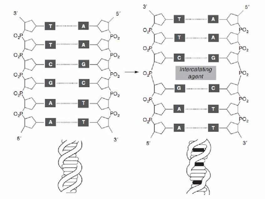
Fig. 1 : General DNA Intercalation model
DNA AS A THERAPEUTIC TARGET: STRUCTURAL AND ENERGETIC CONSIDERATIONS
Deoxyribonucleic acid (DNA) represents a structurally ordered and energetically dynamic macromolecule whose physicochemical properties render it highly amenable to small-molecule recognition. In its canonical B-form,[1,4] DNA consists of antiparallel strands stabilized by hydrogen bonding between complementary bases[1] and extensive π–π stacking interactions along the helical axis. These stacking interactions contribute significantly to duplex stability, generating a hydrophobic aromatic core that favors the insertion of planar polycyclic molecules.[16,39] The major and minor grooves further provide electrostatic and hydrogen-bonding environments that modulate ligand accessibility and sequence preference.
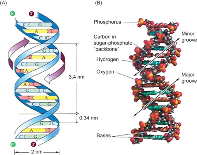
Fig. 2 : Structural features of B-DNA
From a thermodynamic perspective, DNA intercalation is driven predominantly by favorable enthalpic contributions arising from π–π stacking and van der Waals interactions, counterbalanced by entropic penalties associated with helical unwinding and conformational restriction.[10,16] Intercalative binding typically induces measurable structural perturbations, including base pair separation, helix lengthening, and partial unwinding of the double helix. These distortions interfere with replication[6,44] and transcription machinery, thereby forming the mechanistic basis for therapeutic intervention. A rigorous understanding of these structural and energetic determinants is therefore essential for the rational design of phenanthrene-based DNA intercalators with optimized affinity and selectivity.[5]
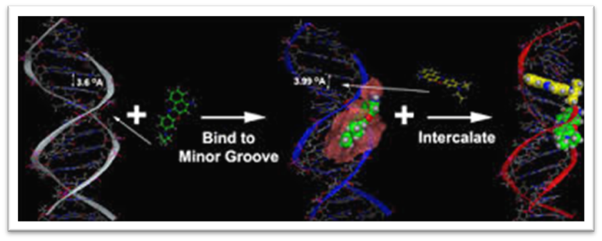
Fig. 3 : Thermodynamic Profile of DNA Intercalation
PHENANTHRENE AS A PRIVILEGED AROMATIC SCAFFOLD
Phenanthrene represents a structurally privileged aromatic framework for the development of DNA-interactive small molecules, owing to its rigid angular topology, extended π-conjugation, and favorable electronic distribution. Composed of three fused benzene rings arranged in a non-linear configuration, phenanthrene differs subtly yet significantly from linear polycyclic systems such as anthracene. The angular fusion imparts enhanced thermodynamic stability and permits distinct substitution patterns that influence steric orientation and electronic density across the aromatic surface. These structural attributes render phenanthrene particularly suitable for π–π stacking interactions[25] within the hydrophobic core of DNA base pairs, a fundamental requirement for effective intercalation.
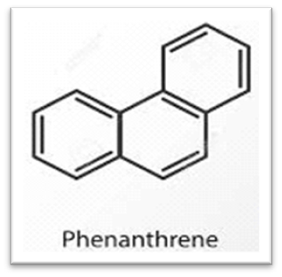
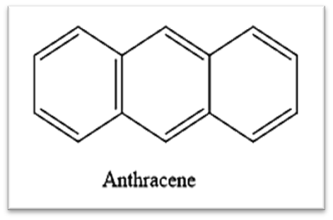
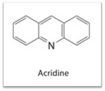
Fig. 4 : Chemical Structure comparison
The planarity of the phenanthrene nucleus facilitates insertion between adjacent nucleobases, while its electronic tunability allows modulation of intermolecular interactions through strategic functionalization. Introduction of electron-donating or electron-withdrawing substituents alters charge distribution and polarizability, thereby influencing binding affinity and selectivity.[27,33] Furthermore, phenanthrene offers synthetic accessibility through well-established cyclization and cross-coupling methodologies, enabling precise structural diversification. In comparison to heteroaromatic intercalators such as acridine[35,50] derivatives, phenanthrene provides a purely carbocyclic scaffold that can be systematically engineered with peripheral cationic or hydrogen-bonding functionalities to enhance electrostatic complementarity with the negatively charged phosphate backbone of DNA. Collectively, these physicochemical and synthetic advantages position phenanthrene as a versatile and rationally adaptable core for the design of next-generation DNA intercalators with optimized therapeutic performance.
RATIONAL DESIGN STRATEGIES OF PHENANTHRENE-BASED INTERCALATORS
The rational engineering of phenanthrene-based DNA intercalators necessitates a multidimensional optimization of structural, electronic, and physicochemical parameters to achieve enhanced affinity, selectivity, and biological efficacy. At the most fundamental level, preservation of aromatic planarity is indispensable for efficient π–π stacking within the DNA base pair environment. Structural rigidification through ring fusion or constrained substitution patterns minimizes conformational flexibility, thereby reducing entropic penalties upon binding and improving thermodynamic stability of the intercalative complex.
Electrostatic complementarity represents a second critical design principle.[29] Given the negatively charged phosphate backbone of DNA, incorporation of cationic side chains—typically tertiary amines or quaternary ammonium functionalities—enhances long-range Coulombic interactions and facilitates pre-association with the nucleic acid surface. Proper spatial orientation of these side chains is essential; excessive steric bulk may hinder intercalation, whereas optimally positioned cationic moieties can simultaneously stabilize the complex and improve aqueous solubility. Electronic modulation through strategic placement of electron-donating or electron-withdrawing substituents further allows fine-tuning of π-electron density, thereby influencing stacking efficiency and binding constants.
Table 1. Representative Phenanthrene-Based DNA Intercalators and Their Biological Profiles
|
Compound |
Structural modification |
DNA binding constant (kb) |
Topoisomerase inhibition |
Cytotoxic ic50 (µm) |
|
Phenanthrene (parent) |
Unsubstituted scaffold |
~10?–10? M?¹ |
Weak/Indirect |
>50 µM |
|
Amino-substituted phenanthrene |
4-amino phenyl substitution |
~10?–10? M?¹ |
Moderate |
10–25 µM |
|
Cationic phenanthrene derivative |
Tertiary amine side chain |
~10? M?¹ |
Enhanced stabilization of cleavage complex |
5–15 µM |
|
Metal-coordinated phenanthrene complex |
Phenanthrene–Cu(II) |
>10? M?¹ |
Strong inhibition |
1–10 µM |
|
Electron-withdrawing substituted derivative |
Nitro/ halogen substitution |
~10? M?¹ |
Moderate |
15–40 µM |
Beyond purely organic modifications, coordination of phenanthrene derivatives with transition metals introduces additional geometric and redox-active dimensions. Metal–ligand complexes can enhance DNA affinity[31,32] through cooperative binding modes while also enabling oxidative DNA cleavage or topoisomerase interference. Importantly, rational design increasingly integrates computational methodologies, including molecular docking and dynamic simulations, to predict binding orientation, free energy contributions, and sequence preference prior to synthesis. Such structure-guided approaches shift phenanthrene-based intercalator development from empirical discovery toward predictive molecular engineering, ultimately facilitating the generation of therapeutically optimized DNA-targeted agents.
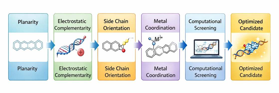
Fig. 5 : Rational design strategies
BIOPHYSICAL AND COMPUTATIONAL CHARACTERIZATION OF DNA BINDING
Comprehensive evaluation of phenanthrene–DNA interactions require integration of complementary biophysical and computational methodologies to establish binding mode, affinity, and thermodynamic parameters. UV–visible absorption spectroscopy remains a primary analytical tool, where intercalative binding is typically evidenced by hypochromism and bathochromic shifts resulting from π–π stacking interactions with nucleobases. Fluorescence spectroscopy further substantiates complex formation[23] through quenching behavior and Stern–Volmer analysis, enabling quantitative determination of binding constants and assessment of static versus dynamic quenching mechanisms.[24] Circular dichroism spectroscopy provides additional structural insight by detecting perturbations in DNA helicity, while viscosity measurements serve as a classical diagnostic for intercalation, as insertion between base pairs induces measurable elongation of the DNA helix.
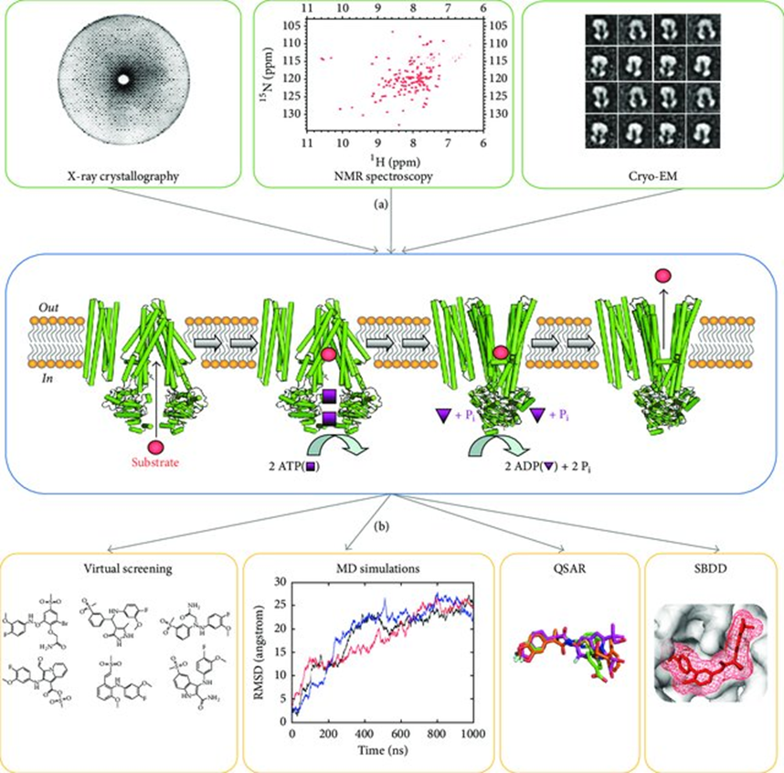
Fig. 6 : Experimental workflow from biophysical characterization of ABC proteins in multiple conformational states to in silico discovery and design of small molecule modulators
Thermodynamic characterization through isothermal titration calorimetry allows direct determination of enthalpic and entropic contributions, thereby distinguishing intercalative binding from groove association. Increasingly, computational approaches complement experimental observations. Molecular docking studies predict binding orientation, stacking geometry, and electrostatic interactions, whereas molecular dynamics simulations evaluate complex stability over time and identify sequence-dependent preferences. Together, these techniques establish a rigorous structure–binding relationship framework, enabling rational optimization of phenanthrene derivatives prior to extensive biological evaluation.
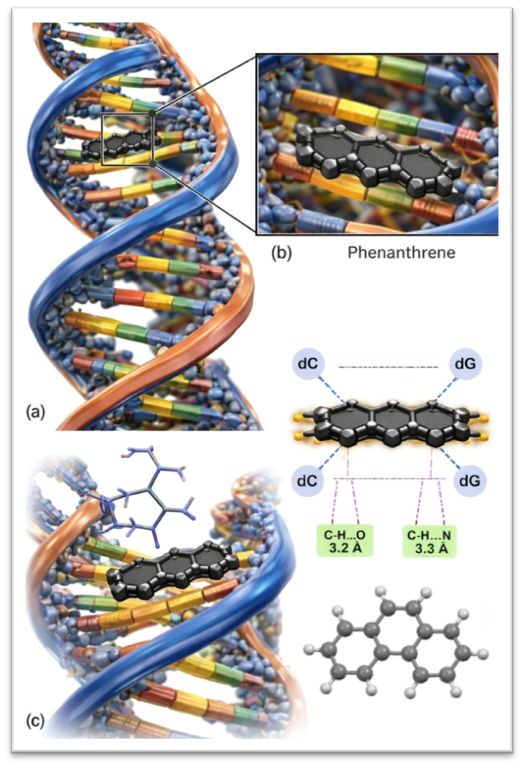
Fig. 7 : Molecular visualization of phenanthrene intercalation into duplex DNA.
(a) Surface and structural representation of double-stranded DNA with phenanthrene intercalated between stacked base pairs. (b) Detailed binding interface showing aromatic π–π stacking between phenanthrene and neighboring nucleobases. (c) Orthogonal view illustrating the planar geometry of phenanthrene and the resulting perturbation of the DNA helical structure.
MOLECULAR DOCKING ANALYSIS WITH TELOMERASE REVERSE TRANSCRIPTASE (TERT)
To further elucidate potential enzyme-level interactions beyond direct DNA intercalation, molecular docking simulations were conducted to evaluate the binding affinity of representative phenanthrene derivatives toward telomerase reverse transcriptase (TERT).[42,43] Telomerase is a ribonucleoprotein reverse transcriptase responsible for maintaining telomere length and genomic stability, and its overexpression is a hallmark of malignant cells.[40]
Docking analysis revealed that phenanthrene-based scaffolds preferentially occupy hydrophobic pockets located near the thumb–TRBD interface of TERT. The planar aromatic core facilitates π–alkyl and hydrophobic interactions with conserved residues within the FVYL motif (including Phe, Val, Tyr, and Leu residues),[40,29] while appropriately positioned substituents may establish additional hydrogen bonding interactions with surrounding amino acids. These interactions support the hypothesis that phenanthrene derivatives may exert dual activity through both DNA intercalation and indirect telomerase inhibition.
The predicted binding orientation and favorable docking scores further reinforce the structure–mechanism relationship underlying their cytotoxic potential.
MOLECULAR MECHANISMS OF CYTOTOXICITY
The cytotoxic activity of phenanthrene-based DNA intercalators arises primarily from their capacity to disrupt essential genomic processes following stable insertion between base pairs. Intercalation induces localized helical unwinding and base pair separation, thereby impairing the progression of replication and transcription complexes. A major downstream consequence of this structural perturbation is the inhibition of DNA topoisomerases,[7,49] enzymes responsible for relieving torsional stress during DNA replication. Stabilization of the DNA–topoisomerase cleavage complex[18] prevents religation of DNA strands, leading to accumulation of double-strand breaks and replication-associated genomic instability.
Beyond topoisomerase interference, sustained intercalative binding can obstruct replication fork progression, triggering activation of DNA damage response pathways and checkpoint signaling cascades.[38,41] These events culminate in cell cycle arrest, typically at the S or G2/M phases, followed by induction of programmed cell death. Apoptotic signaling may proceed through mitochondrial pathways characterized by altered Bcl-2 family protein expression and caspase activation. In certain metal-coordinated phenanthrene systems, redox cycling and reactive oxygen species generation further amplify DNA damage. Collectively, these mechanistic events establish a coherent link between molecular intercalation and cellular cytotoxicity, underscoring the therapeutic relevance of structurally optimized phenanthrene derivatives.
THERAPEUTIC APPLICATIONS
Phenanthrene-based DNA intercalators exhibit potent cytotoxic effects in rapidly proliferating tumor cells. DNA destabilization and replication arrest contribute to apoptosis induction.[8,45]
These compounds interfere with topoisomerase-mediated DNA processing. Stabilization of the cleavage complex enhances double-strand break formation and cell death.[7,19]
Structural optimization of the phenanthrene scaffold improves activity in multidrug-resistant phenotypes. Modulation of efflux susceptibility enhances intracellular retention.[14]
Certain derivatives may interact with telomerase reverse transcriptase (TERT). Dual DNA and enzyme targeting can suppress telomere maintenance in malignant cells.[40]
DNA intercalation increases susceptibility to radiation-induced damage. This may enhance therapeutic efficacy in combination radiotherapy.
Phenanthrene derivatives may act synergistically with platinum agents and other DNA-targeting drugs. Such combinations may allow dose reduction and improved safety.
Intercalative disruption of microbial DNA replication supports antibacterial potential. Membrane-active substituents may further enhance selectivity.[50]
DNA-targeted mechanisms may impair protozoal replication and genomic stability. This provides a basis for exploring antiparasitic applications.
Metal-coordinated phenanthrene systems enhance DNA affinity and oxidative damage potential. Redox activity may amplify cytotoxic outcomes.[31,32]
Nanocarrier incorporation may improve tumor selectivity and pharmacokinetics. Controlled delivery can reduce systemic toxicity.
TOXICOLOGICAL AND PHARMACOKINETIC CONSIDERATIONS
Despite their therapeutic promise, phenanthrene-based DNA intercalators inherently raise concerns related to genomic safety and systemic toxicity. Because intercalation is not intrinsically sequence-specific, off-target interactions with healthy proliferating cells may result in genotoxic effects, mutagenesis, or cumulative tissue damage. Excessive stabilization of DNA–enzyme cleavage complexes can provoke irreversible double-strand breaks, thereby increasing the risk of long-term cytotoxicity. Structural refinement is therefore essential to balance binding affinity with controlled reversibility, minimizing nonspecific genomic perturbation.
From a pharmacokinetic perspective, hydrophobic aromatic frameworks such as phenanthrene often exhibit limited aqueous solubility and variable bioavailability. Strategic introduction of polar or ionizable substituents can enhance solubility and improve systemic distribution, while also influencing plasma protein binding and metabolic stability. Hepatic biotransformation, particularly through oxidative pathways, may alter aromatic integrity or generate reactive intermediates, necessitating careful metabolic profiling.[28] Accordingly, rational scaffold engineering must integrate toxicity prediction and pharmacokinetic optimization to ensure that enhanced DNA-binding potency translates into clinically viable therapeutic performance.
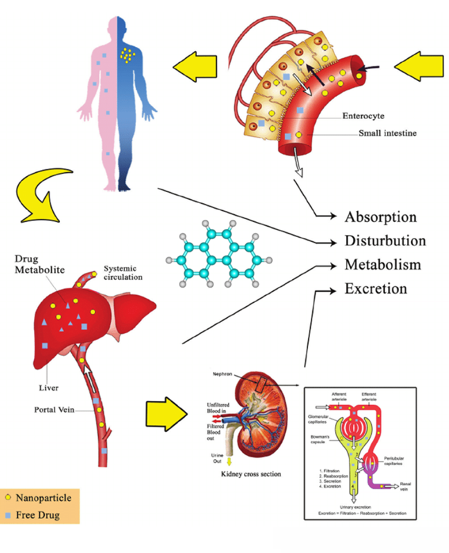
Fig. 8 : ADME Schematic
EMERGING PERSPECTIVES AND FUTURE DIRECTIONS
The future development of phenanthrene-based DNA intercalators is increasingly guided by precision-oriented and technology-integrated approaches. A critical objective is the attainment of sequence[11,29] or structure selectivity, enabling preferential targeting of oncogenic or pathogen-specific genomic regions while minimizing collateral damage to normal cellular DNA. Advances in high-resolution structural biology[47] and biophysical mapping techniques now provide deeper insight into DNA conformational heterogeneity, opening avenues for rationally designing intercalators with improved discriminatory capacity.
Nanotechnology-enabled delivery systems[36] represent another transformative direction, offering controlled release, enhanced tumor accumulation, and reduced systemic toxicity. Encapsulation within liposomal or polymeric carriers may mitigate pharmacokinetic limitations[27] associated with hydrophobic aromatic scaffolds. Concurrently, artificial intelligence and machine learning–driven molecular modeling are accelerating scaffold optimization by predicting binding affinity, ADME properties, and toxicity profiles prior to synthesis. The integration of computational prediction, synthetic chemistry, and mechanistic validation is poised to transition phenanthrene-based intercalator research from empirical development toward precision-engineered therapeutics with enhanced translational potential.
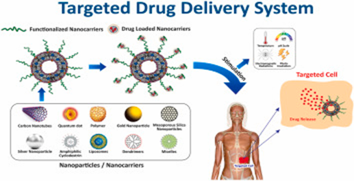
Fig. 9 : Future Strategy integration model
AI-assisted modeling | Nanocarrier delivery | Precision targeting | Structural biology
CONCLUSION
Phenanthrene-based DNA intercalators represent a structurally and mechanistically compelling class of DNA-targeted small molecules, characterized by their rigid aromatic framework, electronic tunability, and synthetic adaptability. The intrinsic planarity of the phenanthrene core enables efficient π–π stacking within the DNA helix, while rational structural modifications—including cationic substitution, electronic modulation, and metal coordination—permit fine control over binding affinity, selectivity, and biological performance. Integration of biophysical characterization techniques with computational modeling has further advanced the transition from empirical discovery to predictive molecular design.[52]
Mechanistically, these compounds exert cytotoxic effects through helical perturbation, topoisomerase inhibition, replication interference,[44] and activation of DNA damage response pathways, underscoring their therapeutic relevance in oncology and beyond. Nevertheless, challenges related to genotoxicity,[46,51] off-target effects, and pharmacokinetic limitations necessitate continued structural refinement and targeted delivery strategies. Future progress will depend upon the convergence of precision molecular engineering, advanced computational tools, and translational pharmacology. Collectively, rationally designed phenanthrene-derived intercalators hold substantial promise as next-generation DNA-directed therapeutics with improved efficacy and safety profiles.
REFERENCES


R. Manikandan, Jemimah S, Krishnan P, Kumaravelan V. M., Ramana Balaji S, Rajalingam D, Rational Design of Phenanthrene - Based DNA Intercalators: Mechanistic & Therapeutic Insights, Int. J. of Pharm. Sci., 2026, Vol 4, Issue 3, 392-404. https://doi.org/10.5281/zenodo.18871396
 10.5281/zenodo.18871396
