Abasaheb Kakade College of Pharmacy Bodhegaon.
Understanding and controlling matter typically in the 1-100 nm dimension range is nanotechnology. The application of Nano technology to medicine, known as Nano medicine. Nanomaterial have unique physicochemical properties such as a ultra-small size large surface area to mass ration, and high reactivity, which are different formula bulk material of the same composition. This review will discuss the formulation nanoparticle method and strategies, as well as various characterization parameters. The review also focuses on important therapeutic applications of nanoparticles for drug delivery effectiveness.
Aren't always made in modern synthesis labs, but they have clearly been around for a long time in nature, so their use dates back to ancient times. While the controlled reinforcement of a ceramic matrix with natural asbestos Nano fibers more than 4500 years ago is more intriguing, the application of clay minerals as natural nanomaterials does not appear to be very sophisticated [1]. Physics, chemistry, biology, materials science, health sciences, and engineering are all utilized in nanotechnology. It can be used in a lot of different areas of science and human life. Nanoparticles can be defined as particulate dispersions or solid particles with a size in the range of 10-1000nm [2]. A nanometer is 250 millionth of a meter, or 1/80,000 of a human hair's diameter or ten times the diameter of a hydrogen atom [3]. For a number of years, gold nanoparticles (AuNPs) have been used in high-end cosmetics, such as skin creams, lotions, hair products, lipsticks, deodorants, and facial masks. These products claim to have anti-aging, hydrating, restorative, and regenerative properties [4]. Using response surface methodologies (RSM), the current study sought to optimize drug carrier formulation parameters for intravenous drug delivery. Dipalmitoyl phosphatidylcholine (DPPC), distearoylphosphatidylcholine (DSPC), and phosphatidylserine (PS) concentrations in the presence of lecithin were the focus of this investigation [5]. Richard Feynman, a Nobel Prize winner, proposed nanotechnology for the first time in 1959[6]. Healthy fibroblast cells and breast cancer (MCF-7) cells were treated with either free phthalocyanine or phthalocyanine bound to either gold nanoparticles or encapsulated in liposomes [7]. Technologies for self-organizing nanoparticle thin film fabrication and surface modification research and development [8]. Customizability of NPs and it becomes clear that NPs have a great deal to offer in clinical research. In addition, the flexibility of nanotechnology allows for the development of safer, yet more effective, diagnostic, therapeutic, and imaging modalities [9, 10] improved bioavailability, chemical and biological reactivity, adsorptive capacity, intracellular accessibility, increased ability to cross cell membranes and even the blood brain barrier, as well as a significantly improved safety profile (due to reduced toxicity) are some of the advantages that Nano doses of medicinal agents offer over crude doses of the same substance[11]. Sustainable and environmentally friendly methods for the synthesis of AgNPs have been developed using extracts from plants. AgNPs synthesized using plant extracts as reducing agents have also shown effective antibacterial activities against clinical pathogens [12].
ADVANTAGES AND DISADVANTAGES: -
Advantages many potential applications and advantages include:
Disadvantages Include: -
Synthesis: -
After several decades of intense research effort, it is apparent that a large number of synthesis approaches to a great variety of nanoparticles are available. Here we will exclusively focus on liquid phase routes. In addition, due to space limitations, we restrict the number of the examples to just a few that are representative for a specific class of materials and that are still frequently used. Ref provides a more in-depth description of the routes that solutions take to reach nanoparticles. [13]
Gas condensation was the first method used to synthesize Nano crystalline metals and alloys. In this method, a metallic or inorganic material is vaporized using thermal evaporation sources such as a Joule heated refractory crucibles, electron beam evaporation devices, in an atmosphere of 1-50 m bar. In this method a high residual gas pressure causes the formation of ultra-fine particles (100 nm) by gas phase collision. The ultrafine particles are formed by collision of evaporated atoms with residual gas molecules. This gas requires pressures greater than 3mPa (10torr). Vaporization sources used may be resistive heating, high energy electron beams, low energy electron beam and inducting heating. Clusters form in the vicinity of the source by homogenous nucleation in the gas phase grew by incorporation by atoms in the gas phase. It consists of an evaporation source fitted with an ultra-high vacuum (UHV) system, a cluster collection device filled with liquid nitrogen, a cold finger scrapper assembly, and a compacting device. During heating, atoms condense in the super saturation zone close to Joule heating device. The nanoparticles are removed by scrapper in the form of a metallic plate. Evaporation is to be done from W, Ta or Mo refractory metal crucibles [14].
Colloidal particles are very larger than normal molecules or nanoparticles. However, upon mixing with liquid colloids appear bulky whereas the Nano sized molecules always look clear. This technique involves the evolution of networks through the formation of colloidal suspension (sol) and gelatin to form a network in continuous liquid phase (gel). Aloxysilane and metal alkoxide ions make up the precursor for the production of these colloids. The most widely used are tetramethoxysilane (TMOS), and tetraethoxysilanes (TEOS) which form silica gels. Alkoxides are immiscible in water. They are organometallic precursors for silica, aluminum, titanium, zirconium and many others. Alcohol serves as a mutual solvent. [15-17]

Fig - Image of the nanoparticle
Using extracted bio reagents or directly in the body of bacteria, fungi, or plants, green synthesis is possible. In general, the relatively simple process involves mixing a plant extract or bacterial culture with a metal ion solution at a certain temperature and pH, which affects the final shape, size, and morphology of the nanoparticles [18].AuNPs Suspension Synthesis The AuNPs were produced with a custom USP device from Zlatarna Celje d.o.o., Slovenia. The device uses a specially designed aerosol generator with a 1.6 MHz piezoelectric transducer (Liquifog II, Johnson Matthey Piezo Products GmbH, and Germany) for aerosolization of the initial Au precursor solution. With a concentration of 1.0 g Au/L (2.0 g HAuCl4/L), the utilized Au precursor solution was dissolved Au chloride (HAuCl4, Glentham Life Sciences, UK) in deionized water. Aerosolized precursor solution droplets were transferred into a tube furnace with a nitrogen carrier gas at a flow of 6 L/min. The tube furnace with a 35 mm diameter quartz tube used three heating zones, each with a length of 40 cm. The heating zone temperatures were set at 200, 400, and 400 ?C. The first heating zone was used for precursor droplet evaporation. After the first heating zone, the reaction gas, hydrogen, was introduced into the tube furnace at a gas flow of 5 L/min. The second and third heating zones were used for reactions of dried precursor particles with hydrogen and formation of AuNPs. The gas flow then carries the AuNPs into three consecutively connected gas washing bottles with a collection medium. The collection medium used was deionized water with an added 2.5 g polyvinylpyrrolidone/L (PVP; average molecular weight, 50,000 g/mol; stabilizing agent; Sigma Aldrich, Germany) for preventing the AuNPs’ Agglomeration. The synthesis was performed for 12 h. afterwards,the produced AuNPs suspension was concentrated further with a rotary evaporation device, Büchi Rotavapor R-300 (Büchi Labortechnik AG, Flawil, Switzerland). Vapor pressure was set at 25 mPa, with a 195 rpm rotation speed and bath temperature of 35 ?C. The obtained suspension had a final volume of 6.3 L. ICP-MS (Agilent 7500 CE, Santa Clara, CA, USA) confirmed that the final AuNPs suspension’s gold concentration was 148.1 mg/L, yielding a total mass of 933.03 mg of AuNPs [19].
It was predicted that using magnetic nanoparticles approximately 3 nm in size, which contain hundreds of atoms, one could make recording media of up to 1 Tb/in2 in recording density, by correctly organizing the particles. FePt nanoparticles could be used to create magnetic media with an extremely high density. The FePt nanoparticles have been synthesized by mixing two precursor liquids: ferric acetyl acetonate, Fe(acac)3, and platinum acetyl acetonate, Pt(acac)2 in polyol solution of sodium hydroxide at high temperatures[20]. The morphology did not agglomerate and the particle size distribution was monodisperse. Magnetic evaluation of samples annealed at high temperature indicated the high coercivity values close to 10 kOe at room temperature, even though the size of the particle is as low as a single nanometer (below 10 nm). The continuous mixing method has been used to mass produce FePt nanoparticles. Biosensors, drag delivery systems (DDS), and a catalyst for fuel cells are just a few of the many uses for the FePt nanoparticles.
TEM bright field image of as.prepared Fept nanoparticles

Figure 2. Left: Polyol-Process-made Fe-Pt Particles (After Annealed).
4. Effect of Fe-Fraction on the Coercivity and Crystallinity: -
Chemical Vapor Deposition (CVD) and Control of Particle Agglomeration Chemical vapor deposition (CVD) is one of the commonly used gas-phase aerosol processes for producing high-purity nanoparticles though generated nanoparticles are in the form of aggregates due to their coagulation at the high temperatures used. No agglomerated spherical oxide (SiO2, TiO2, and ZrO2) nanoparticles having diameters in the range of 10–40 nm has been prepared using an electrospray assisted chemical vapor deposition (ES-CVD) technique
5. Preparation of Luminescent Nanoparticles (Quantum Dots): -
Phosphor nanoparticles of CdSe/ZnS (core/shell) were synthesized using a continuous hot-soap reactor as well as a batch process [21].
Nanoparticle Formulation Strategies:
1. Precipitation `Method:
Since 1980, this method has been used to make nanoparticles. This straightforward procedure involves the solubilization of API in an organic solvent and the dissolution of excipients such as stabilizer and polymer surfactant in a miscible inorganic solvent. Following this, the addition of organic solvent to the inorganic solvent via spontaneous agitation results in the precipitation of particles. This technology is simple and need law cost so compare with other techniques this process is easy and need less time for fulfillment. For performing this technique, we require the solubility of API at least in one organic solvent and that organic solvent have properties to miscible with inorganic solvent so this is limits of this technique [22].
2. Milling Method
This technology is developed in 1990. In this technique API and surfactant are charged with milling pearls and fill in chamber of milling and then high rotation is applied by using a motor and nanoparticles are form in suspension form. This process need long time for production because drug hardness and quantities like factor may affect this process. This technique needs high energy and sometimes erosion of pearls leads product degradation and gives risk of bacterial and micro logical contamination. The media milling chamber is produce by using zirconium oxide, glass or polystyrene resin. The milling process is used for aqua and organic medium. It can use in both situation to produce diluted as well as concentrated suspension formulation. This method have disadvantage is it consume long time and milling may give instability to suspension [23].
3. Homogenization Method
In 1990, this method was developed for the production of nanoparticles and Nano suspension. With this method, a Nano suspension of API and excipients is passed through the homonizer gap at high pressure, resulting in cavitation and a high kinetic force nanoparticle. In high pressure homogenizer both pressure and mechanical force can apply for produce unique product. Homogenizers also have option to change the force and pressure so one can adjust it for producing desire size of particle. Additionally, it has a temperature adjustment option. In hot homogenization process high temperature is used which melt the lipoids phase and give aqua phase. This method is not use for drug which can degreed at high temperature. For that type of drug we can use cold homogenization techniques. This technology is also use for Parenteral formulation and in food and cosmetic production. For this technique one should need to observe the cycle number applied for homogenization and temperature and pressure which is applied. The pressure ranges from 100 to 1500 bar for the lab experiment. Because of pressure small particle size is obtained which have better bioavailability and have large surface area so improvement of dissolution can be achieved. Using these methods, Albendazole, ibuprofen, Spironolactone, Nifedipine, and Omeprazole are produced [24].
Preparation of Liposomes
Lecithin vesicles were prepared using the thin film hydration method described by Bangham in 1965 [25].
A] AuNPs Suspension Synthesis:-
The AuNPs were produced with a custom USP device from Zlatarna Celje d.o.o., Slovenia [26].
Preparation of Liposomes:
B] Spray Drying Method
This method is used for tablets and powder production. First the macro suspension is produce which contained API and excipients with proper solvent and then this suspension is passed from homonizer and it give Nano suspension. The second step is removing solvent. For solvent removing two methods are used like freeze drying and spray drying. The freeze-drying method is costly so spray drying method is first choice for industries and it gives dry powder form nano powders and crystals [27].
C. Tollens’s Method
A simple one-step process, Tollens’s method, has been used for the synthesis of silver NPs with controlled size. Tollens' reagent, Ag (NH3)2, is reduced by an aldehyde in this green synthesis method. In the modified Tollens procedure, silver ions are reduced by saccharides in the presence of ammonia, yielding silver nanoparticle films (50-200 nm), silver hydrosols (20-50 nm) and silver NPs of different shapes. Ammonia concentration is increased using this method. And the nature of the reducing agent plays an important role in controlling the size and morphology of silver NPs. It was revealed that the smallest particles were formed at the lowest ammonia concentration [28].
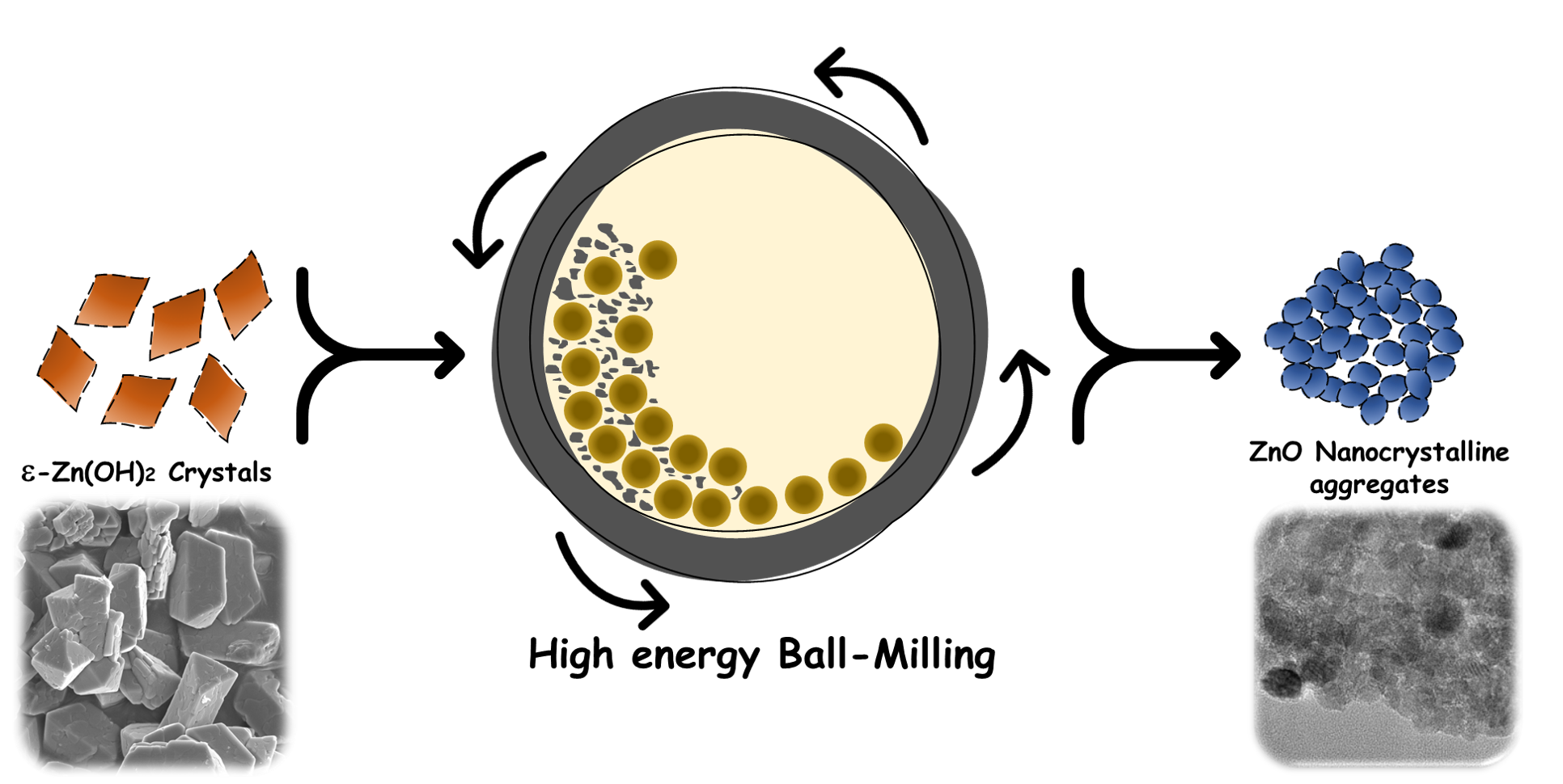
Synthesis of Nanomaterial’s by High Energy Ball Milling:
A ball mill is a type of grinder that is used to mix or grind materials for a variety of purposes. The impact force allows the feed material to be reduced in size. The horizontal axis of the cylindrical shell rotates [29].
Salting Out Method:
This method separates the water-miscible solvent by salting it out of an aqueous solution [30].
1. Emulsions Diffusion Method:
Some drawbacks of this method are: the high volumes of water to be eliminated from the suspension and reduced encapsulation efficiency during emulsification because in the saturated-aqueous external phase leakage of water-soluble drug [31].
Examples of some drug- loaded nano particles which were produced by this technique? Cyclosporine (cy-A-)? Loaded sodium glycolate nanoparticles [32]. Nanoparticles containing mesotetra (hydroxyphenyl) porphyrin and PLGA (p-THPP).[33] and Nano particles of doxorubicin-loaded PLGA[34].
2. Irradiation Methods:
Silver NPs can be synthesized by using a variety of irradiation methods. Laser irradiation of an aqueous solution of silver salt and surfactant can produce silver NPs with a well-defined shape and size distribution. Furthermore, the laser was used in a photo-sensitization synthetic method of making silver NPs using benzophenone. At short irradiation times, low laser powers produced silver NPs of about 20 nm, while an increased irradiation power produced NPs of about 5 nm. In visible light irradiation studies, photo-sensitized growth of silver NPs using thiophene (a sensitizing dye) and silver nanoparticle formation by illumination of Ag (NH3) + in ethanol have been performed. Mercury lamps and lasers can also be used as light sources for the production of silver NPs [35].
Applications
1. A completely different application of nanoparticles is in food products. Amorphous silica nanoparticles are used as anti-caking agent to maintain the flow properties in powder products (e.g., instant soups) and to thicken pastes [36].
2. In addition to these mass applications, selected nanoparticle have found their way in to more sophisticated, high-techproducts. However, the timeline from the discovery in the laboratory to a commercial product is typically very long. For example, from the first report on the use of titanium dioxide nanoparticles in a dye sensitized solar cell [37].
3. Nanotechnology by employing nanoparticles gains researchers and scientific interest in medicinal and medical field as nanoparticles are core form of Nano-biomaterials. Nanoparticles in medical applications are used for cancer [38].
4. Drug delivery is one of the most common medical applications for nanoparticles.[39].
5. Therapy techniques [40].
6. Diagnostic techniques [41].
7. Anti-microbial techniques [43].
8. Targeting tumor cells, cell repair, and gene transfer [44].
Dark Field Microscopy
GNPs have been extensively utilized in dark field microscopy6, in which only an outer ring of illumination is provided by blocking some of the light that enters the microscope. This ring is condensed and focused on the sample, where it either transmits through the sample or is scattered by it. The scattered light reaches the objective lens while the transmitted light is blocked and not collected. The collected light is used to produce an image of the sample with a dark back ground. In 2005, El-Slayed et al. demonstrated the ability to use dark-field microscopy to label cancer cells in vitro.97 Figure 3 shows the light scattering images of HaCaT noncancerous cells (left column), HOC cancerous cells (middle column) and HSC cancerous cells (right column) without NPs[45] .Targeting ,imaging agent[46] Controlled and systemic Delivery of water insoluble Drugs[47] .contain titanium dioxide and zinc oxide nanoparticles, because they are colorless and reflect/scatter ultraviolet light more efficiently than larger particles[48]. Nanoparticles are used, or being evaluated for use, in many fields and list below introduces several fields where nanoparticles can be potentially applied [49].
CONCLUSION –
The following conclusions can be drawn from the characterization study of AuNPs embedded in cosmetic skin care creams:
AuNPs with a PVP stabilizer are present as individually dispersed nanoparticles and as groups of physically separated primary nanoparticles, which are not hard agglomerates that cannot be split up. These groups of AuNPs are present in the freeze-dried form, as well as in the cosmetic cream. Nanoparticles have a wide spectrum of application in different areas of science and technology, which represents a highly attractive platform for research and innovative studies [50]. In addition to the already approved nanoparticles, numerous other nanoparticle platforms are currently under various stages of preclinical and clinical development, including various liposomes, polymeric micelles, Dendrimers, quantum dots, gold nanoparticles, and ceramic nanoparticles. With continued research and development efforts, nanotechnology is expected to have a tremendous impact on medicine for decades to come. Nanotechnology is definitely a medical boon for diagnosis, treatment and prevention of cancer disease. A number of approaches for successful applications of nanomaterial-based cancer drugs will be possible because of their unusual characteristics. Areas of greatest clinical impact likely include novel, targeted drug-delivery vehicles, molecularly targeted contrast agents for cancer imaging, targeted thermal tumor ablation, and magnetic field targeting of tumors.
REFERENCES


Vanve Kanchan*, Mokate Rajesh, Overview of Nanoparticle, Int. J. of Pharm. Sci., 2025, Vol 3, Issue 7, 3998-3908. https://doi.org/10.5281/zenodo.16561020
 10.5281/zenodo.16561020
