Abasaheb kakade college of B.pharmacy at Bodhegaon
Over the past few decades, there has been significant research interest in drug delivery systems employing particulate carriers for molecules of varying sizes. Particulate systems, such as nanoparticles and liposomes, have been utilized to enhance the pharmacokinetic and pharmacodynamic properties of drugs. They function in vivo to shield drugs in circulation, regulate access to target sites, and enable controlled, sustained release at the site of action. Various polymers have been employed in nanoparticle formulations to optimize therapeutic efficacy while minimizing side effects. Liposomes, specifically, are effective due to their structural resemblance to cell membranes, facilitating penetration to target tissues where free drugs may not reach effectively. Additionally, other delivery systems like niosomes, microparticles, resealed erythrocytes, and pharmacosomes have been explored. Medical implantable devices are intricate, and their physicochemical properties, including surface wettability, functional groups, charge, and topology, are crucial for acceptance by the body. Textile materials are favored for implantable devices due to possessing these essential properties
Nanotechnology has gained significant traction in recent years, with nanoparticles emerging as its cornerstone. These minute particles, smaller than 100 nm, encompass various materials like carbon, metals, metal oxides, or organics, each exhibiting distinct physical, chemical, and biological characteristics compared to their larger counterparts. [1] Their properties, including heightened chemical reactivity or stability and increased surface area-to-volume ratio, make them invaluable for numerous applications. Nanoparticles, spanning diverse sizes, shapes, and compositions, range from zero-dimensional nanodots to one-dimensional nanotubes and two-dimensional materials like g-C3N4 thin films. Additionally, liposomes have emerged as effective carriers for sensitive active compounds, boasting attributes such as biocompatibility, high loading capacity, entrapment efficiency, and sustained release. Meanwhile, implantable devices, crucial for diagnostic, monitoring, or therapeutic purposes, have seen advancements through the utilization of responsive polymers. These polymers enable tailored responses to clinical needs, minimizing tissue damage during deployment or removal, supporting device functionality, and facilitating tasks such as drug delivery, infection[1] control, or physiological monitoring."
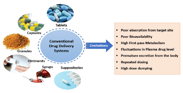
Fig.No 1. Conventional drug Delivery systems
The conventional method of drug delivery is commonly utilized to administer drugs swiftly into the body, especially when rapid absorption is essential. Techniques such as simple oral, topical, inhalation, or injectable methods are examples of this approach. However, these methods often fail to maintain a consistent plasma concentration over an extended period, necessitating multiple doses at regular intervals. Consequently, fluctuations in blood plasma drug levels occur, and patients frequently miss doses, emphasizing the necessity for more advanced drug delivery systems.[7]
A Novel Drug Delivery System (NDDS) represents an innovative approach integrating advanced development, formulations, and cutting-edge technologies to efficiently deliver pharmaceutical compounds within the body to achieve desired pharmacological effects safely. [2]
These systems offer formulation scientists an opportunity to address numerous challenges associated with antiretroviral (ARV) drug therapy, thus enhancing the management of HIV/AIDS patients. Existing anti-HIV drugs fall into three categories: nucleoside reverse transcriptase inhibitors, non-nucleoside reverse transcriptase inhibitors, and protease inhibitors. However, many of these drugs exhibit significant limitations such as short half-life, low bioavailability, poor permeability, and adverse side effects. Consequently, endeavors have been made to develop drug delivery systems for anti-HIV agents aiming to reduce dosing frequency, enhance bioavailability, minimize degradation/metabolism in the gastrointestinal tract, improve CNS penetration and inhibit CNS efflux, and selectively target cells with minimal side effects. The primary objective of these new drug delivery systems is to administer drugs at a rate suitable for the body's requirements during treatment and to swiftly deliver the active ingredient to the site of action. Drug delivery systems (DDS) capable of precisely controlling drug release rates or targeting specific body sites have revolutionized the healthcare sector. The ideal drug delivery device ensures a consistent delivery pace tailored to the body's needs over the treatment duration, effectively delivering the active ingredient to the intended site of action. This is achieved by binding the drug to a carrier particle, thereby optimizing therapeutic outcomes.
Characteristics of Novel Drug Delivery System:
Increase the bioavailability
Provide controlled delivery of drug
Transport the drug intact to the site of action avoiding the non-diseased tissue.
Stable and delivery be maintained under various physiological variables.
Easy to administer, safe and reliable.
Cost-effective.[2]
Advantages of NDDS:
1.Protection from physical and chemical degradation.
2. Sustained delivery.
3. Improved tissue macrophages distribution.
4. Enhancement of stability.
5. Enhancement of pharmacological activity.
6. Enhancement of solubility [3]
7. Protection from toxicity.
8. Increased bioavailability.
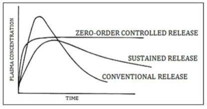
Fig.No 2. Plasma conce of drug after administration of conventional and novel drug graph
Controlled and Novel Drug Delivery which was only a dream or at best a possibility is now a reality. During the last decade and half pharmaceutical and other scientists have carried out extensive and intensive investigations in this field of drug research.[7]
Need of Novel Drug Delivery System:- [NDDS]
Conventional dosage forms often result in an abrupt release of drugs, leading to plasma drug level oscillations. To maintain drug concentrations within the therapeutic window and minimize undesired effects while enhancing therapeutic efficacy, novel drug delivery systems are imperative. Various medications, including vaccines, peptides, proteins, antibodies, and gene-based drugs, typically necessitate the utilization of conventional drug delivery systems due to potential enzymatic degradation, poor bioavailability, and limited penetration.
A novel drug delivery system represents an innovative approach to sustained drug delivery within the body, ensuring a steady-state plasma concentration over an extended period. These systems offer advantages such as targeted drug delivery, enhanced potency, and improved patient compliance, among others.
Recent developments in novel drug delivery system of herbals
1. Liposome
2.. Nanoparticles
4. Emulsions
5. Microsphere
6. Ethosome
7. Solid lipid nanopartical
8. Niosomes
9. Proniosomes
10. Transdermal Drug Delivery System
11. Dendrimers
12. Liquid Crystals
13. Hydrogels [3][14]
1.Nanopartical
Nanotechnology is science of matter and material that deal with the particle size in nanometers. The word ?Nano? is derived from Latinword, which means dwarf (1nm=10-9m). Nanoparticles are defined as particulate dispersions or solid particles with a size in the range of 10-1000nm. The drug is dissolved, entrapped, encapsulated or attached to a nanoparticle matrix. Nanoparticles offer some specific advantages such as they help to increase the stability of drugs/proteins and possess useful controlled release properties. It can be modified to achieve both active and passive targeting; drug loading is very high and can be administered by various routes such as parenteral, nasal, Nanotechnology refers to an emerging field of science that includes synthesis and development of various nanomaterials.Nanoparticles can be defined as objects ranging in size from 1-100 nm that due to their size may differ from the bulk material.Presently, different metallic nanomaterials are being produced using copper, zinc, titanium, magnesium, gold, alginate and silver. Nanoparticles are being used for diverse purposes, from medical treatments, using in various branches of industry production such as solar and oxide fuel batteries for energy storage, to wide incorporation into diverse materials of everyday use such as cosmetics or clothes .
Properties of nanoparticles
Nanoparticles exhibit a wide range of sizes, shapes, and structures, spanning from spherical, cylindrical, tubular, conical, hollow, spiral, flat, to extraordinary shapes, with lengths varying from 1 nm to 100 nm. The surface of nanoparticles can be uniform or irregular, featuring variations in surface texture. Some nanoparticles possess a crystalline or amorphous structure, consisting of single or multi-crystal solids that may agglomerate. The material properties of nanoparticles are influenced by their shape and composition, which can be engineered or modified by adjusting the relative impact of interfacial or interphase properties and macroscopic bulk properties through the size or dimension of components and domains. This approach, dating back centuries with steel alloys, has been highly effective, leading to the widespread use of composite engineering materials with micro to nanoscale component sizes today. The relationship between the structure and composition of nanoparticles is complex, with intricate interrelations between physical and chemical characteristics. This interrelation extends to bulk and surface properties of each component, as well as newly emerging characteristics localized at interfaces. Selective chemical reactivity is common in nanoparticle composites, enabling material disintegration into constituent elements. The behavior of nanoparticle composites is governed by complex processes, including nanoparticle release into the surrounding environment.
Formulation of nanoparticle
1.Precipitation system
The system for manufacturing nanoparticles was developed around 1980. This method involves solubilizing the active pharmaceutical ingredient (API) in a natural solvent, while excipients such as polymers, surfactants, and stabilizers are dissolved in a miscible inorganic solvent. Through spontaneous agitation, organic solvent is then added to the inorganic solvent, leading to the precipitation of nanoparticles. This technique is straightforward and cost-effective compared to other methods, requiring less time for successful outcomes. However, it relies on the solubility of the API in at least one organic solvent, and the natural solvent must be miscible with the inorganic solvent, which are the limitations of this approach.
2.Milling system
This technology emerged in the 1990s, involving the charging of the active pharmaceutical ingredient (API) and surfactant into a milling chamber filled with milling pearls. Subsequently, high-speed rotation is applied using a motor, resulting in the formation of nanoparticles suspended in a liquid. However, this process is time-consuming, influenced by factors such as the hardness of the drug and the quantity of components involved. Additionally, it requires significant energy input, and the erosion of milling pearls can lead to product degradation and pose risks of bacterial and microbial contamination. [10] The milling chamber in this method is typically made from materials such as zirconium oxide, glass, or polystyrene resin, and the milling process can be conducted in both aqueous and organic mediums. It is suitable for producing both diluted and concentrated suspension formulations. However, a notable drawback is the extended duration required for the process, and milling may introduce instability to the suspension.
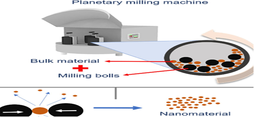
Fig.No 3. Milling Systems
1. Method of Pelletization
In general, nanosuspensions are stable; however, in particular situations, such as oral administration, and stability issue we need solid formulation so, this technique is usable for this problem solution. By applying techniques like lyophilization, spray drying, extrusion, and spheronization, or by layering on sugar pellets, nanosuspension is transformed into solid form. Using an extrusion technique, the final product is produced using this procedure, which involves mixing a suspension with the matrix excipients. The substance is forming free-flowing, small, spherical pellets in the shape of the material
2.Production Process for Hot Melted
This approach involves applying homogenization at a high temperature to melted material. Temperature is a decision based on the melting point of the material matrix being employed. Because the container is covered with temperature control jackets, the Micro Lab 40 homonizer is typically utilized for this approach. This process moves on to solidification, which is accomplished at room temperature by cooling once the desired particle size has been attained .
3. Direct Compression Method
The nanoparticle powder is obtained from nano suspension by using spray drying or any other method and which can use orally by filling it into capsule. In case of acid sensitive drug nanoparticle are fill into hard gelatin capsule. The other way of applying nanoparticle as orally is convert it into tablet formulation. For that drug nano suspension is mixed with matrix forming material like micro size polymer powder or lipid and lactose powder and then spray drying is apply. By this process the liquid phase is go into API-matrix compound and convert into free flowing powder than direct compression is apply and which give long release tablet formulation
APPLICATION
1.Therapeutic applications of Polymeric nanoparticles
They create novel medication delivery systems for the treatment of neurodegenerative and brain-related diseases using polymeric nanoparticles.
? Drugs are protected by polymeric NPs by being encapsulated, trapped inside the core, or adsorbing on to the particle surface.
? Polymeric NPs use the endocytosis and transcytosis pathways to transport molecules with across through the BBB.
? This polymeric covering is expected to lessen immunogenicity and restrict the reticuloendothelial system's ability to phagocytose nanoparticles, leading to increased blood levels of the drug in organs like the brain, intestines, and kidneys.
? These have been used in gene therapy to have an antiproliferative effect on breast cancer cells
2.Nanoparticles for gene delivery
Polynucleotide vaccines operate by delivering relevant antigen-coding genes into host cells, where they are expressed, leading to the production of antigenic proteins close to expert antigen-presenting cells, thus triggering an immune response. This mechanism stimulates both the humoral and cell-mediated immune systems, as intracellular protein synthesis is involved. DNA, the primary component of polynucleotide vaccines, offers advantages such as easier manufacturing, better storage, and handling characteristics compared to protein-based vaccine components. Additionally, nanoparticles loaded with plasmid DNA can serve as effective sustained release gene delivery vehicles, owing to their rapid transition from the degradative endo-lysosomal compartment to the cytoplasmic compartment.
Furthermore, the utilization of poly(lactic-co-glycolic acid) (PLGA) nanoparticles carrying therapeutic genes, such as those involved in bone repair, showcases the potential of this gene delivery technique. These nanoparticles can deliver morphogenic proteins for bone regeneration, highlighting their versatility in various biomedical applications.
3. Nanoparticles for drug delivery into the brain
The blood-brain barrier (BBB) is the primary barrier preventing the creation of novel medications for the central nervous system network of nerves. Endothelial cells with tight connections, enzymatic activity, and active efflux transport mechanisms, which are relatively impermeable, characterize the BBB. It successfully blocks the entry of water-soluble molecules from blood circulation into the CNS and, through the action of enzymes or efflux pumps, can also lower the concentration of lipid-soluble molecules in the brain. As a result, the BBB only allows for the selective transit of chemicals that are necessary for brain activity
4.Antibody targeting of nanoparticles
Numerous studies have shown the use of antibody-mediated nanoparticles to create targeted drug delivery systems, particularly in the context of cancer treatment. Antibodies that target drug substances have the potential to enhance the therapeutic efficacy of the drug substance as well as the concentration and distribution of the medication at the intended site of action[12]
Liposome
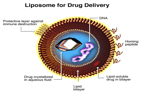
Fig.No 4. Liposome
Liposomes were first discovered by Alec Douglas Bangham, a British hematologist, in 1961 at the Babraham Institute in Cambridge, England. While testing a new electron microscope with dry phospholipid and gram-negative stain, Bangham and R.W. Horne observed a "bag-like" arrangement forming automatically, which was initially termed "multilamellar smectic mesophase" or "Banghasomes" by Bangham himself. It was Bangham's colleague Gerald Weissman who suggested the more user-friendly term "liposome." The term "liposome" originates from the Greek words "lipos" meaning "fats" and "somas" meaning "body." A.D. Bangham and colleagues first described liposomes in 1964. Liposomes are microscopic vesicles composed of one or more lipid layers. They are colloidal particles formed when phospholipids are hydrated in water, resulting in liposomes ranging from 0.01 to 0.5 µm in diameter.Liposomes have become one of the most important novel drug delivery systems due to their biocompatibility and biodegradability. They consist of an aqueous core entrapped by one or more phospholipid bilayers composed of natural or synthetic phospholipids. Liposomes can carry both hydrophilic and lipophilic drugs, and drug targeting can be achieved by surface modification, making them more localized to target disease tissues.Naturally occurring phospholipids form bilayers, where hydrophilic polar heads face water inside and outside the membrane, while the lipophilic hydrocarbon tails face each other to form the bilayer. Disruption of the phospholipid bilayer forms spheres smaller than normal cells, which can be monolayer (micelles) or bilayer (liposomes). Liposomes are composed of naturally derived phospholipids such as egg phosphatidylethanolamine or pure surfactant components like dioleoyl-phosphatidylethanolamine (DOPE).
•Advantages of Liposomes
There are many drugs in the market, which have good therapeutics activities, but they are used in the dearest situation, because of their poor pharmacokinetics and pharmacodynamics activities. Drugs encapsulated in liposomes can be used regularly, as its pharmacokinetics and pharmacodynamics can be controlled. Some of the advantages of the liposome are as follows:
1. Provides selective passive targeting to tumor tissues (Liposomal doxorubicin).
2. Increased efficacy and therapeutic index.
3. Increased stability via encapsulation.
4. Reduction in toxicity of the encapsulated agents.
5. Site avoidance effect.
6. Improved pharmacokinetic effects (reduced elimination, increased circulation lifetimes).
7. Flexibility to couple with site-specific ligands to achieve active targeting. [4]
Disadvantages Of Liposomes
All drug delivery system has faults, same is the case of liposomes. As liposomes are required to enhance and increase the efficacy of drugs, the cost as well as all the other implications thereof must be taken into account. Cost is an issue when it comes to phospholipid preparation. This preparation is expensive to produce because of the costly raw material and equipment required for preparation. Liposomes are non-toxic but in the case of cationic liposomes, it tends to be toxic at higher concentrations.
Other problems related to liposomes are as following:
1. Sterilization:
Sterilization of liposomes is a complicated process. Because it is unstable in heat and certain methods of radiation. Sterilizing with chemicals may affect stability problems. The only sterilization method is a membrane filter that is capable to filter liposomes of size <0>
2. Short self-life and stability:
It is very difficult to achieve the stability of liposomal formulation due to chemical and physical degradation. Chemically, they are prone to oxidation and hydrolysis and they can physically fuse forming larger vesicles. It can be prevented by the addition of anti-oxidant such as tocopherol and the addition of cholesterol to avoid fusion.
3. Entrapment efficacy:
The amount of drug a liposome can entrap is often low and sometimes leakage of drugs takes place.
4. Removal from circulation by the reticuloendothelial system (RES):
The major drawback of liposomes as a drug carrier is that they are rapidly cleared by a phagocytic cell of the Mononuclear Phagocytic System (MPS). Larger liposomes are eliminated from circulation faster than smaller liposomes. PEGylation can increase the self-life of liposomes
Types of Liposomes
Liposomes are classified based on their structural properties, methods of preparation and composition, and application. Their properties such as the size of liposomes, number, the position of lamellae depend widely on the method of preparation, types of lipids used, and preparation condition of liposomes. This parameter, influence the in-vitro and in-vivo characteristics of liposomes. The classification of liposomes based on structural properties is mentioned in Table 1, classifications based on liposomes preparation are mention in Table 2, and based on composition and application are following: [5]
Based on structural parameters
MLV Multilamillar large vesicle->0.5um
OLV Oligolamellar vesicles -0.1-1um
UV Unilamillat vesicles -(all sizes range)
SUV Small unilamellar vesicles -20-100um
MUV Medium size unilamellar vesicles
LUV Large unilamellar vesicles->100nm
GUV Giant unilamellar vesicles-->1um
MV Multivesicular vesicles->1um[5]
Based on the method of Liposomal preparation
REV Single Oligolamellar vesicles made by reverse phase method
MLV-REV Multilamillar vesicles made by reverse phase method
SPLV Stable plurilamellar vesicles
FATMLV Frozen and thawed MLV
VET Vesicle prepared by extrusion technique
DRV. Dehydration rehydration vesicles
Based on the composition and Application
It is classified as follows:
Conventional Lyposome
PH sensitivive Liposome
Cationic Liposome
Long circulatory Lyposomal(LCL)
Immuno Lyposome [5]
Mechanism of Liposomes Formation
basic understanding of the physicochemical properties of phospholipids is needed to understand the liposome formation. Phospholipids are amphiphilic (having both aqueous and polar moiety affinity), it has two fatty acid chains containing 10-24 carbon atoms and 0-6 double bond in each chain, which are the non-polar tail of phospholipids. The polar end is mainly phosphoric acid bond to the water-soluble molecule when phospholipids are hydrated, they are arranged in such an orientation that the polar portion of the phospholipids remain in contact with the polar environment and at the same time shield the non-polar part. The most common natural polar phospholipids are phosphatidylcholine (PC). [6]
METHOD OF PREPARATION
The conventional method for the preparation of liposomes includes the solubilization of lipids in the organic solvent, drying down the lipids from organic solution, dispersion of lipids in aqueous media, purification of resultant liposomes, and analysis of the final product. All the method for the preparation of liposomes involves four steps:
1. Drying down lipids from an organic solvent.
2. Dispersing the lipid in aqueous media.
3. Purifying the resultant liposome.
4. Analyzing the final product Techniques used for the preparation of liposomes are described below;
1. Hand Shaking method: In this method, the lipid is solubilized in an organic solvent (mainly ethanol) in a round bottom flask with constant shaking in a circular manner, when the organic solvent evaporates, it forms a thin film of lipid on the RBF which on hydrated with purified water, with constant shaking, form a liposome. This method is useful for the preparation of MLV liposomes. Nowadays, a Rotary evaporator machine is used for the formation of lipid film and hydration as it is more reliable than the handshaking method. [6]
2. Sonication Method: This is the most widely used method for the preparation of SUV from MLV, prepared from the handshaking method and rotary evaporator method. There are two types of sonication methods used in the preparation of SUVs.
a) Probe Sonication method: In this method, the tip of the titanium probe is directly dispersed into liposome dispersion for the production of SUVs. In this method, the energy input is high due to which there is the generation of heat. For controlling heat, liposome dispersion is kept in the ice bath. The main disadvantage of this method is that the titanium fragment is sludge in a solution and contaminate it.[11][13]
b) Bath sonication: In this method, liposome dispersion in a container is placed on the sonication bath. This method is more convenient as compared to probe sonication for the production of SUVs because the temperature can be controlled easily. The sterilized liposome can be obtained, there is no titanium contamination.
3. French Press method: In this method, unstable MLVs are converted to SUVs and LUVs bypassing then through a small orifice of equipment. Liposomes produced through this method are more reliable, as it has good stability as compared to those prepared by sonication method. The drawback of this method is that it has a small working volume of a maximum of 50 ml and a high temperature is hard to manage. [11]
4. Freeze Thawed liposomes: Here, SUVs formed by the sonication method is frozen and thawed slowly and continuously, resulting in the formation of LUVs due to aggregation of SUVs during the thawing process. By this method, the encapsulation efficacies increase by 20%-30%.
5. Solvent Dispersion method
a) Ether injection (solvent evaporation): In this method, lipid dissolved in a diethyl-ether or ether-methanol mixture is gradually injected in an aqueous medium containing drug at the temperature of 50 to 65 ?c or reduced pressure. The removal of ether under vacuum results formation of liposomes. The main drawback of this technique is the formation of a heterogeneous population of liposomes (70-200 nm) and exposure of liposomes in high temperatures during encapsulation which can hamper the stability of liposomes.
b) Ethanol injection: To a buffer a solution of lipid and ethanol is injected, resulting in the formation of MLVs. The drawback is the formation of a heterogeneous population of liposomes (30-110 nm). It is also difficult to remove ethanol from a solution consequently increasing the chances for the inactivation of biologically active macromolecules.
c) Reverse Phase evaporation method: This method has brought a breakthrough in the history of liposomes. The aqueous and lipid ratio used in this method is high, about four times higher than the handshaking method or MLVs. This method is based on the formation of reverse micelle where an aqueous medium is sonicated. The aqueous medium contains a water-soluble molecule to be encapsulated, lipids, and an organic phase. The slow elimination of organic solvent results in the formation of a gel-like consistency. point, the gel-like structure collapses to form liposomes. [11]
6. Detergent removal method (removal of non-encapsulated material)
a) Dialysis: In this method, detergent is used to dissolve lipids at Critical Micelles Concentration (CMC). When it is removed by dialysis, using a commercial device such as LipoPrep (Diachema AG, Switzerland) which is a version of the dialysis method.
b) Detergent (cholate, alkyl glycoside, Triton X-100) removal of mixed micelles (absorption): In this method, removal of detergent in achieved by shaking mix micelle with beaded organic polystyrene absorbers such as XAD-2 beads (SERVA Electrophoresis GmbH, Heidelberg, Germany) and Bio-beads SM2 (Bio-Rad Laboratories, Inc., Hercules, USA).
c) Gel-permeation chromatography: Sephadex G-50, Sephadex G-l 00 (Sigma-Aldrich, MO, USA), Sepharose 2B-6B, and Sephacryl S200-S1000 (General Electric Company, Tehran, Iran) can be used for gel filtration as packing material for a column. Liposomes cannot penetrate through this packing which makes it easier to obtain liposomes. The physical and chemical characteristics of liposomes have a direct impact on the properties of a liposome, in-vivo, and in-vitro. The characterization of liposomes should be done immediately after the formation of liposomes by analysis methods (such as GLC, TLC, and HPTLC). Size, number of lamellae, internal morphology charges, and bilayer fluidity play a direct role in in-vivo properties. Sterilizing of liposomes is difficult as it is sensitive to heat.[ 6]
Application of Liposomes as Drug Delivery System:
Liposomes, a novel drug delivery system, have various applications aimed at achieving therapeutic efficacy and safety:
1. Site avoidance delivery: Liposomal formulations of cytotoxic drugs can minimize adverse effects on normal cells due to their low therapeutic index (TI). For instance, Doxorubicin, known for causing cardiac toxicity, exhibits reduced toxicity when formulated in liposomes.
2. Site-specific targeting: Liposomal formulations enable site-specific targeting by encapsulating drugs and attaching specific ligands to the liposomes. These ligand-attached liposomes selectively target specific cells, crucial for achieving therapeutic effects at desired sites of action.
3. Intra-cellular drug delivery: Liposomal formulations facilitate the delivery of drugs into the cytosol of cells. For instance, N-(phosphonacetyl)-L-aspartate (PALA), which is poorly taken up by cells, demonstrates enhanced activity against ovarian tumor cell lines when encapsulated in liposomes.
4. Sustained release drug delivery: Liposomal formulations can retain drugs within the system for prolonged periods, enabling sustained release effects in the body. Drugs like cytosine Arabinoside can be encapsulated in liposomes to achieve optimized release rates for sustained drug delivery in vivo.
5. Reduced toxicity: Liposomal formulations release drugs over an extended period within the therapeutic index, resulting in reduced toxicity compared to free drugs. Liposomal formulations have been shown to reduce the toxic effects of drugs by up to 50%.[6]
Overall, liposomal formulations offer versatile and effective strategies for drug delivery, enhancing therapeutic outcomes while minimizing adverse effects.
Implabtabl Device
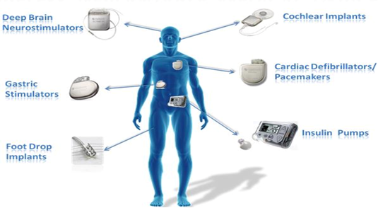
Fig.No 6. Implabtabl Device
Implantable medical devices are introduced into the human body through surgical procedures or other medical interventions to fulfill specific functions. Examples of such devices include artificial joints used in joint replacement surgeries, breast implants for augmentation or reconstruction, cochlear implants to restore hearing function, intraocular lenses for vision correction after cataract surgery, pacemakers and other cardiac implants to regulate heart rhythm, and intrauterine contraceptive devices for birth control purposes. These devices play crucial roles in improving patient health and quality of life by addressing various medical conditions and enhancing bodily functions.
What are implantable medical devices?
Implantable medical devices serve variouspurposes within the human body, whetherpermanently or temporarily, to support organ functions, monitor physiological activities, or administer medications. Some implants are prosthetics designed to replace damaged body parts. These devices may consist of human tissues or materials like metals, plastics, and ceramics.
Active implantable medical devices operate using an electrical energy source external to the human body or gravity. These battery-powered devices are frequently employed to support cardiac functions and are meant to remain implanted in the body following the surgical or medical procedure.
Common examples of implantable medical devices include:
1. Cardioverter Defibrillator: A battery-powered device implanted in the heart tissue to monitor heart rate rhythms and deliver electrical shocks to restore normal heartbeat rhythms in patients with recurrent ventricular tachycardia.
2. Pacemaker: A small battery-operated device implanted under the collarbone to send electrical impulses to the heart through wires, maintaining normal heartbeat rhythms in individuals with irregular heartbeats due to damage to the heart's electrical conduction system.
3. Left Ventricular Assist Device (LVAD): A mechanical pumping device implanted surgically to support cardiac circulation by assisting the left ventricle in pumping blood, particularly beneficial for end-stage heart failure patients who cannot undergo heart transplantation.
4. Breast Implants: Placed under the breast tissue or chest muscle for breast augmentation or reconstruction, typically composed of a silicone outer shell filled with saline or silicone gel.
5. Cochlear Implants: Electronic hearing devices consisting of an external microphone, sound processor, transmitter system, and internally implanted receiver and electrode system, providing effective hearing sensations in individuals with severe to profound hearing loss by stimulating the inner ear with electrical currents. [9]
CONCLUSION:
The conclusion of, the exploration of advancements in liposomes, nanoparticles, and implantable devices has studied their immense potential to revolutionize drug delivery systems. Liposomes offer targeted and controlled release capabilities, nanoparticles provide enhanced bioavailability and tissue specificity, while implantable devices offer sustained and localized drug delivery. These advancements hold great promise for improving the efficacy, safety, and patient compliance of pharmaceutical treatments across a wide range of diseases. Continued research and development in these areas are crucial for unlocking further innovations and ultimately transforming the landscape of modern medicine.
REFERENCES


Shivkanya Jadhav*, Amol Supekar, Kanchan Jawale, Aarti Garje, Vaishnavi Jaygude, Sanika Sarode, Novel Drug Delivery System: Exploring advancements in drug delivery systems such as Nanoparticles, Lyposome and Implantable devices, Int. J. of Pharm. Sci., 2025, Vol 3, Issue 2, 1569-1581. https://doi.org/10.5281/zenodo.14889397
 10.5281/zenodo.14889397
