Rasiklal M. Dhariwal Institute of Pharmaceutical Education and Research, Late Dhodiba Detter Patil Marg, MIDC, Chinchwad, Pune, Maharashtra 411019
Niosomes are novel, non-ionic surfactant-based vesicular drug delivery systems that have gained significant attention in recent years due to their ability to improve the bioavailability, stability, and therapeutic performance of various pharmaceutical agents. Structurally similar to liposomes, niosomes consist of an aqueous core surrounded by a bilayer of non-ionic surfactants and cholesterol, offering enhanced biocompatibility and controlled drug release. They are capable of encapsulating both hydrophilic and lipophilic drugs, thereby providing targeted and sustained delivery with reduced side effects. Several methods such as thin-film hydration, ether injection, reverse-phase evaporation, and microfluidization are employed for their preparation, each influencing vesicle size, entrapment efficiency, and stability. Niosomes have found wide applications in transdermal, ophthalmic, and anticancer drug delivery, as well as in vaccine adjuvants and cosmetic formulations. Their advantages over conventional systems, including chemical stability, low production cost, and easy scale-up, make them promising candidates for future pharmaceutical innovations. This review highlights the composition, types, methods of preparation, characterization parameters, and diverse applications of niosomes, emphasizing their potential as an efficient and versatile drug delivery system.
The concept of selectively transporting therapeutic agents to specific tissues dates back to 1909, when Paul Ehrlich proposed the idea of a “magic bullet” capable of targeting diseased regions without affecting healthy areas. This laid the foundation for modern targeted drug-delivery systems, which aim to deliver active molecules precisely to their site of action while minimizing systemic exposure and unwanted effects. (Umbarkar, 2021)
To improve drug performance, it is essential not only to reach the desired site but also to sustain therapeutic levels for an extended duration. Vesicular drug-delivery systems emerged as a transformative approach in this direction. These systems are composed of lamellar arrangements formed by amphiphilic molecules, creating a unique architecture with both aqueous and lipid components. This enables them to encapsulate a wide variety of drugs hydrophilic agents within their internal aqueous core and lipophilic substances within the surrounding lipid bilayer.
Among the vesicular carriers, liposomes have been widely investigated and are regarded as a benchmark system. However, their practical use is restricted by several drawbacks, including a short shelf-life, susceptibility to oxidative degradation, high production costs, and challenges in achieving consistent purity, size, and morphological uniformity. These limitations have driven the search for more robust vesicular alternatives. Niosomes have emerged as a promising solution. Composed of non-ionic surfactants and cholesterol, these bilayered structures exhibit improved stability, lower formulation cost, and reduced risk of degradation compared with liposomes. Their composition provides a fundamental distinction from liposomes and enables better manipulation of physicochemical properties, positioning niosomes as a more versatile and accessible platform for advanced drug-delivery applications. (N.K. Jain PAGE NO :230)
NIOSOMES:
Niosomes are nanosized, layered vesicular systems that form when non-ionic surfactants are combined with cholesterol and, in some cases, a charge-modifying agent, followed by hydration in an aqueous environment. These components self-assemble into a bilayer structure composed of non-ionic surface-active molecules, which is characteristic of niosomes. Their unique architecture allows efficient encapsulation of therapeutic agents, making them highly effective in enhancing transdermal drug transport as well as facilitating targeted drug delivery. Noisome made of alpha, omega-hexadecyl-bis-(1-aza-18-crown-6) (Bola-surfactant)-Span 80-cholesterol (2:3:1 molar ratio) is named as Bola-Surfactant containing noisome (Kaur IP et.al 2004)
NOISOME: A SUPERIOR DRUG-DELIVERY SYSTEM COMPARED WITH LIPOSOMES:
Niosomes possess a bilayer structure, which is similar to liposomes. However, the materials used to prepare niosomes confer better stability on them. (Diljyot K. 2012)
Niosomes are typically formulated using non-ionic, single-chain surfactants in combination with cholesterol, whereas liposomes are composed of neutral or charged double-chain phospholipids. Although both systems contain cholesterol, its proportion is generally higher in liposomes compared to niosomes. This compositional distinction affects drug-loading behavior, resulting in niosomes exhibiting greater entrapment efficiency than liposomes.
From a manufacturing perspective, niosomes are more economical and easier to produce on an industrial scale, as they do not require stringent storage conditions. In contrast, the phospholipids used in liposome preparation are chemically unstable and prone to oxidative degradation, making their formulation more expensive and demanding in terms of handling and storage requirements. Consequently, liposomes require specialized processing to maintain stability.
Overall, niosomes demonstrate improved stability and a longer shelf-life compared to liposomes (Kazi KM et al. 2010) Niosomes possess a longer shelf life than liposomes. (Reddy BS 2012)
THE APPLICATION OF NIOSOMES FOR THERAPEUTIC PURPOSE MAY OFFER SEVERAL ADVANTAGES:
Niosomes present several noteworthy benefits when used for therapeutic drug delivery. Because they are formulated in an aqueous medium, they offer greater patient comfort compared to oily dosage forms. Their bilayer architecture allows them to encapsulate a wide range of drug molecules, including both water-soluble and lipid-soluble agents. One of the key strengths of niosomes is the ability to modify their physicochemical properties. Parameters such as vesicle size, number of layers (lamellarity), surface charge, composition, and drug concentration can be easily adjusted to tailor the formulation for specific therapeutic needs. These vesicles can also function as depot systems, ensuring gradual and controlled release of the encapsulated drug. Niosomes exhibit good osmotic behavior and help enhance the stability of the entrapped drug, protecting it from degradation. Their formulation does not require stringent storage conditions since the surfactants used are relatively stable. Furthermore, niosomes can enhance the oral bioavailability of poorly absorbed drugs and improve the transdermal penetration of therapeutic agents. These carriers are versatile and can be administered through multiple routes, including oral, parenteral, and topical pathways, allowing targeted delivery to the site of action. Additionally, the surfactants used in niosomal preparations are generally biocompatible and biodegradable, making them safe for medical applications.
DISADVANTAGES OF NIOSOMES:
Although niosomes are promising carriers for drug delivery, they possess certain limitations. Their physical stability is a major concern, as vesicles may lose integrity over time. In some cases, variations in surface charge can cause oppositely charged vesicles to attract each other, resulting in fusion or structural alteration. If proper formulation techniques are not followed, niosomes may also undergo aggregation, leading to changes in size distribution and reduced performance.
Another drawback is the possibility of hydrolytic degradation of the encapsulated drug, which can compromise therapeutic efficacy. Additionally, some formulations exhibit limited drug-loading capacity, restricting the amount of active ingredient that can be delivered. The preparation processor niosomes can also be labor-intensive and time-consuming, requiring careful optimization of multiple parameters to achieve a stable and effective formulation. (Umbarkar 2021)
STRUCTURE:
A non-ionic surfactant of the alkyl or dialkyl polyglycerol ether class, which is usually stabilized by the addition of cholesterol and a small amount of anionic surfactant, such as dicetyl phosphate, which also helps in stabilizing the vesicle. The surfactant molecules tend to orient themselves in such a manner that the hydrophilic ends of the non-ionic surfactant point outwards, however, the hydrophobic ends face each other to form the bi-layer. In the bilayer structure, hydrophobic parts are oriented away from the aqueous solvent, whereas the hydrophilic heads remain in contact with the aqueous solvent. Vesicles properties may be affected by factors, such as vesicles composition, size, lamellarity, tapped volume, surface charge and concentration. A number of forces inside the niosomes helps in maintaining its vesicular structure, such as Vander Waals forces among surfactant molecules, repulsive forces emerging from the electrostatic interactions among charged groups of surfactant molecules, entropic repulsive forces of the head groups of surfactants, short-acting repulsive forces, etc. However, factors such as type of surfactant, nature of encapsulated drug, storage temperature, detergents, use of membrane spanning lipids, the interfacial polymerization of surfactant monomers in situ and inclusion of charged molecule can affect the stability of niosomes. (N.K. JAIN PAGE NO. 230)

Figure 1 Structure of Niosomes
COMPOSITION:
1. Surfactants: A variety of non-ionic surfactants, used alone or in different molar proportions, can be employed to encapsulate diverse drug molecules within niosomes. The choice of surfactant influences key properties such as vesicle size, entrapment efficiency, and stability. (Giddi H.S. et al., 2007)
2. Ether-linked Surfactants: This category includes polyoxyethylene alkyl ethers, which contain hydrophilic and hydrophobic segments joined via an ether linkage. Their general structure is represented as CnEOm, where n typically ranges from 12–18, and m from 3–7. Surfactants containing polyhydroxyl head groups and ethylene oxide units have also been explored in niosomal systems. A notable example is C16 monoalkyl glycerol ether, consisting of an average of three glycerol units. Brij-type surfactants are commonly used in niosome preparation are Polyoxyethylene-4-lauryl ether (Brij 30) has an HLB value of 9.7 and a phase transition temperature below 10°C, but it is unsuitable for preparing formulations containing iodides, mercury salts, phenolic compounds, salicylates, sulfonamides, or tannins, as it may undergo oxidation and cause discoloration , Polyoxyethylene cetyl ethers (Brij 58) and Polyoxyethylene stearyl ethers (Brij 72, Brij 76) are also used extensively. (Baillie A.J. et al., 1985; Gannu P.K. & Pogaku R., 2011)
3. Ester-linked Surfactants: These surfactants feature an ester bond between hydrophilic and hydrophobic regions. They have been explored in the delivery of drugs such as sodium stibogluconate for the treatment of experimental visceral leishmaniasis. (Hunter C. et al., 1988)
4. Sorbitan Esters (Spans): Sorbitan esters are among the most common ester-linked surfactants used in pharmaceutical and food applications. They are produced as mixtures of partial esters of sorbitol and its anhydrides with oleic acid. Various sorbitan esters have been utilized to encapsulate multiple therapeutic agents, including doxorubicin. (Uchegbu I.F. et al., 1996)
5. Cholesterol: Cholesterol plays a crucial role in modifying the bilayer’s fluidity and permeability, contributing to niosome stability. While it does not form the bilayer independently, it can be incorporated in significant molar amounts into the membrane. As an amphiphilic molecule, cholesterol aligns its hydroxyl group toward the aqueous environment while its hydrophobic chain orients alongside the surfactant tails. Its inclusion increases membrane rigidity and reduces bilayer leakage by suppressing the gel-to-liquid phase transition, thereby improving vesicle integrity and drug-retention characteristics. (Dahiya N.K. et al., 2011)
6. Charge Inducers: Charge-inducing agents are added to impart surface charge to the vesicles, enhancing stability by generating electrostatic repulsion between similarly charged vesicles. This prevents vesicle fusion and increases zeta potential. Common negative charge inducers are Dicetyl phosphate, Dihexadecyl phosphate, Lipoamine acid.
Examples of positive charge inducers are Stearyl amine, Cetyl pyridinium chloride (Bandyopadhyay P. & Johnson M., 2007; Shan W. et al., 2008)
TYPES OF NIOSOMES:
1. Bola-Surfactant–Based Niosomes:
These niosomes are formulated using bola-type surfactants, which contain hydrophilic groups at both ends of a long hydrophobic chain. A typical composition includes omega-hexadecyl-bis-(1-aza-18-crown-6) along with Span-80 and cholesterol in a 2:3:1 molar ratio.
Because of the dual hydrophilic ends, bola surfactants impart excellent structural rigidity, stability and resistance to degradation, making these niosomes more robust. These systems are useful for controlled drug delivery. (Yoshida H.Et .Al 1992)
2. Proniosomes:
Proniosomes are dry, free-flowing granular or powder formulations composed of a water-soluble carrier (such as sorbitol) coated with non-ionic surfactant.
Upon hydration with an aqueous phase, the coating reorganizes to form niosomal vesicles.
Proniosomes offer several benefits are Reduce problems of fusion, leakage, and aggregation commonly seen with niosomes, Show improved storage stability, Simple to transport and re-hydrate before use. Thus, proniosomes act as a convenient precursor for efficient and stable niosome formation. (Jadon P.S Et Al 2009)
3. Aspasomes:
Aspasomes are tiny vesicles made by combining ascorbyl palmitate (a Vitamin-C derivative), cholesterol and a charged lipid called diacetyl phosphate. When this mixture is hydrated with water and then sonicated, it forms small niosome-like vesicles.
These systems are useful for delivering drugs through the skin more effectively. Because ascorbyl palmitate has strong antioxidant properties, aspasomes can also help protect tissues by reducing damage caused by reactive oxygen species (ROS) (Bhaskaran S & Lakshmip.K 2009)
4. Niosomes in Carbopol Gel:
In this system, niosomes formulated from drug, Span surfactants and cholesterol are incorporated into a Carbopol-934 gel base containing propylene glycol and glycerol.
Studies using diffusion cells and human cadaver skin showed the niosomal gel significantly reduces the flux and diffusion coefficient compared to plain drug gels. This demonstrates a controlled and sustained release profile, improving therapeutic benefit. Such systems are especially useful for topical drug delivery. (Yashida H.et .al 1992)
5. Vesicles in Water-in-Oil (v/w/o) System
In this type, niosomes are dispersed within an oil phase, forming a water-in-oil (v/w/o) emulsion.
To prepare this system, aqueous niosome suspension-made from sorbitol monostearate, cholesterol and solulan C24 is mixed with oil at around 60 °C.
Upon cooling, the emulsion transforms into a gel-like structure (v/w/o gel).
This configuration Enhances entrapment and provides sustained release of the drug. It is particularly useful for delivering lipophilic or hydrophilic drugs over an extended period. (Hu C & Rhodes D.G 1999)
6. Niosomes in Hydroxypropyl Methylcellulose (HPMC) Base
Here, niosomes are incorporated into an HPMC base containing about 10% glycerin.
This formulation helps for Improved drug bioavailability & Enhanced therapeutic activity
For example, in anti-inflammatory models, this system demonstrated better reduction of carrageenan-induced paw edema compared to conventional formulations. This delivery method is particularly compatible with topical or localized treatment applications. (Uchegbu I.F& Vyas S.P 1998) (Verma A.K.& Bindal J.C 2011)
FACTORS AFFECTING THE PHYSICOCHEMICAL PROPERTIES OF NIOSOMES:
1. Nature of Surfactants:
For niosome formation, the selected surfactant must possess both a hydrophilic head and a hydrophobic tail. The hydrophobic segment may be composed of one or two alkyl or perfluoroalkyl chains or sometimes a single steroidal group.
Ether-linked surfactants with single hydrophobic chains generally exhibit greater toxicity than their dialkyl ether counterparts. Conversely, ester-linked surfactants are less stable than ether-based ones but tend to be less toxic because they are broken down enzymatically in vivo to yield fatty acids and triglycerides. Surfactants with alkyl chains between C12–C18 are considered optimal for forming stable niosomal structures. (Madhav N.V.S. & Saini A., 2011)
2. Membrane Additives:
The addition of membrane-modifying agents influences the stability, morphology, and permeability of niosomal vesicles. Components such as cholesterol are frequently incorporated to enhance membrane rigidity and minimize drug leakage by reducing bilayer permeability. For example, niosomes prepared using C16G2, cholesterol and MPEG-Chol have been reported to form spherical vesicles with sizes between 20–200 nm. (Rajendra et al., 1994)
3. Resistance to Osmotic Stress:
Niosomes exhibit changes in structure when exposed to different osmotic environments. Under hypertonic conditions, vesicle diameter decreases due to water efflux. In hypotonic media, vesicles initially exhibit slow release accompanied by slight swelling, likely due to restricted fluid movement. This may be followed by accelerated release caused by loosening of the bilayer under osmotic stress. (Malhotra M. & Jain N.K., 1994)
4. Influence of Drug:
Drug incorporation can affect vesicle dimensions. Entrapment often increases vesicle size due to interactions between drug molecules and surfactant head groups, which enhance electrostatic repulsion between bilayers. Furthermore, the hydrophilic-lipophilic balance (HLB) of the drug strongly influences entrapment efficiency. (Gayatri D.S. et al., 2000; Silver B.L., 1985)
5. Cholesterol Content:
Cholesterol plays a key role in strengthening the niosomal bilayer by reducing membrane permeability and improving rigidity. Its integration into the vesicle membrane enhances drug-retention capacity and increases entrapment efficiency. For instance, the permeability of vesicles toward 5,6-carboxyfluorescein was found to decrease tenfold with cholesterol incorporation. (Weissman G. et al., 1975)
METHODS OF PREPARATION OF NIOSOMES:
Various methods are reported for the preparation of niosomes such as:
In this technique, a surfactant solution prepared in diethyl ether (a volatile organic solvent) is gradually injected into warm water maintained at around 60°C using a fine needle (usually 14-gauge). As the ether vaporizes, it leads to the formation of single-layered niosomal vesicles. The resulting vesicle size can vary from about 50 nm to 1000 nm, depending on the preparation conditions. (Rogerson A. et al., 1988; Baillie A.J. et al., 1986)
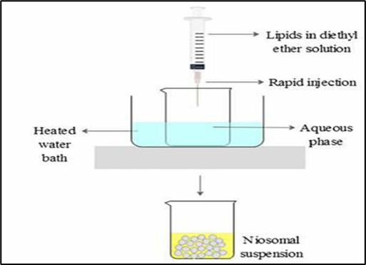
Figure 2 Ether Injection method
2. Thin film hydration technique:
Here, all vesicle-forming components typically surfactant, cholesterol and charge-inducing agents are dissolved in a volatile organic solvent inside a round-bottom flask. The solvent is then removed under reduced pressure using a rotary evaporator, leaving behind a thin dry film of the mixture on the flask walls. This film is subsequently hydrated with an aqueous phase under gentle agitation to produce niosomes. Hydrophilic drugs can be added to the aqueous phase, whereas hydrophobic drugs are dissolved along with the lipid components in the organic solvent. (Palozza P. et al., 2006; Arunothayanun P. et al., 2000)

Figure 3 Thin film hydration technique
In this method, a dispersion of surfactant and cholesterol in the aqueous phase is first prepared. The mixture is then probe-sonicated at 60°C for about 10 minutes to produce multilamellar vesicles (MLVs). These MLVs are further processed by ultrasonication, either using a probe or bath sonicator to yield smaller unilamellar vesicles. (Bhaskaran S. & Panigrahi L., 2002)

Figure 4 Sonication Method
Surfactant and cholesterol in a 1:1 ratio is dissolved in a mixture of ether and chloroform. The aqueous phase containing the drug is added to this mixture, followed by sonication at a low temperature (4–5°C). After forming a clear gel, a small amount of phosphate-buffered saline (PBS) is added, and the mixture is sonicated again. The organic solvents are then removed under low pressure at 40°C, resulting in a viscous niosomal suspension. This suspension is diluted with PBS and heated at 60°C for 10 minutes to obtain stable niosomes. (Raja Naresh R.A. et al., 1994)
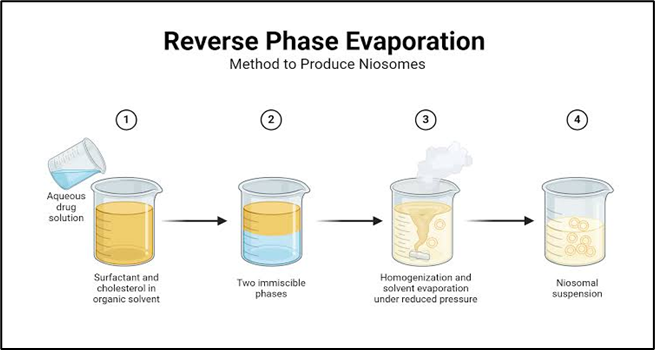
Figure 5 Reverse phase evaporation technique (REV)
This modern method employs a microfluidizer to prepare small multilamellar vesicles. The fluid is pumped under high pressure (approximately 10,000 psi) through microchannels where two fluid streams collide at right angles, ensuring efficient energy transfer and uniform vesicle formation. The process may be repeated until niosomes of the desired spherical size and uniformity are obtained. This technique offers high reproducibility and produces small, homogeneous vesicles. (Cook E.J. & Lagace, 1994)
In this approach, niosomes are passed through polycarbonate membrane filters to reduce their size and achieve uniformity. Depending on the applied conditions, this method can yield either multilamellar or large unilamellar vesicles. It is particularly effective for controlling the particle size distribution of niosomes. (Junyaprasert V.B. et al., 2008)
Surfactant and cholesterol are dissolved in chloroform and evaporated under reduced pressure to form a thin lipid film. This film is hydrated with 300 mM citric acid solution (pH 4.0) using vortex mixing, followed by three cycles of freezing and thawing. The suspension is then sonicated. To this niosomal dispersion, an aqueous drug solution (10 mg/mL) is added and vortexed. The pH is adjusted to 7.0–7.2 using 1 M disodium phosphate, and the mixture is heated at 60°C for 10 minutes, producing the final niosomal formulation. (Mayer L.D. et al., 1985)
8. The “Bubble” Method:
This novel, solvent-free technique allows one-step preparation of liposomes and niosomes. The apparatus consists of a three-necked round-bottom flask immersed in a water bath to control temperature. One neck holds a water-cooled condenser, another a thermometer, and the third a nitrogen gas inlet. Surfactant and cholesterol are dispersed in phosphate buffer (pH 7.4) at 70°C, mixed for about 15 seconds using a high-shear homogenizer and then bubbled with nitrogen gas at the same temperature to for niosomes. (Chauhan S. & Lawrence M.J., 1989)
9. Formation of niosomes from proniosomes:
In this method, a water-soluble carrier (such as sorbitol) is coated with a thin layer of surfactant, resulting in a dry, free-flowing powder known as proniosomes. Upon hydration, these proniosomes rapidly form niosomal vesicles. This method enhances stability and simplifies storage and transportation. (Blazek-Walsh A.I. & Rhodes D.G., 2001)
CHARACTERIZATIONS OF NIOSOMES:
1. Entrapment efficiency: After niosomes are prepared, the encapsulated drug is separated using techniques such as dialysis, centrifugation, or gel filtration. The drug that remains entrapped within the vesicles is then quantified by disrupting the niosomes completely, typically using 50% n-propanol or 0.1% Triton X-100, followed by analysis with an appropriate assay for the drug.
The entrapment efficiency is calculated using the formula:
% EF = (Amount of drug entrapped/ total amount of drug) x 100. (Yoshida H. et.al 1992)
2. Vesicle diameter: The size of niosomes can be measured using light microscopy, photon correlation spectroscopy, or freeze-fracture electron microscopy. Repeated freeze-thaw cycles (e.g., freezing the vesicle suspension at –20°C for 24 hours, then warming to room temperature) can increase vesicle diameter, likely due to fusion of vesicles during the process. (Malhotra M. & Jain N.K., 1994)
3. In-vitro release: Drug release from niosomes is commonly studied using dialysis tubing. The tubing is washed, soaked in distilled water, and filled with the vesicle suspension, then sealed. This bag is placed in a beaker containing buffer solution (200 mL) at a controlled temperature (25°C or 37°C) with constant shaking. At specific intervals, samples of the buffer are collected and analysed to determine the amount of drug released. (Yoshioka T. et al., 1992; Keservani R.K. et al., 2011)
4. Membrane rigidity: The rigidity or fluidity of niosomal membranes can be assessed by studying the mobility of a fluorescent probe incorporated into the membrane, usually as a function of temperature. (Mokhtar M. et.al 2008)
5. Bilayer formation: The formation of a bilayer structure in niosomes can be confirmed using polarized light microscopy, where the characteristic cross pattern (X-cross) indicates proper bilayer assembly of non-ionic surfactants. (Madhav N.V.S. & Saini A 2011)
6. Stability study: Niosomal formulations are typically stored at two different temperatures, commonly 4±1°C and 25±2°C, to evaluate their stability. Vesicle size, shape and the number of vesicles per cubic millimeter are measured before and after storage, often at intervals of 15 and 30 days. The size is determined using light microscopy and the number of vesicles is counted using a hemocytometer:
Number of niosomes/mm3 = Total number of niosomes counted × Dilution factor × 400 ÷

Number of small squares counted.
Residual drug content is also evaluated after the storage period. (Erdogan S. et.al 2005)
7. Vesicular surface charge: Niosomes are often formulated with charged molecules in their bilayers to prevent vesicle aggregation. Incorporating molecules like dicetyl phosphate reduces aggregation and enhances stability. The surface charge is measured in terms of zeta potential, which can be calculated using Henry’s equation: ζ= ?μEηΣ

(Desai A.R. et.al 2010)
Where:
APPLICATIONS OF NIOSOMAL DRUG-DELIVERY SYSTEMS:
Niosome-based delivery systems have shown promise in improving the therapeutic performance of many drugs. Their ability to encapsulate both hydrophilic and lipophilic compounds makes them useful across multiple medical fields. Key applications are summarized below.
Niosomes have been explored for delivering medications to the lungs, particularly for asthma and chronic obstructive pulmonary disease (COPD). Terzano and colleagues developed polysorbate-20 niosomes containing beclomethasone dipropionate and found that the formulation enhanced drug penetration through the mucus layer, prolonged drug release, and improved therapeutic action in COPD patients. These non-ionic vesicles significantly increased the permeation rate of the drug across a model mucosal barrier, offering improved targeting of corticosteroids. (Rajeswari, 2011; Moazeni et al., 2010)
Oral delivery of peptides and proteins is limited by enzymatic degradation, pH variation, and poor epithelial permeability. Encapsulation of insulin within niosomal bilayers has been shown to shield it from digestive enzymes such as trypsin, chymotrypsin, and pepsin. Similarly, vasoactive intestinal peptide (VIP), a molecule investigated for Alzheimer’s disease, typically fails to cross the blood–brain barrier and is rapidly eliminated from circulation. Studies by Dufes and coworkers demonstrated that glucose-modified niosomes could transport VIP to specific brain regions, suggesting their potential as a brain-targeting system. (Pardakhty et al., 2007; Paolino et al., 2008)
Niosomes have been used to transport a variety of pharmacological agents: They can encapsulate haemoglobin without altering its oxygen-binding properties. Methotrexate-loaded niosomes showed promising pharmacokinetics in tumor-bearing mice. Surface-modified niosomes containing doxorubicin demonstrated sustained release and increased plasma concentrations compared to free drug. Ciprofloxacin and norfloxacin showed improved absorption when delivered through niosomal inclusion complexes. Sodium stibogluconate delivered via niosomes accumulated effectively in organs such as the liver, spleen, and bone marrow, enhancing its antiparasitic effect. Indomethacin-loaded niosomes improved therapeutic activity while reducing toxicity in animal models. (Moser et al., 1989; Moser et al., 1990)
The skin’s barrier limits drug penetration, but niosomes have been used to enhance transdermal transport. Jayaraman and colleagues studied erythromycin-loaded niosomes and observed improved skin penetration and selective targeting of pilosebaceous units, as confirmed through confocal microscopy. (Jayaraman et al., 1996)
Doxorubicin is an effective anticancer drug but causes dose-related cardiac toxicity. When delivered as a niosomal formulation to mice with S-180 tumors, the drug showed prolonged circulation, reduced metabolism, lower toxicity, and increased survival in treated animals. Methotrexate-loaded niosomes also produced complete tumor regression in experimental models and maintained higher plasma levels with slower elimination. (Cummings et al., 1984; Suzuki & Sokan, 1990)
Because Leishmania parasites reside in the liver and spleen, niosomes—which naturally accumulate in the reticuloendothelial system-are useful for targeted delivery. Hunter and colleagues demonstrated enhanced liver uptake of antimonials delivered via niosomes, while Baillie reported that sodium stibogluconate-loaded niosomes increased therapeutic efficacy, with additive effects observed upon repeated dosing. (Mujoriya et al., 2011)
Niosomes have been evaluated as vaccine adjuvants. Brewer and Alexander showed that they can enhance immune responses while maintaining low toxicity and good stability, making them suitable carriers for antigens. (Brewer & Alexander, 1992)
Niosomal formulations coated with bio adhesive polymers such as chitosan have demonstrated improved ocular drug delivery. For example: Span-60-based niosomes containing acetazolamide reduced intraocular pressure more effectively than a marketed formulation. Chitosan coated timolol maleate (0.25%) niosomes exhibited enhanced activity with fewer cardiovascular side effects compared to conventional products. (Nasir et al., 2012)
9. Targeted Delivery of Bioactive Molecules:
a. Targeting the Reticuloendothelial System (RES): Niosomes are naturally taken up by RES cells in the liver and spleen. This property can be exploited for treating diseases involving these organs, including certain tumors and parasitic infections.
b. Targeting Beyond RES: Niosomes can also be directed to specific organs or cells by attaching targeting ligands such asantibodies or carbohydrates to their surface. This enhances selective drug delivery to tissues expressing matching receptors. (Sankhyan & Pawar, 2012)
10. Diagnostic Imaging:
Niosomes have been investigated as carriers for imaging agents. Formulations such as C16C12G7 and C16G3 niosomes loaded with the radiopaque compound iopromide showed preferential accumulation in the kidneys, likely due to their positive surface charge. Although the niosomes enhanced the agent’s contrast properties, low encapsulation efficiency prevented clinically meaningful imaging improvement. (Erdogan et al., 1996)
CONCLUSION
Noisome, non-ionic surfactant vesicles, provides a novel approach towards drug delivery system. The concept of incorporating the drug into niosomes for a better targeting of the drug at appropriate tissue destination is widely accepted by researchers and academicians. It is obvious that niosome appears to be a well preferred drug delivery system over liposome as niosome being stable and economic. Also, niosomes have great drug delivery potential for targeted delivery of anti-cancer, anti-infective agents. Drug delivery potential of niosomes can enhance by using novel concepts like proniosomes, discomes and aquasome. Niosomes also serve better aid in diagnostic imaging and as a vaccine adjuvant. Thus, these areas need further exploration and research to bring out commercially available niosomal preparation.
REFERENCES


Rohan Kaswa, Rutuja Kadam, Aboli Kasar, Suyog Kamble, Sakshi Kashid, Manisha Khaire, Niosome: as a Novel Pharmaceutical Drug Delivery System, Int. J. of Pharm. Sci., 2026, Vol 4, Issue 2, 1551-1566. https://doi.org/10.5281/zenodo.18596625
 10.5281/zenodo.18596625
