Konkan Gyanpeeth Rahul Dharkar College Of Pharmacy And Research Institute Karjat.
Poor aqueous solubility and low oral bioavailability remain major challenges in the development of many therapeutic agents, particularly drugs belonging to Biopharmaceutics Classification System (BCS) Class II and Class IV. Liposomal drug delivery systems have emerged as an effective nanocarrier platform to overcome these limitations by improving drug solubility, stability, and therapeutic performance. Liposomes are spherical vesicular structures composed of phospholipid bilayers that can encapsulate both hydrophilic and hydrophobic drugs within their aqueous core and lipid membrane, respectively. Their structural similarity to biological membranes, along with excellent biocompatibility and biodegradability, makes them promising carriers for controlled and targeted drug delivery.This review provides a comprehensive overview of liposomal drug delivery systems and their potential in enhancing the bioavailability of poorly soluble drugs. It discusses the structure, composition, and classification of liposomes, including conventional, charged, stealth, actively targeted, and stimuli-responsive liposomal systems. Various preparation methods such as thin-film hydration, reverse-phase evaporation, solvent injection, microfluidic technology, and supercritical fluid methods are also summarized. In addition, important characterization parameters, evaluation techniques, and post-preparation handling processes are described. The review further highlights clinically approved liposomal formulations and their therapeutic applications. Despite significant advancements, problems including medication leakage, stability problems, and scale-up constraints still exist. Future advancements in formulation design and surface modification strategies may further enhance the clinical potential of liposomal drug delivery systems
Important factors that control the rate and degree of drug absorption and, consequently, its overall bioavailability are solubility, dissolution rate, and gastrointestinal permeability. Many recently created chemical entities have limited permeability or poor aqueous solubility, which can limit their therapeutic efficacy. In order to get around these restrictions, the creation of sophisticated drug delivery systems has become crucial. Achieving the appropriate systemic drug concentration required to generate an ideal pharmacological response depends critically on drug solubility and bioavailability. [1]
The liposomal drug delivery system is a potential method for enhancing medication breakdown and subsequent absorption in the gastrointestinal (GI) tract. The primary causes of this are its remarkable biocompatibility and ability to encapsulate hydrophobic medicinal molecules within the lipid bilayer region. These characteristics make liposomes an effective carrier system for boosting the solubility and bioavailability of drugs with low water solubility.
The current review focuses on the in vivo data showing that liposomal formulations improve the bioavailability (BA) of water-insoluble medication. Additionally, it seeks to address the issues surrounding liposomal oral delivery and offer insights by showcasing the different strategies used to get around these difficulties.
The Biopharmaceutics Classification System (BCS) states that Class II and Class IV drugs have low oral bioavailability (BA). The primary reason for the lower BA of BCS class II drugs is their poor solubility in gastrointestinal fluids. Conversely, the low BA of BCS class IV medications is caused by a combination of poor solubility and limited intestinal permeability. [2]
Among the several nanoparticulate drug delivery technologies, liposomes have been extensively studied due to their great biocompatibility, which has facilitated their approval for parenteral administration. [3] In addition to their exceptional solubilizing capacity and biocompatibility, liposomes' structural and compositional resemblance to biological membranes has promoted its usage as a non-invasive oral administration strategy for weakly permeable drugs. In addition to their exceptional solubilizing capacity and biocompatibility, liposomes' structural and compositional resemblance to biological membranes has promoted its usage as a non-invasive oral administration strategy for weakly permeable drugs. [4]
The number of publications indexed in PubMed has rapidly increased in recent years, indicating a resurgence of interest in liposomal oral delivery. The development of sophisticated modification technologies is the main cause of this growth. The number of publications indexed in PubMed has rapidly increased in recent years, indicating a resurgence of interest in liposomal oral delivery. The development of sophisticated modification technologies is the main cause of this growth. However, research on liposomal oral delivery still only makes up 5–6% of all publications pertaining to liposomes.
Liposomes' strong biocompatibility, biodegradability, and minimal immunogenicity have made them one of the most popular nanocarriers for delivering hydrophobic and hydrophilic bioactive compounds.
Apart from enhancing the solubility of drugs, liposomal agents can control drug distribution and bottom surface modifications and allow targeted, prolonged, and sustained drug release. Liposome systems have progressed from classical formulations to stealth, targeted, and immunoliposomes based on their composition and functionalization, while more recently the advancements towards stimuli-responsive liposomal systems and active ligand-targeted liposomal systems. Multiple liposomal drug delivery formulations have been developed and marketed as clinically approved products for the treatment of cancer, as well as fungal and viral infections; many more are in various advanced stages of clinical trials.
Liposomal membranes are very similar to cellular membranes and mainly consist of phospholipid bilayers. Phospholipids are amphiphilic molecules with hydrophilic phosphatidyl head groups and hydrophobic fatty acid tails. Their molecular structure enables them to self-assemble into vesicular structures when they are hydrated in aqueous conditions. Similar to its presence in the plasma membrane, where it aids in membrane stability and regulates drug release, cholesterol (CH) may be easily absorbed into the liposomal bilayer.[5]
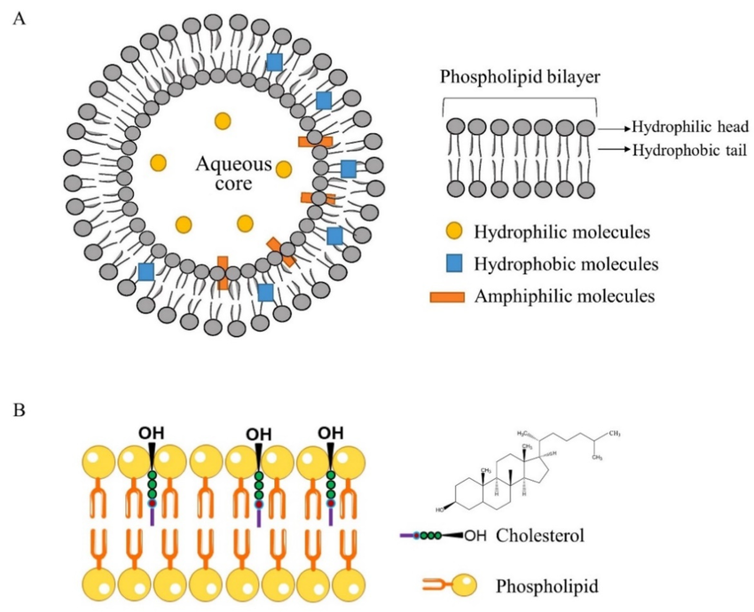
According to their structure, hydrophobic medications are integrated into the lipid bilayer in the hydrophobic fatty acid tail region, whereas hydrophilic pharmaceuticals can be enclosed within the interior watery core of liposomes. Similar to biological membranes, liposomal membranes are fluid, which ultimately allows the drugs inside to seep out of the vesicles. The rate of drug release is influenced by the nature of the liposomal membrane, which includes components like the presence of cholesterol (CH) and the kind of fatty acid acyl chains of the phospholipids. [6].
Small unilamellar vesicles (SUVs), large unilamellar vesicles (LUVs), multilamellar vesicles (MLVs), and multivesicular vesicles (MVVs) are the four primary groups of liposomes based on their size and the quantity of lipid bilayers.
Unilamellar liposomes have a single phospholipid bilayer, while multilamellar liposomes have an onion-like structure made up of several concentric lipid bilayers. The multilamellar architecture of multivesicular vesicles (MVVs) is made up of many unilamellar vesicles encased in a larger liposomal structure.
The encapsulation effectiveness of liposomes for hydrophilic substances typically rises with vesicle size and falls with an increase in bilayer count. Furthermore, one important factor affecting the circulation half-life of liposomes is vesicle size. The quantity of medication that can be encapsulated within the vesicles is greatly influenced by the size and quantity of bilayers. Liposomes are usually made between 50 and 150 nm in size for drug delivery applications in order to maximize therapeutic efficacy.
B) Liposomes Compositions:-
Liposomes are structurally spherical or multilayered vesicular systems that are created when diacyl-chain phospholipids self-assemble into lipid bilayers in aquatic conditions. The hydrophilic head groups and hydrophobic fatty acid tails that make up the phospholipid bilayer provide an amphiphilic structure that promotes vesicle formation. Lipid-soluble drugs are directly incorporated into the lipid membrane, whereas water-soluble drugs are usually contained inside the aqueous core's center. Both synthetic and natural phospholipids may be used to form liposomes.. [7].
The physical characteristics of liposomes, such as particle size, stiffness, membrane fluidity, stability, and surface charge, are also largely determined by the lipid makeup.
Lipids' hydrophilic head groups can be positively, negatively, or zwitterionally charged (having both positive and negative charges within the same molecule).
Through electrostatic repulsion between vesicles, the surface charge of these hydrophilic groups supports liposomal stability. The following groups of lipids can be employed to make liposomes:
1. Natural lipids :-
The membrane bilayer of normal cells are mainly composed of glycerophospholipids.
Natural phosphatides the most used to produce liposomes:
A) Phosphatidylcholine,
B) Phosphatidylethanolamine,
C) Phosphatidylserine,
D) Phosphatidylinositol,
E) Phosphatidylglecerol,
F) Phosphatidic acid.
These lipids are chosen because they can form bilayer structures that resemble the structure of natural cellular membranes, having hydrophilic head groups and hydrophobic tails. Because of their natural biocompatibility and biodegradability, which make them ideal for therapeutic applications, the use of natural lipids in liposomal formulations is very beneficial.
2. Synthetic lipids :-
The polar and non-polar portions of natural phospholipids are modified chemically to create synthetic phospholipids.
A) 1,2-Dipalmitoyl-sn-glycero-3-phosphorylethanolamine,
B) 1,2-Dipalmitoyl-sn-glycero-3-phosphatidic acid sodium salt,
C) 1,2-Distearoyl-sn-glycero-3-phosphorylglycerol sodium salt,
D) 1,2-Distearoyl-sn-glycero-3-phosphocholine.
Furthermore, unsaturated fatty acids in either one hydrocarbon chain or both can be used to create synthetic phospholipids.
A) 1,2-dioleoyl-sn-glycero-3-phosphoethanolamine,
B) 1,2-dioleoyl-3-trimethylammonium-propane
3. Steroid:-
Since cholesterol is integrated into the liposomes' lipid bilayer, it is the primary steroid that is typically utilized in the synthesis of liposomes at a ratio of less than 30% of the total lipids to increase the stability and rigidity of the liposomes. [8]
4. Surfactants :-
Surfactants were employed in liposome formulations to change the encapsulation and release properties of liposomes by lowering the surface tension between different immiscible phases. In liposome formulations, surfactants including sodium cholate, Span 60, Span 80, Tween 60, and Tween 80 are commonly utilized. [9].
C) Liposomes types :-
Liposomes can be divided into the following categories according to their compositions and uses:
Conventional liposomes, which are regarded as the initial generation of liposomal systems, are usually made from synthetic or natural phospholipids, either with or without cholesterol. In order to increase membrane fluidity, adjust bilayer rigidity, and improve liposome stability overall, cholesterol is frequently incorporated into the lipid bilayer.
According to Wu et al., adding cholesterol to liposomes made of DSPE-PEG2000 and hydrogenated soybean phospholipids (HSPC) decreased the membrane's stiffness. Additionally, liposomes with a somewhat stiff membrane showed increased anticancer efficacy and better tumor penetration. The average liposome size has also been linked to an increase in cholesterol content, along with a morphological change from irregular structures to regular, spherical, nanosized vesicles. Moreover, cholesterol reduces bilayer fluidity and alters the release of hydrophilic molecules from lipid vesicles.
Conventional liposomes show comparatively less stability in vitro despite these benefits. In order to improve the in vivo stability of liposomal drug delivery systems and extend the duration of blood circulation, stealth liposomes were subsequently created. [10][11].
Lipid vesicles known as charged liposomes have a net positive or negative surface charge as a result of charged lipids being incorporated into the phospholipid bilayer. Liposomes are typically spherical vesicles made of phospholipid bilayers; however, the addition of particular charged lipids results in a surface charge that is either positive (cationic) or negative (anionic).
Surface charge improves liposomal stability during storage by reducing aggregation through electrostatic repulsion between similarly charged particles. Gene therapy makes extensive use of cationic liposomes because of their capacity to efficiently encapsulate nucleic acids via electrostatic interactions. [12].
However, anionic liposomes frequently exhibit poorer stability in the circulation because they are removed faster than neutral and cationic liposomes. However, anionic liposomes are widely utilized in transdermal drug delivery systems because they enhance drug penetration through the stratum corneum of the skin. [13]
Second-generation liposomes are identified by surface modification with synthetic polymers, glycoproteins, polysaccharides, or specific receptor ligands to provide a more targeted dispersion and enhanced accumulation at the intended site of action. Polymers such as hyaluronic acid, polyethylene glycol (PEG), and polyvinyl alcohol (PVA) have been investigated in several research as effective steric stabilizers for liposomal systems. PEGylated or stealth liposomes are terms used to describe liposomes that have been changed using polyethylene glycol. By decreasing recognition and clearance by the reticuloendothelial system and increasing circulation time, the surface PEG layer offers steric protection. Doxil was the first pharmaceutical product based on PEGylated liposomes that proved effective. Stealth-stabilized liposomes show better accumulation at target sites and longer systemic circulation than normal liposomes. [14].
The third generation of liposomal drug delivery devices consists of actively targeted liposomes. By encouraging receptor-mediated endocytosis and improving the selectivity of liposome contacts with sick cells, active targeting makes it easier for the liposome and its therapeutic payload to be internalized into particular cellular targets. By adding molecular recognition moieties, which increase the effectiveness of medication delivery while reducing side effects, liposomal targeting can be further enhanced.
Both passively targeted (non-conjugated) and actively targeted (ligand-conjugated) liposomes first enter the target tissue via passive distribution mechanisms. Because of their strong affinity and specificity for target molecules, aptamers and aptamer-functionalized nanoparticles have garnered significant interest in targeted medicine administration in recent years. Targeting ligands can be covalently or non-covalently conjugated to the drug molecule or the nanocarrier's surface to enable selective binding to overexpressed biomarkers on tumor cells, hence enabling active targeting of nanocarriers.
Nevertheless, after cellular internalization, conventional and stealth liposomes frequently show limited fusion with endosomal membranes and sluggish drug release. Stimuli-responsive liposomal systems, which allow regulated drug release in response to particular physiological or environmental triggers, have been created to overcome these constraints. [15]
Advanced liposomal systems called stimuli-responsive liposomes are made to release drugs quickly and under control in response to particular physicochemical or biochemical stimuli, such as changes in pH, temperature, redox potential, enzyme concentration, ultrasound, and external electric or magnetic fields. By reacting to stimuli found within target tissues, such as variations in pH, redox conditions, or enzyme activity, these devices allow targeted medication release. [16].
pH-responsive liposomes are one of these systems that has been extensively studied for site-specific drug delivery. This method takes advantage of the pH variations between normal and diseased tissues. Tumor microenvironments, for instance, are usually more acidic than healthy tissues, with extracellular pH values routinely falling to as low as 5.7 and commonly ranging from roughly 6.8 to 7.0. [17].
|
Stimuli liposomes |
Stimuli |
Principle |
Advantages |
Reference |
|
Light-sensitive liposomes |
UV, near-infrared, or visible light exposure, |
Photoactivable liposomes have been produced by adding light-sensitive functional groups to the fatty acyl chains of phospholipids. |
Regulating wavelength, intensity, exposure, and time |
[18,19] |
|
Thermosensitive (temperature-sensitive) liposomes |
Radiofrequency or microwave ablation |
These liposomes have been created using lipids having a transition temperature of 40–45 °C, such DPPC. |
Drug release in locations with high temperatures |
[20,21] |
|
Redox-sensitive liposomes |
Singlet oxygen, hydroxyl radicals, and reactive oxygen species (ROS) peroxides |
depends on the redox potential difference that arises during biological activity between the oxidizing extracellular area and the intracellular reducing space. |
ROS causes tumor cells to produce large concentrations of glutathione (GSH), which cleaves the liposomal formulations. |
[22,23] |
|
Enzyme-responsive liposomes |
Protease, amidase, and esterase enzymes |
Drugs are released when protease or esterase enzymes hydrolyze amides or esters. |
reduces the negative effects of hazardous medications and makes prodrug encapsulation possible. |
[24,25] |
|
pH-sensitive liposomes |
pH change |
pH-sensitive liposomes were made using 1,2-dioleoyl-sn-glycero-3-phosphoethanolamine (DOPE) and cholesterol hemisuccinate (CHEMS). |
pH-dependent release characteristics of liposomes |
[26,27] |
D) Methods of preparation:-
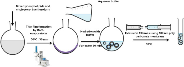
In a round-bottom flask, lipids and hydrophobic medications are first dissolved in an appropriate organic solvent. After that, a thin lipid coating is formed on the flask's inner wall by carefully evaporating the organic solvent under low pressure. The thin film is then hydrated with an aqueous buffer solution at a temperature greater than the phase transition temperature (Tm) of the lipids. The hydration medium may contain hydrophilic medications intended to be contained in the watery core of the liposomes.
Drug encapsulation efficiency is largely dependent on the rate of hydration; generally speaking, slower hydration rates result in higher encapsulation efficiency. Liposome size, lamellarity, and particle size distribution can be regulated by bath or probe sonication or extrusion utilizing polycarbonate membranes with specific pore diameters. When opposed to sonication, the extrusion method usually yields liposomes that are more stable and have better encapsulation efficiency. On the other hand, sonication frequently produces small unilamellar vesicles (SUVs) and may lead to the hydrolysis or destruction of medicines and/or lipids that are encapsulated. Furthermore, liposomal suspensions may be exposed to possible metal contamination during probe sonication. [28]
Thin-film hydration is often replaced with reverse-phase evaporation, which produces a water-in-oil emulsion. Lipids are dissolved in an organic solvent and then mixed with an aqueous buffer solution containing the hydrophilic drug. Lipid vesicles are then dispersed throughout the aqueous phase after the organic solvent is evaporated under low pressure using a rotary evaporator.
Extrusion can further decrease the average vesicle size and polydispersity of the resultant liposomes. High molecular weight molecules are especially well-suited for encapsulation using this technique. However, exposure to organic solvents and the sonication conditions frequently employed during the production procedure may cause denaturation of therapeutic peptides. [29,30]
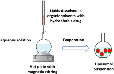
Liposome preparation injection techniques are categorized according to the kind of organic solvent used. These techniques cause liposomes to spontaneously form by dissolving lipids and hydrophobic active compounds in an organic solvent, followed by a quick injection into an aqueous phase. The use of diethyl ether as the organic solvent permits direct solvent evaporation at temperatures higher than its boiling point during the mixing operation
On the other hand, a 10- to 20-fold excess of aqueous solution is needed for ethanol injection. The ethanol can then be extracted under vacuum using a rotary evaporator, dialysis, or filtration.
Nonetheless, injection-prepared liposomes frequently show comparatively high polydispersity index (PDI) values, suggesting a wider size distribution. Additionally, the stability of the encapsulated medication and the lipid components may be negatively impacted by extended exposure to high temperatures and organic solvents during the preparation process. [31,32]
This technique uses a round-bottom flask to dissolve lipids and a surfactant with a high critical micelle concentration (CMC) in an appropriate organic solvent. A thin layer of lipid forms at the bottom of the flask when the organic solvent is gradually evaporated. An water solution containing the drug molecules is then used to hydrate the resultant lipid film, creating a mixed micellar solution.
Then, using methods like dialysis, size-exclusion chromatography, adsorption onto hydrophobic beads, or dilution, the surfactant is eliminated. Large unilamellar vesicles (LUVs) are created when the surfactant and solution concentration are eliminated. However, a major drawback of this approach is that the detergent removal phase may result in a large loss of hydrophilic medicines. [33]
Using sonication, this organic solvent-free technology creates large unilamellar vesicles (LUVs). Its foundation is the direct dispersion of lipids at low concentrations in a drug-containing aqueous solution, which is then sonicated to promote the formation of vesicles. In order to create a multilayered lipid film that traps the drug molecules, a dehydration phase is first carried out to eliminate water under a nitrogen stream. The medication is then encapsulated in big vesicles by a hydration phase.
Despite being very straightforward and avoiding the use of chemical solvents, this process frequently produces liposomes with a high degree of size variation. [34]
Another organic solvent-free method for liposome synthesis is this one. This method involves immediately hydrating lipids with an aqueous solution and heating them for at least an hour at a temperature higher than the phospholipids' phase transition temperature (Tm) while adding three to five percent of a hydrating agent, such as propylene glycol or glycerin. The suspension can be heated to temperatures of up to 100 °C when cholesterol is present in the formulation.
In order to stop nanoparticle coagulation and sedimentation, the hydrating agents serve as stabilizing and isotonizing additives. Furthermore, the heating procedure is especially appropriate for the manufacture of powder inhalable liposomal formulations because these agents have a cryoprotective action. [35]
The pH-jump approach is another solvent-free method for manufacturing liposomes. This method involves rapidly increasing the pH of an aqueous solution containing phosphatidic acid and phosphatidylcholine, usually by about four times in a brief amount of time. This causes multilamellar vesicles (MLVs) to disintegrate and transform into small unilamellar vesicles (SUVs).
The ratio of phosphatidic acid to phosphatidylcholine plays a crucial role in determining the proportion of SUVs and large unilamellar vesicles (LUVs) formed during the process [36].
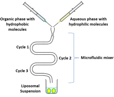
A new method for preparing liposomes has recently surfaced: the microfluidic channel method. Controlled mixing at the microscale is made possible by the exact manipulation of fluids within small channels made possible by microfluidic technology. This technique involves dissolving lipids in an organic solvent, like ethanol or isopropanol, and then injecting the resultant solution either in the same direction or in the opposite direction to an aqueous phase inside the microchannels. The organic and aqueous phases are continuously mixed axially, which promotes liposome spontaneous production.
In order to stabilize the liposomes and stop phase separation or aggregation, surfactants are frequently added. The synthesis of liposomes with repeatable properties, such as controlled average size, polydispersity, morphology, and lamellarity, is made possible by the microfluidic approach's exact control over the mixing process. [37,38].
Instead of using traditional organic solvents to dissolve lipids, this approach uses a supercritical fluid, usually carbon dioxide (CO?). This method facilitates the phase transition of the dissolved phospholipids by delivering a continuous flow of the aqueous phase into a chamber holding the supercritical lipid solution using a high-performance liquid pump. Liposomes are created when carbon dioxide is entirely eliminated by a sudden drop in pressure.
When compared to traditional approaches, this technology has been claimed to yield encapsulation efficiencies that are up to five times higher. However, this technology has some drawbacks, such as high operating costs, low manufacturing yield, and the need for specialized infrastructure and equipment, even if it uses cheap and environmentally safe carbon dioxide. [39,40].
E) Post preparation handlings:-
This method is frequently used to improve liposomal lamellarity and encapsulation efficiency during liposome production. The process entails repeated freeze-thaw cycles, usually alternating between temperatures below the phase transition temperature (Tm) of the phospholipids utilized and −196 °C in liquid nitrogen. These cycles facilitate better drug encapsulation within the vesicles by encouraging the fusing and restructuring of lipid bilayers. [41].
This procedure is frequently used to improve the shelf stability of liposomal compositions. In freeze-drying (also known as lyophilization), the liposomal suspension is deep frozen with the addition of a cryoprotective substance, usually 5–10% sucrose or trehalose. The frozen liquid sample is then transformed into a porous, dry solid powder via a sublimation process carried out at extremely low temperatures under decreased pressure. For liposomal compositions containing thermosensitive biomolecules, lyophilization is especially crucial since it preserves their stability throughout storage. [42].
|
Liposomes characteristics |
Characterization technique |
References |
|
Average particle size |
Atomic force microscopy (AFM), cryogenic-TEM (Cryo-TEM), scanning and transmission electron microscopy (SEM/TEM), and dynamic light scattering (DLS) |
[43,44] |
|
Zeta potential/Surface charge |
Electrophoretic mobility, DLS |
[45] |
|
Particle shape/morphology |
TEM, Cryo-TEM, and AFM |
[46] |
|
Lamellarity |
Cryo-TEM and ?31P-NMR |
[47] |
|
Phase behavior |
X-ray diffraction (XRD), thermogravimetric analysis (TGA), and differential scanning calorimetry (DSC) |
[48,49] |
|
Encapsulation efficiency/Drug release |
Centrifugation, dialysis, and then chromatographic or spectrophotometric techniques to determine the drug content |
[50,51] |
F) Marketed clinical liposomes:-
|
Usage |
Trade name |
Active ingredient(s) |
Liposome platform (Molar Ratio) |
Manufacturer |
Year Approved |
Administration Route |
References |
|
Anti-Cancer |
Doxil® |
Doxorubicin |
HSPC:Cholesterol:PEG 2000-DSPE (56:38:5) |
Sequus Pharmaceuticals |
1995 |
I.V |
[51] |
|
DaunoXome® |
Daunorubicin |
DSPC:Cholesterol (2:1) |
NeXstar Pharmaceuticals |
1996 |
I.V |
[51] |
|
|
Depocyt® |
Cytarabine |
DepoFoam™ |
SkyPharma Inc. |
1999 |
Spinal |
[51] |
|
|
Myocet® |
Doxorubicin |
Cholesterol:EPC (45:55) |
Elan Pharmaceuticals |
2000 |
I.V |
[51] |
|
|
Mepact® |
Mefamurtide |
DOPS:POPC (3:7) Multilamellar liposome |
Takeda Pharmaceutical Limited |
2004 |
I.V |
[51] |
|
|
Lipodox® |
Doxorubicin |
DSPC:Cholesterol:PEG 2000-DSPE (56:39:5) |
Sun Pharma |
2012 |
I.V |
[51] |
|
|
Marqibo® |
Vincristine |
SM:Cholesterol (60:40) |
Talon Therapeutics |
2012 |
I.V |
[51] |
|
|
Onivyde™ |
Irinotecan |
DSPC:Cholesterol:MPEG-2000-DSPE (3:2:0.015) |
Merrimack Pharmaceuticals |
2015 |
I.V |
[51] |
|
|
Lipusu® |
Paclitaxel |
NA |
Luye Pharma Group |
2006 |
I.V |
[51] |
|
|
Vyxeos® |
Cytarabine:Daunorubicin 5:1 |
DSPC:DSPG:Cholesterol (7:2:1) |
Jazz Pharmaceuticals |
2017 |
I.V |
[51] |
|
|
Anti-Fungal |
Ambisome® |
Amphotericin B |
HSPC:Cholesterol:DSPG (2:1:0.8) |
Astellas Pharma |
1997 |
I.V |
[51] |
|
Fungisome® |
Amphotericin B |
PC:Cholesterol (7:3) |
Lifecare Innovations |
2003 |
I.V |
[51] |
|
|
Photodynamic therapy |
Visudyne® |
Verteporphin |
Verteporphin:DMPC&EPG (1:8) |
Novartis AG |
2000 |
I.V |
[51] |
|
Analgesic |
DepoDur™ |
Morphine sulfate |
DepoFoam™ |
SkyPharma |
2004 |
Epidural |
[51] |
|
Exparel® |
Bupivacaine |
DepoFoam™ |
Pacira pharmaceuticals |
2011 |
I.V |
[51] |
G) Evaluation Test :-
|
Parameter |
Acceptable Limit |
|
Particle size |
50–200 nm (nano liposomes) |
|
PDI |
≤ 0.3 |
|
Zeta potential |
≥ ±30 mV ideal |
|
Entrapment efficiency |
≥ 70% |
|
Drug content |
90–110% |
|
pH |
6.0–7.5 (injectable) |
|
Sterility |
Pass |
|
Residual solvent |
As per ICH Q3C |
|
Stability |
No major change |
|
Leakage |
< 10% |
|
|
|
CONCLUSION
Liposomes have been effectively used as effective drug delivery methods to treat a variety of illnesses, including pain management and cancer therapy. Liposomal formulations can greatly enhance the pharmacokinetic and pharmacodynamic profiles of poorly water-soluble, low bioavailability, and extremely toxic medications due to their biocompatibility, biodegradability, and low immunogenicity. To get around the drawbacks of early formulations, liposomal systems have seen significant improvements in composition and production techniques over time. More than 500 liposomal compounds are now being studied in various stages of clinical trials, and some liposomal formulations are officially approved for clinical usage in the treatment of various disorders.
Despite these developments, liposomal systems still face significant difficulties due to their chemical and physical instability. Consequently, the creation of extremely stable liposomal formulations continues to be a crucial element affecting their clinical suitability. In this regard, by calculating appropriate lipid compositions and three-dimensional structural features, in silico simulations and computational methods may offer insightful information for forecasting ideal liposomal formulations.
REFERENCES


Harsh Niwatkar, Dr. Swapnil Phalak, Next-Generation Liposomal Drug Delivery Systems: Design Strategies, Preparation Techniques, and Therapeutic Applications., Int. J. of Pharm. Sci., 2026, Vol 4, Issue 3, 4130-4144, https://doi.org/10.5281/zenodo.19356977
 10.5281/zenodo.19356977
