1,2,3 Dr. Shivajirao Kadam College of Pharmacy, Kasabe Digraj, Miraj, Sangli, Maharashtra, 416305.
4 Shri Gajanan Arts Commerce and Science College, Jadra Boblad
Amyloid-? (A?) plaque buildup, tau protein hyper phosphorylation, chronic neuroinflammation, and logical impairment are Primary signs of Alzheimer's disease (AD), a progressive neurodegenerative illness. Even with a great deal of study, early diagnosis is still quite difficult, and current treatment approaches simply alleviate symptoms. Nanotechnology has become a viable strategy for getting around these limitations in recent years. Because of their customizable size, surface characteristics, and functionalization potential, nanoparticles (NPs) have special benefits for AD diagnosis and treatment. This article concentrates on significant advancements in targeted delivery of pharmaceuticals using nanoparticles, which enable the possibility of effectively transfer medication via the BBB, or blood-brain barrier. To help with early and precise analysis of AD. NPs have also been used for imaging and biomarker detection. Nanoparticles of several kinds, such as metal-based NPs, polymeric NPs, liposomes, and dendrimers, have shown promise in lowering oxidative stress, reducing neuroinflammation, and regulating A? aggregation. Theranostics is the term for the development of intelligent, multipurpose nanoparticle systems that combine therapeutic and diagnostic capabilities into a single platform. This article addresses the reasons, strategies, and possibilities of nanoparticle-based approaches that could transform AD treatment soon enough.
The particularly prevalent type of dementia, Alzheimer's disease (AD), affects people globally
and a gradually developing neurological illness. Memory loss, cognitive decline, and neuropathological features such external amyloid-β (Aβ) plaques and intracellular neurofibrillary tangles of hyperphosphorylated tau, neuroinflammation, and oxidative stress are its defining characteristics [1]. Timely intervention depends on early diagnosis, but traditional diagnostic approaches include Magnetic resonance imaging (MRI), Positron emission tomography (PET), and cerebrospinal fluid (CSF) Evaluation of biomarkers, are frequently intrusive, costly, or insensitive in the preclinical stages [2].
Because of the large proportion of surface to volume, capacity to cross blood-brain barrier (BBB) and potential for functionalization with ligands for targeted delivery, nanoparticles (NPs) have become innovative methodology in AD. In areas of diagnostics, nanoparticles (NPs) such Superparamagnetic Iron oxide nanoparticles (SPIONs), Quantum dots, and gold nanoparticles have been developed to improve imaging techniques and facilitate early detection by identifying biomarkers [3,4].
In a therapeutic context, NPs decrease systemic toxicity while enhancing drug solubility, stability, and targeted delivery. In order to deliver anti-amyloid medicines, antioxidants, siRNA, and gene treatments directly to the affected brain regions, advanced nanotechnologies are being developed, including polymeric nanoparticles, liposomes, dendrimers, and lipid-based carriers [5,6]. Targeting ligands, such as aptamers, peptides, or antibodies, improve NP selectivity for tau or Aβ aggregates, providing site-specific treatment [7]. Additionally, real-time monitoring and individualized therapy of AD are made possible by multifunctional "theranostic" nanoparticles that combine diagnostics and treatments [8].
With these properties, nanoparticles offer an innovative approach to Alzheimer's disease diagnosis, treatment, and targeted delivery, resolving significant issues in contemporary clinical practice and providing a route to more efficient and customized therapies.
For therapeutic and/or diagnostic reasons, it has been proposed to target the Aβ1–42 peptide in every form of agglomeration [9–14]. The Blood and central nervous system (CNS) are separated by BBB, a dynamic biological and physical barrier. The capillary endothelial cells in the brain, which limit transcellular transit, and firm and tight connections between the cells, which limit paracellular flux, are primarily responsible for functional complexity of blood-brain barrier [15].
Nanomedicines (NMs), which aim to engage in molecular interactions with BBB cells, that are currently the most sought-after approaches. In order to transfer NM for therapeutic purposes, these NMs take advantage of the physiological processes already present in the BBB. The transportation of NM is accomplished by a variety of transport techniques. The most significant processes that enable transcellular transfer of NM from Blood to the brain are receptor- and adsorptive-mediated transcytosis [16). Specific inhibition can be achieved by taking advantage of the NPs' differential binding to Aβ monomers and oligomers once they have entered the brain. Furthermore, blocking the oligomers by NPs would be crucial from the biological point of since it is thought that they are a type of Aβ agglomerates that are most neurotoxic.[17]. Due to the fact that they influence their toxicity, targeting capacity, and in vivo distribution, Particle size and charge, core and surface characteristics, shape and flexibility, multivalency, and controlled synthesis are the most significant characteristics of NP formulations. The uses of Polymeric nanoparticles loaded with AD drugs shown in fig. 1. As a result, these characteristics significantly affect the stability of nanoparticles as well as their ability to load and release drugs (18).
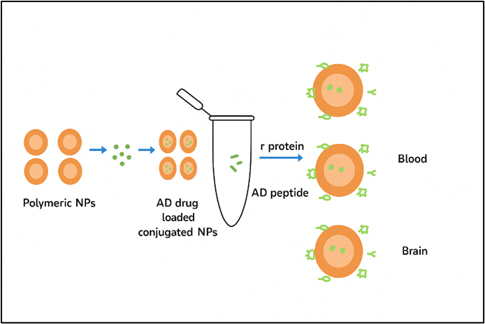
Fig 1 Polymeric nanoparticles containing drugs to treat AD.
Nanotechnology Could assist in early identification of Alzheimer's disease through very effective signal transduction techniques. The process of transforming and amplifying a biological signal for recording is known as signal transduction. Target-based treatment (TBT) advancement is currently of interest because the traditional medications used to treat AD can only slow its progression and not cure it [19,20]. Nanocarriers or nano-formulations offer many benefits over traditional treatment for AD, including avoiding hepatic metabolism, lowering dosage, improving medication stability, bioavailability, and targeted administration at area of the action. The most difficult aspect of treating neurological illnesses is getting medications into the brain. The physical barrier known as the blood–brain barrier (BBB) divides circulation in the periphery from CNS., prevents molecules from moving freely within the brain parenchyma [21]. Among the most difficult physiological hurdles is this one. The BBB can be penetrated by lipophilic molecules that are less than 400 Da. The mobility of larger hydrophilic molecules or lipid-insoluble molecules is restricted [22, 23]. Some methods to improve medication transport across BBB such as Liposomes, polymeric nanoparticles (PNPs), Solid lipid nanoparticles (SNPs), along with gold nanoparticles [24,25,26]. These potential alternatives, such as drug-loaded PNPs, can enhance the drug's distribution throughout brain. A broad spectrum of ligands can be effectively incorporated into surface of PNPs to enhance their stability, high loading capacity, regulated kinetics of drug release, and drug encapsulation. (27,28) Through targeted medication administration, metallic nanoparticles have been shown to be a helpful therapeutic method in Alzheimer’s disease therapy. These elements, which include iron, cerium, selenium, gold, & silver, are recognized to have strong anti-AD characteristics [29]. Because gold nanoparticles travel by transcytosis movement through the brain's endothelial cells without altering their surface, they are designated for AD.
These positively charged nanoparticles have the ability to transport bioactive substances and aid in the targeted delivery of brain regions [30]. Gold nanoparticles (Au-NPs) show both neuroprotective and excellent BBB penetration capabilities [30]. Because it prevents Aβ aggregation, Au-NPs conjugated utilizing glutathione has been found to have an anti-Alzheimer's action [31].
The steps involved in creating nanoparticles:
Nanoparticles (NPs) can be created chemically, physically, or biologically. The different components of NPs and its functions are listed in table 1 and methods involved are mentioned in fig 2. The uses, surface properties, and desired size all influence the process selection. The following are typical methods:
Mostly used for nanoparticles of polymers. A surfactant is used to emulsify a polymer in an aqueous phase after it has been dissolved in an organic solvent. After then, the solvent evaporates, producing NPs.
Applicable to drug carriers based on PLA, PLGA, and PCL.
A polymer dissolves in water when it is solubilized in a solvent that is organic and is introduced to water phase while being stirred, solvent diffusion causes the production of nanoparticles (33).
Benefit: Easy to use, quick, and prevents excessive heat.
Used for chitosan nanoparticles, in which a polyanion (such as tripolyphosphate) combines with cationic chitosan. (44)
Utilized to produce magnetic and metallic nanoparticles. Agents such as NaBH? convert metal salts to form nanoparticles. (35)
To produce homogeneous nanoparticles, a surfactant-stabilized emulsion is employed as a nanoreactor. (36)
Suitable for: oxide and metallic nanoparticles.
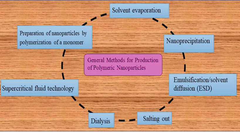
Fig 2 Some methods for production of Nanoparticle
Table No. 1 Ingredients Used in Nanoparticles and Their Significance
|
Ingredient |
Function |
Examples |
Significance |
|
Polymers |
Matrix or carrier material |
PLGA, PLA, PCL, chitosan |
Biodegradable, biocompatible; controls drug release [37] |
|
Lipids |
Form lipid nanoparticles or liposomes |
Phospholipids, cholesterol |
Good for encapsulating hydrophobic drugs [38] |
|
Surfactants/ Stabilizers |
Prevent aggregation, stabilize particles |
PVA, Tween 80, Poloxamer 188 |
Improve stability and dispersion [39] |
|
Crosslinking agents |
Solidify polymer or protein structure |
Glutaraldehyde, TPP |
Essential in ionic gelation or protein NPs [34] |
|
Solvents |
Dissolve polymers/ drugs |
Acetone, ethanol, dichloromethane |
Aid in nanoparticle formation; must be removed completely [33] |
|
Drugs/ Active Agents |
Therapeutic function |
Donepezil, curcumin, siRNA |
Incorporated for targeted delivery [37] |
|
Targeting Ligands |
Enable specific cell binding |
Antibodies, peptides, aptamers |
Enhance cellular uptake and selectivity [40] |
|
Metal Precursors |
For metallic nanoparticles |
FeCl? (iron), HAuCl? (gold) |
Provide core material for inorganic NPs [35] |
Alzheimer's Disease Causes:
A mixture of genetic, biochemical, and environmental factors that contributes to etiology of AD, a complex neurodegenerative disorder shown in fig 3
1. Accumulation of Amyloid-β (Aβ) Plaque
Aβ peptides build up extracellularly to form plaques, which impair synaptic function, induce neuroinflammation, and neuronal death [41].
2. Hyperphosphorylation of Tau Protein
Tau proteins that are abnormally phosphorylated produce neurofibrillary tangles, which disrupt microtubule activity and cause cell death [42].
3. Neuroinflammation
Pro-inflammatory cytokines released by activated microglia and astrocytes enhance neuronal damage and aid in the development of illness [43].
4. Oxidative Stress and Dysfunction of the Mitochondrion
While mitochondrial malfunction affects how neurons use energy, excessive reactive oxygen species (ROS) damages cellular components [44].
5. Genetic Variables
AD that begins early: brought on by changes in the genes PSEN1, PSEN2, APP as well.
Late-onset AD: Associated with APOE ε4 allele, one of main genetic hazardous factors, [45].
6. Lifestyle and Environmental Aspects
Cardiovascular illness, poor diet, smoking, low cognitive stimulation, social isolation, and physical inactivity are risk factors [46].
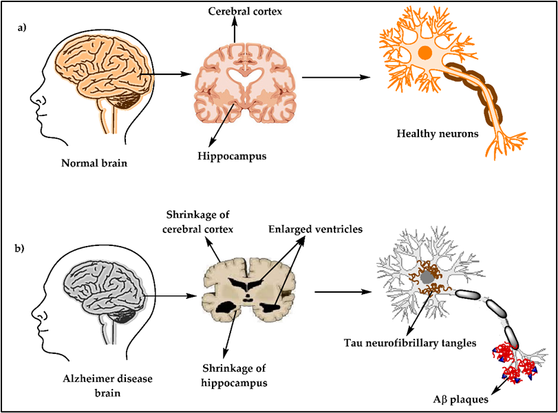
Fig 3 Causes of Alzheimer's disease
Prevention of Alzheimer's Disease:
Although at moment, there is no solution to completely avoid AD, changes in lifestyle can reduce the risk:
1. Nutritional Therapy
Slower cognitive decline is linked to Omega-3 fatty acids and antioxidants are abundant in Mediterranean or MIND diet. [47].
2. Exercise
Frequent exercise lowers Aβ levels, increases cerebral blood flow, and promotes neurogenesis [48].
3. Stimulation of the Cognition
Mentally demanding activities can postpone the development of symptoms and improve cognitive reserve [49].
4. Management of Cardiovascular Risk
Vascular contributions to cognitive impairment are decreased by managing blood pressure, cholesterol, diabetes, and obesity [50].
5. Hygiene of Sleep
Aβ elimination through the glymphatic system is facilitated by getting enough good sleep [51].
Alzheimer's Disease Treatment:
1. Pharmaceutical Interventions
Cholinergic function is enhanced by Cholinesterase inhibitors, including rivastigmine and donepezil.
Neurons are shielded from glutamate-induced toxicity by NMDA receptor antagonists, like memantine [52].
Aβ plaques are targeted and decreased by anti-amyloid monoclonal antibodies, for example lecanemab and aducanumab [53].
2. Alternative Medicine Methods
Quality of life is enhanced and functional deterioration is postponed through behavioral therapy, cognitive training, physical exercise, and caregiver support [54].
3. Emerging Nanotechnology-Based Therapies
In order to reduce Aβ buildup, oxidative stress, and neuroinflammation, nanoparticles are being investigated for the passage of specific medications via BBB.
Importance of nanoparticles in diagnosis of Alzheimer's Disease:
Early diagnosis of AD is essential to avoiding dementia and irreversible neural damage. Developing techniques to identify AD in its early stages is essential because investigating a real human brain is challenging and takes a long time.
Magnetic Nanoparticles as MRI (Magnetic Resonance Imaging) contrast agents:
Most efforts are focused on employing contrast-agent-doped nanoparticles in MRI to detect & identify amyloid plaques, as it is generally acknowledged in the field of science that development of senile plaques results in degeneration of the neurofibrilla (56). A contrast agent containing Nanoparticles that are magnetic and Aβ peptide was created using co-deposition and surface modification approaches. An MRI machine was used to image the brains of AD transgenic mice in order to investigate in vivo imaging. The findings showed that the contrast agent has good MRI enhancement of senile plaque and is about 5 nm in size (57).
It has been revealed that amyloid oligomers (AOs) are the only ones that have a sensitive contrast probe for molecular MRI. Antibodies that are unique to magnetic nanostructures with AOs showed that complex is stable; they attach to AOs on brain tissues as well as cells to generate an MRI signal. The probe immediately arrived in hippocampus of AOs in an AD mouse model after being administered intranasally. To differentiate AD from controls in separate samples of human brain tissue, an MRI signal was present. in isolated human brain tissue samples. These neurotoxic AO-targeting nanostructures should be helpful for assessing effectiveness of new medications and, potentially, for early diagnosis plus in addition to AD therapy (58). Furthermore, according to reports, development of Nanoparticles of monocrystalline iron oxide (MIONs) has advanced for coupled targeting and imaging of senile plaques and that these particles form a covalent link with amide linking to the A1-40 peptide's N-terminus.
Nanogels
Drug delivery treatments for AD have used nanogels, an efficient method for delivering pharmaceuticals that provides improved cellular absorption, decreased toxicity, enhanced medication loading and controlled release at the intended location. A recent study showed that chitosan and tripolyphosphate nanogels are effective in delivering deferoxamine in order to treat AD. By stopping formation of Aβ amyloid, Cholesterol-modified pullulan backbones serve as synthetic chaperones that decrease the pathophysiology of AD. Nanogels, which have non-toxic, stable, hydrophilic, and biodegradable qualities, improved the distribution of insulin, a possible AD medication, to the brain in preclinical animal studies, particularly when mixed with polysaccharides (60).
To overcome the drawbacks of existing AD treatments that are lacking ability to cross the BBB, researchers are investigating nanomaterials for precision medicine.
Nanotechnology in AD therapy:
Innovative drug development techniques are now essential to overcoming the restriction or obstacle that CNS medications experience in attempting to cross BBB. This need is satisfied by several methods based on nanotechnology that increase entrapped drug's effectiveness and prolonged release. Examples of techniques based on nanotechnology comprises polymers, solid lipid carriers, Nanoparticles made of lipoprotein & curcumin, optical imaging, metal-based carriers, magnetic, inorganic and antibody-tethered nanoparticles, as well as nanocomposites, dendrimers, and emulsion.
Polymeric nanoparticles:
The purpose of Nanoparticles of polymers are to improve bioavailability of hydrophobic medications while protecting them through deactivation of the environment & destruction.
Use of chitosan NPs in conjunction with rivastigmine for AD was investigated by Wilson et al. (2011). By emulsifying rivastigmine, the researchers synthesized chitosan nanoparticles (NPs) with mean size of 47 ± 4 nm. According to a zeta potential investigation, coating chitosan nanoparticles with polysorbate 80 reduced their positive charge. Drug release from NPs was shown to follow a biphasic, Fickian diffusion release pattern. Additionally, it was shown that covering NPs with 1% polysorbate 80 changed how the various organs absorbed the NPs. (61)
Liposomes:
Using a mediator of BBB transport with an anti-transferrin antibody (TrF) and a metabolite of curcumin, Mourtas et al. (2014) developed multifunctional liposomes. According to study of Liposomes containing either a curcumin derivative and anti-TrF shown significant attraction for amyloid plaques in post-mortem brain tissues from AD patients. Additionally, the authors found that liposomes containing curcumin derivatives did not stop Aβ aggregation or block Aβ deposit staining. However, brain-targeting ability was unaffected by the existence of a lipid-curcumin-PEG conjugate, indicating that these multifunctional NLs may be effective AD theranostics. (62)
Nanoparticles based on lipids or lipoproteins:
It is well known that lipid-based nanoparticles have a significant attraction for Aβ., which facilitates their breakdown and this enables them to be applied to diagnostic and therapeutic purposes. The system of apolipoprotein E3–reconstituted high-density lipoprotein (ApoE3–rHDL) nanoparticles was created by Song et al. (2014). in order to remove Aβ. Over four weeks of treatment every day, the amount of Aβ deposits, microgliosis, neurological changes, and memory issues diminished., indicating potential therapeutic application of ApoE3–rHDL in AD. Approximately 0.4% ID/g of ApoE3–rHDL was found in brain of a mouse one hour after intravenous delivery. Its toxicity is still unknown, though. (63)
According to Loureiro et al. (2017), extracts from skin of grape and grape seeds prevented AD patients' Aβ from aggregating. because resveratrol, which is found in grapes, is known to have neuroprotective characteristics., it was selected for the study. The authors found that bioactive extract was transported to an in vitro model of human BBB, An antibody that is monoclonal to the transferrin receptor was employed to functionalize solid lipid nanoparticles (SLNs).(OX26 mAb). OX26 SLN cellular absorption was considerably greater than that of SLNs that were not functionalized and SLNs that were functionalized using an unspecific antibody, according to experiments conducted on human brain-like endothelial cells. (64)
NPs loaded with curcumin:
Researchers have studied curcumin in recent decades and found that it has a variety of biological activities, including neuroprotective potential. Curcumin's effectiveness does not reach the biological system despite this potential. It has a limited bioavailability and is known to be sensitive to biodegradation and oxidation. Therefore, by encapsulating it in nanocapsules, this limitation can be removed., which will make the BBB easier to traverse. (65)
Metal-based carriers:
Recently generated Gold-based Magnetically sensitive carriers or metal-based nanoparticles, For instance Yb-doped or Ce-doped maghemite NPs (MNPs), have been employed as vehicles for drugs that have photosensitizing properties. These nanoparticles exhibit stability and increased ability to enter tissues, even ones that are cancerous.
Conjugates of nanoparticles:
Even very small concentrations of Up to 10–18 moles of protein biomarkers per liter, can be detected using DNA–nanoparticle complexes. The bio-barcode technique, another name for this detection method, use an antibody that is specific to a protein and has been tagged with gold nanoparticles. (65).
A dual-purpose, spherical delivery of A medication that contains β-sheet breaker peptides H102 (TQNP/H102) was designed by Zhang et al. (2014). To facilitate Aβ42 targeting and BBB transit, respectively, two targeting peptides—QSH & TGN—were attached to surfaces of nanoparticles. A unique dual-functional material was presented by Zhang and colleagues, and it was discovered to be a promising nanocarrier for proteins and peptides as well as for the selective administration of drugs into CNS and, later, brain AD lesions.
A very specialized form of AD therapy may be possible with this kind of technology. (67)
Delivery of nanoparticles intranasally:
In order to treat AD, Elnaggar et al. (2015) explored selective intranasal administration of NPs of chitosan laden with piperine (PIP). These chitosan NPs substantially decreased piperine-induced nasal trouble in a rat study without negatively affecting the brain. This work was first to show that PIP's benefits in AD were caused by its anti-apoptotic and anti-inflammatory effects. Additionally, the authors produced mucoadhesive chitosan nanoparticles (NPs) with success, which offered a way to circumvent PIP's negative effects by delivering PIP to the brains at 5% of the oral dosage, but at the same concentrations as oral delivery.
Alzheimer's Disease (AD) Nanoparticle Applications (69,70)
By overcoming the drawbacks of traditional therapeutics, such as inadequate BBB permeability, non-specific targeting, & limited drug bioavailability, nanoparticles (NPs) offer revolutionary potential in diagnosis, treatment, monitoring of AD as well. An extensive summary of uses of NPs in Alzheimer's Disease is provided below, along with relevant citations:
1. Systems for Drug Delivery
Transporting therapeutic medications crossing the BBB is a significant obstacle to AD treatment which NPs can help with.
Biodegradable polymeric nanoparticles (like chitosan and PLGA):
These can be altered with ligands to improve brain targeting.
For instance, in AD mice models, PLGA nanoparticles loaded with donepezil demonstrated enhanced memory.
Solid lipid nanoparticles and other lipid-based nanoparticles and liposomes:
Improve stability and solubility of medications.
As an illustration, liposomes laden with curcumin have demonstrated promise in lowering amyloid-beta (Aβ) plaques in AD mice.
Gold nanoparticles (AuNPs):
Photothermal treatment and targeting can be accomplished by functionalizing their surface.
Example: AuNPs conjugated employing antibodies against Aβ were able to bind Aβ and reduce toxicity.
2. Imaging and Diagnosis:
The early diagnosis of AD's sensitivity and specificity are enhanced by nanoparticles.
In order to identify Aβ plaques, magnetic nanoparticles, such as SPIONs, are employed as MRI contrast agents.
For instance, in transgenic mice with AD, imaging of amyloid plaques was improved by SPIONs coupled with an Aβ antibody.
Fluorescent nanoparticles known as quantum dots (QDs) are utilized to image Aβ aggregates in real time. Aβ peptide-tagged QDs, for instance, assisted in tracking the development of plaque.
3. Removal of Aβ Plaque
Aβ plaques, a defining feature of AD, can be broken down or removed with the help of NPs.
Immunotherapy with nanoparticles:
Provide peptides or antibodies that specifically target Aβ.
For instance, polymeric nanoparticles that delivered anti-Aβ antibodies enhanced cognition and decreased Aβ load.
Metal chelation:
Metal ions (such as Cu and Zn) linked to Aβ aggregation can be bound by chelators carried by NPs.
4. Gene Therapy:
To control genes linked to AD, genetic material (such as siRNA and miRNA) can be delivered by nanoparticles.
siRNA delivery: NPs facilitate effective siRNA transport to neurons while shielding it from degradation.
For instance, chitosan NPs loaded with siRNA decreased expression of the BACE1 gene, which decreased the generation of Aβ.
5. Anti-inflammatory and Antioxidant Effects:
Antioxidant substances carried by NPs can lessen AD's neuroinflammation and oxidative stress.
For example, CeO? NPs, or cerium oxide nanoparticles, mimic antioxidants.
CONCLUSION
In the fight to prevent Alzheimer's, nanoparticles have proven to be an incredibly adaptable platform, providing cutting-edge approaches to combination therapy, tailored medication administration, and early diagnostics. They are very appealing for both therapeutic and diagnostic applications because of their capacity to get beyond conventional obstacles including inadequate drug bioavailability and poor BBB permeability. Every type of nanoparticle, from metallic and hybrid nanostructures to polymeric and lipid-based carriers, offers distinct advantages for AD research. Even if there are still issues with long-term safety, targeted delivery, and clinical translation, continuous developments in molecular biology, materials science, and nanomedicine are opening up possibilities for Alzheimer's treatments of the future. In the nearer future., nanoparticles could completely change how we identify, treat, and eventually prevent Alzheimer's disease should interdisciplinary cooperation and translational research continue.
ACKNOWLEDGEMENTS
The authors wish to acknowledge the support and contributions that facilitated the completion of this work
Conflict of Interest Statement: The authors declared no conflict of interest.
REFERENCES


Tejaswini Chavan, Komal Shinde, Neha Irsur, Aishwarya Chavan, Nanoparticles in Alzheimer's Disease: Advances in Diagnosis, Therapy, and Targeted Delivery., Int. J. of Pharm. Sci., 2025, Vol 3, Issue 10, 492-504. https://doi.org/10.5281/zenodo.17278033
 10.5281/zenodo.17278033
