Department of Pharmaceutics, Channabasweshar Pharmacy College (Degree), Latur-413512, Maharashtra, India.
A nanofiber is a type of fiber with at least one of its dimensions in the nanometer range, typically having a diameter of less than 100 nanometers (nm). This nanofiber has unique properties such as high surface area to volume ratio, high aspect ratio, and high porosity. Due to their high surface-area-to-volume ratio, nanofibers often demonstrate enhanced strength, flexibility, and conductivity compared to their larger counterparts. The fabrication of nanofibers can be accomplished through various techniques, such as Electrospinning, Template synthesis, Self-assembly, Phase separation, and Drawing, each providing specific advantages tailored to the desired material properties and intended applications. Depending on their intended use, nanofibers are manufactured using a variety of polymers. Polymers encompass a range of materials, including natural polymers, semi-synthetic polymers, synthetic polymers, metals, metal oxides, ceramics, carbon-based materials, non-porous materials, mesoporous materials, hollow structures, core-shell structures, biocomponents, and multi-component materials. The primary function of this review paper is to provide a comprehensive knowledge of nanofiber characteristics, synthesis techniques, morphological, structural, thermal, mechanical, surface and chemical characterization of nanofiber, as well as their applications in biomedical, chemistry, defense, and environmental field.
Nanofibers are defined as nanomaterial’s with at least one dimension of 100 nm or less, where the length can exceed diameter by 100-times. However, all fibers with diameter less than 1 µm are considered nanofiber. nanofiber technology is a branch of nanotechnology whose primary function is to create a material in the form of nanoscale fibers to achieve superior function. They are ultra-fine fibers with diameter typically in the range of 1 to 1000 nanometer(nm), significantly smaller than those of conventional fibers. It is a new class of material used in various value-added applications, including medical, filtration, barrier, personal care, wipes, garments, composites, energy storage, and insulation. one of the most striking features of nanofiber is high surface are to volume ratio, high porosity, and high aspect ratio making them an attractivecandidate for many applications. Nanofibers possess a high length-to-diameter ratio, imparting superior mechanical properties, including flexibility and strength. many nanofibers, along with their resultant nanofibrous 3D structures, it exhibit a porous configuration that can be precisely controlled and customized for specific applications. these include low-density or lightweight materials, filtration systems, medical devices, and tissue scaffolds. At the nanoscale, nanofiber has two identical exterior dimensions but a third dimension that is much larger. they are classified according to their structure, rigidity, composition, and nature. nanofiber can make pharmaceutical compound from BCS class II and IV more soluble and permeable. Nanofibers, due to their extended-release profile, high loading capacity, and enhanced encapsulation efficiency, contribute to improved therapeutic efficacy, reduced toxicity, and adverse effect, and facilitate alternative administration methods. The fabrication of nanofibers is accomplished using various methods, including, electrospinning, template synthesis, centrifugal spinning and drawing, with the choice of technique depending on the desired morphology and specific application requirements. due to its adaptability, simplicity, and the inherently high ratio of surface area to volume in electrospun fibers, electrospinning technology has emerged as the most practical technique for the fabrication of nanofiber. a wide range of polymers and compounds, including natural polymers like collagen, cellulose, and gelatin, as well as polyurethane and lactic acid, are used to create nanofibers. These materials enhance technologies such as regenerative tissue, batteries, fuel cells, and fluid filtration.
2. Characteristics of nanofiber4
3. Fabrication method of nanofiber5-12
There are many ways or techniques used to fabricate nanofiber, such as, showed in below figure 1.

Fig.1. Fabrication method of nanofiber
3.1. Electrospinning:
Electrospinning is a highly versatile and straight forward technique used to produce nanofibers by subjecting a polymer solution or melt to a high voltage (typically ranging from 30 to 50 kV).it is the most versatile and cost effective for producing filament of the congaing drug-polymer solution when exposed to high-voltage electric field. these tiniest fibers are a thousand times smaller than the human hair in diameter. The electrospun method of making electrostatic fiber is one of the most advanced and straight-forward techniques of nanotechnology and has tremendous potential as a drug carrier for the delivery of therapeutics. Electrospun nanofibers can be designed in various morphologies, including core-shell, hollow, and porous structures. They can be aligned or randomly oriented in a nanofiber mesh, using single or blended copolymers, with optional additive materials.
An electrospinning unit consists of three essential components
The high voltage on the metal syringe provides a usual electric charge on the solution's surface, which induces the formation of nanofibers. using a needle of small diameter that shoots out a tiny stream of polymer solution, this charge attracted to an electrically grounded collector enclosed by a piece of aluminum foil. as a syringe is charged with high voltage, nanofibers are formed on the solution’s surface and this charge attracts an electrically grounded collector enclosed by aluminum foil. due to the voltage differential, the solvent is driven to evaporate as it emerges from the syringe needle.a schematic representation of the electrospinning setup is shown in figure 2.

Fig.2. Fabrication Of Nanofiber by Electrospinning
The electrospinning technique offers numerous advantages over its limitations, including high production rates, ease of setup, and superior cost-effectiveness. It is widely regarded as one of the most efficient methods for fabricating nanofibers. due to its exceptional properties, electrospinning is extensively employed in various applications, including wound dressing, tissue engineering, and drug delivery, among others.
3.2.Template synthesis
Template synthesis techniques are a well-established method for fabricating nanofibers, where a template or mold is used to guide the formation of the nanofiber structure. This method can be performed in collaboration or alone with another fabrication method such as chemical vapor depositions and sol-gel to fabricate nanomaterial of a wide variety, like semiconductors, metals, polymers with electro conductivity and carbon nanotubes. Given the wide range of templating methodologies available, other techniques, such as layer-by-layer assembly and lithographic synthesis, are also briefly addressed. However, many newer and innovative methods may not be explored in detail. there are two different synthesis method such as,
The majority of nanoparticles are produced using hard and soft templating methods, which can be broadly categorized into deposition onto colloidal suspensions and emulsions, respectively. in the hard template synthesizer method, former materials such as polystyrene nanoparticles are used as the core for target material deposition, while in the letter emulsions are a straightforward method to similarly deposit material onto micelles or phases within a solution. The fundamental principle of emulsion systems involves the phase separation of two media, from which preferential micelles or nanoparticles are formed. As the target material deposits onto the micelles of one phase, typically either water or oil, the nanostructure progressively develops and is finalized upon removal of the template. due to this mechanism, colloidal solutions can also serve as emulsion templates, such as those composed of polystyrene latex, a commonly used template for this method. The synthesis process typically involves utilizing a metal oxide or polymer template containing cylindrical pores with a uniform diameter. In one approach, the template is directly applied to a current collector. the nanoscale cylindrical pores are typically filled with material via any of many process (e.g. sol-gel, electrodeposition, or chemical vapor deposition). after the nanofiber have formed, the template is removed leaving the nanofiber adhered to the current collector the main limitation of this method is the removal of the template after synthesizing, which involves chemical and physical processes like calcinations and dissolution depending on the nature template synthesis method have been identified to be one of the apt method to fabricate porous carbons for supercapacitor applications with accept able shape and adjustable pore size distribution(7).

Fig.3. Fabrication of nanofiber by template synthesis
3.3. Self-assembly
Self-assembly is a bottom-up process in which smaller molecular units spontaneously organize themselves into defined, structured materials, such as nanofibers, through intermolecular interactions. Itinvolves the organization of individual components spontaneously into an ordered and stable structure through non covalent bond interaction. The method relies on the organized assembly of building blocks in a concentric manner, facilitating intermolecular interactions through non-covalent forces, such as hydrogen bonding, hydrophobic interactions, electrostatic forces, and specific bimolecular interactions. this method is advantageous to making small sized nanofibers (>100 nm) with several micrometer length. however, self-assembly is time consuming with low yield and limited control over fibers morphology and diameter. this method is advantageous in making small-size nanofiber with several micrometer length8.
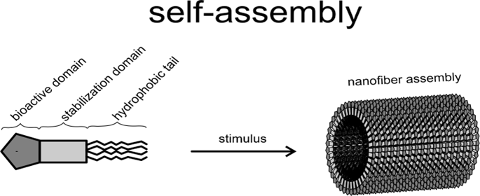
Fig.4. Fabrication of nanofiber by self-assembly
3.4. Phase separation:
In the phase separation process, a polymer is initially dissolved in a solvent before undergoing gelation. The primary mechanism driving this process is the phase separation resulting from the physical incompatibility of the components. one phase, typically the solvent, is subsequently extracted, leaving behind the other phase. the fabrication of nanofibers using the phase separation method entails the controlled manipulation of a polymer or material solution to induce a phase transition under specific conditions.
This technique involves five basic step such as,
In this method, the polymer solution is prepared and maintained at the gelation temperature, where solvent extraction takes place. The sample is then frozen and freeze dried to generate porous nanofiber scaffolds. in this method to avoid nucleation of polymer crystals at higher temperatures low gelatin temperature is required.

Fig.5. Fabrication of nanofiber by phase separation
3.5. Drawing
Drawing is a method similar to dry spinning used to produce single nanofibers of longer length(11).In the drawing process, contact is made with the sharp tio of a micropipette or a glass rod with a previously deposited polymer solution droplet. After that the micropipette or a glass rod is then withdrawn slowly, thus producing nanofibers. While drawing the micropipette or glass rod, the solvent evaporate from the liquid fibers and ultimately solid nanofibers are formed. The rate of withdrawal is based upon polymer nature. solution of polymer used for drawing is selected based on its viscoelastic properties, i.e. it should overcome the stresses developed during pulling and undergo a higher range of deformations. the solvent used to prepare the polymer solution is evaporated during the drawing process, resulting in the formation of dry, solid nanofibers.
Limitations of this process are as follows:
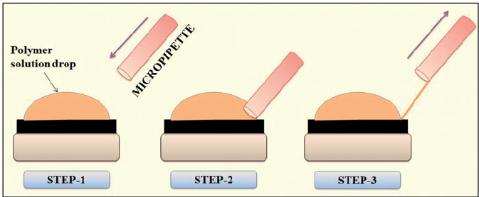
Fig.6. Fabrication Of Nanofiber by Drawing
4.Characterization methods of nanofiber:13-18
Table no. 1: Various Characterization methods used for nanofiber formulation
|
Analytical technique |
Application |
Information provided |
|
|
Morphological characterization |
Scanning electron microscopy (SEM) |
Detail imaging or characterization of the structural features and surface characteristics of nanofibers |
Surface structure, fiber diameter and alignment |
|
Diameter and size distribution of nanofiber |
Determination of fiber diameter and size distribution |
Fiber diameter, size distribution, and uniformity |
|
|
Confocal laser microscopy (CLSM) |
3D imaging of nanofiber structure |
Spatial distribution, layer thickness |
|
|
Structural analysis |
Transmission electron microscopy (TEM) |
High resolution imaging of nanofibers |
Internal structure, crystallinity and defects |
|
X-ray Diffraction |
Crystallinity and phase identification |
Crystal structure, degree of crystallinity |
|
|
Mechanical characterization |
Atomic force microscopy |
Surface topography and mechanical properties |
Surface roughness, mechanical stiffness and elasticity |
|
Dynamic mechanical analysis (DMA) |
Mechanical properties characterization |
Viscoelastic behavior, storage, and loss modulus |
|
|
Universal tensile machine |
Mechanical properties characterization |
Tensile strength and Young’s modulus |
|
|
Thermal properties |
Thermo gravimetric analysis (TGA) |
Thermal stability and composition analysis |
Decomposition temperature and compositional analysis |
|
Differential scanning colorimetry (DSC) |
Chemical transitions and stability |
Glass transition, melting point and crystallization |
|
|
Chemical characterization |
Fourier-transform infrared spectroscopy (FTIR) |
Chemical composition analysis |
Molecular structure and Functional group |
|
Raman spectroscopy |
Chemical and structural characterization |
Molecular vibrations and chemical bonding |
|
|
Nuclear magnetic resonance (NMR) |
Molecular structure and dynamic |
Chemical environment and molecular interactions |
|
|
Surface |
X-ray photoelectron spectroscopy (XPS) |
Surface chemical analysis |
Element composition, oxidation states, and functional groups |
|
Surface Plasmon resonance (SPR) |
Surface binding interactions |
Bimolecular interactions and affinity measurements |
|
|
Contact angle measurement |
Wettability characterization |
Surface energy and contact angle |
|
|
Dynamic light scattering (DLS) |
Particle size and size distribution analysis |
Hydrodynamic diameter and aggregation state |
|
5. Application of nanofiber19-30
Nanofiber is a branch of nanotechnology; it has unique properties such as a high surface area to volume ratio, high aspect ratio, and high porosity these properties enable nanofibers to serve a wide range of functions and applications in different areas, such as biomedical applications, applications in the environmental field, applications in the defense field, and applications in the chemistry field. Fig. 5 shows the application of nanofiber in different areas.
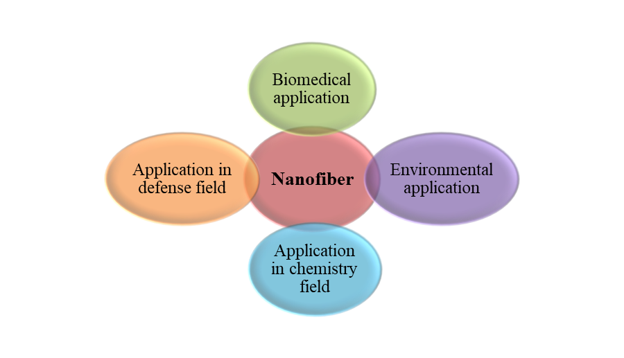
Fig.7. Application Of Nanofibers
5.1. Biomedical application 19-25
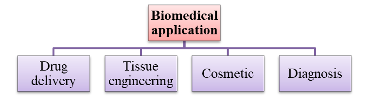
Fig.8. Biomedical Application
5.1.1 Drug delivery
Nanofibers are widely utilized for the delivery of various types of drugs, because nanofibers impart distinctive favor in the field of drug delivery by acting as biodegradability, easily fabricating at a large scale, attaining control release of drugs, extensive surface-area-to-volume ratio, etc. Various forms of controlled-release drugs are integrated into electrospun nanofibers, such as Avandia, carvedilol, hydrochlorothiazide, aspirin, naproxen, nifedipine, indomethacin, ketoprofen, etc.Depending on the nanofiber, drug delivery is classified into two types: drug loading and drug release mechanisms. Drug loading means the entrapment of a drug into the nanofiber to control the release of that drug and to provide site specificity to the drug. While the drug release mechanism is known about the demagnetization from the fiber surface, fiber diffusion through solid-state and in vivo fiber degradation.
5.1.2. Tissue engineering
Nanofibers play a crucial role in tissue engineering due to their ability to mimic the natural extracellular matrix (ECM), providing a scaffold for cell growth and tissue regeneration. tissue engineering is an interdisciplinary field that integrates cell biology, chemistry, and biomaterials to construct three-dimensional tissues that closely resemble the structure of biological membranes or the extracellular matrix, incorporating various nanofibrous matrices.this technique is used for the formation nanofiber matrices, which include polymer self-build, phase segregation, and electrospinning.Nanofibers have significantly broadened their applications in the field of tissue engineering, including bone, cartilage, tendon and ligament, neural, and cardiovascular tissue engineering, among others.
5.1.3. Wound dressing
Wound dressings are one essential type of medical device to cover an open wound to protect the wound against external risk factors while providing an appropriate microenvironment to support the wound-healing process.electrospun nanofiber materials have indeed garnered significant attention in the field of wound healing and skin regeneration due to their unique properties. The high porosity and permeability to air and moisture allow for an optimal environment that supports healing, facilitating the necessary exchange of gases and moisture. This is crucial because proper moisture levels can enhance the healing process and prevent wound dehydration or scab formation.Electrospun nanofiber materials represent highly promising candidates for wound dressing applications, offering the potential for tailored solutions that address specific requirements such as infection prevention, moisture regulation, and cellular support.
5.1.4. Cosmetics
Many preparations of nanofiber have been marketed recently as face masks; they are also used in treating skin defects or various types of skin problems and are also used as skin cleansers by adding different ingredients with different medicinal properties.these nanofiber-based face masks exhibit antioxidant properties. Prior to application, the masks are moistened, and their use promotes a healthier appearance of the skin. the silver nanoparticle-loaded nanofiber is widely used for the prevention and treatment of different diseases, such as bacterial infection, fungi, etc. nanofibers can be incorporated into hair care formulations, such as shampoos and conditioners, to optimize the delivery and deposition of active nourishing ingredients. additionally, they can form a protective film around the hair shaft, contributing to enhanced hair health, improved shine, and increased resistance to environmental damage.
5.1.5. Diagnosis
Nanofiber have demonstrated significant potential in the fields of detection, recognition, and therapeutic applications. They have been employed in a variety of devices, including ultrasonic biosensors for cancer detection, malaria diagnosis, identification of circulating tumor cells in cancer patients, and the detection of biomarkers such as glucose, cholesterol, urea, and bacteria. These biosensors offer numerous advantages, including cost-effectiveness, rapid detection, and excellent portability, making them highly effective in diagnostic and monitoring systems. Electrospun nanofibers in addition to their simplistic production also provides a range of morphological characteristics to elevate sensitivity and rapidness of the detection.The biosensor was developed by immobilizing negatively charged bacteriophages onto positively charged carbon-based nanofibers (NFs). The concentration of C-reactive protein (CRP) in a patient's blood serves as an indicator of tissue damage, infections, and inflammation.
5.2. Application of nanofiber in chemistry field26-27

Fig.9. Application In Chemistry Field
5.2.1. Catalyst and enzyme carrier
Nanofibers play a crucialrole in enhancing the catalytic activity of enzymes by significantly increasing the surface area of catalyst. it can be loading a higher number of catalysts and provide good solid support for the enzymes.they are also used as biological catalysts because of their high loading capability and large specific surface area.
5.2.2. Release control
nanofiber provides a useful tool to control the delivery of many drugs, increase patient compliance, and reduce side effect. It enables controlled and sustained drug release, preventing the occurrence of a burst release. Various types of nanofibers are utilized to regulate the delivery of a diverse array of pharmaceutical compounds. stimuli-responsive nanofibers, fabricated through electrospinning, are designed to control drug release in response to specific signals detected by the body. These signals can include thermal, pH, light, magnetic fields, electric fields, and other forms of stimuli, allowing for precise and targeted drug delivery.
5.2.3. Recovery of metal ions and membrane affinity
Water pollution remains a significant environmental challenge, with heavy metals and soluble inorganic contaminants being primary pollutants in aquatic ecosystems. These pollutants contribute to adverse effects such as premature aging of organisms and eutrophication in lakes, rivers, and ponds. To address these issues, various remediation strategies are being explored, including the use of non-adsorbents, magnetic materials, nanocomposites, and nanofibrous arrays, which are effective in removing large quantities of metals and metalloids from contaminated water sources.
5.2.4. Energy storage
The nanofiber can be used in energy storage in many ways, such as for producing carbon nanofiber network lignin combined with nanofiber. They have a very large surface area that produces great potential for nanofibers, which will be used in energy storage applications.
5.3. Application in defense field28
Fig.10. Application In Defense Field
5.3.1. Protective clothing
they are also used to define the requirements of defense systems and industrial applications through which textile industries are used to manufacture bulletproof clothing, clothes that must protect humans from external chemical or biological agents, fire, cold, heat, etc.when manufacturing protective clothing, it is essential to prioritize key factors such as lightweight construction, cost-effectiveness, and comfort. These elements are crucial to ensuring that the clothing delivers optimal performance while maintaining ease of use and wearability.
5.3.2. Sensors
According to recent research, a sensor-integrated nanofiber was developed using carbon nanotubes, resulting in a fabric that is exceptionally strong, durable, and flexible, with ballistic protection properties. This advanced sensor is incorporated into wearable materials, typically made from lightweight cotton-based fabrics, and has demonstrated bulletproof capabilities. The unique combination of carbon nanotubes' light weight, toughness, and remarkable strength contributes to the enhanced protective performance of the material.
5.4. Environmental application29-30

Fig.11. Environmental Application
5.4.1. Filtration
Nanofibers have numerous applications in the environmental field, leveraging their unique properties such as high surface area, porosity, and reactivity to address various environmental challenges.nanofiber is used to create highly efficient filtration membranes to remove contaminants, such as heavy metals, bacteria, and viruses, from water. their large surface area allows for enhanced filtration, leading to improved water quality and safety.
5.4.2. Removal of toxic waste
Nanofiber containing alumina, which helps to adsorb heavy metal from their imidate environment.This nanofiber formulated by method, which contains PVP solution (10% w/v), was prepared by using ethanol as a solvent in which PVP polymer powder was dissolved in it under constant stirring.An aluminum acetate solution was then incorporated into the preceding solution, with aluminum acetate serving as the aluminum precursor. The ratio of the precursor to the polyvinylpyrrolidone (PVP) solution was carefully controlled. The resultant alumina nanofibers were subsequently employed to adsorb chromium (VI) and fluoride ions from an aqueous environment.
CONCLUSION:
This review paper concludes that, Nanofiber represents a highly versatile class of materials with unique properties such as high surface area to volume ratio, and high porosity, enhanced strength and flexibility which make them suitable for wide range of application. Their unique properties such as, high surface area enhanced mechanical strength and the ability to mimic biological structures, make them ideal for use in field like medicine, filtration, energy storage, and environmental protection. While challenges like cost-effective production, scability, and material consistency persist, ongoing advancements in nanofiber technology and fabrication methods continue to drive innovation. With continued research and development, nanofibers are set to play a crucial role in shaping the future of various technological and medical fields, offering novel solutions to complex problems.
REFERENCES


Akshay Babhulkar, Dr. Shivappa Nagoba*, Rachita Malshette, Shripal Kolsure, Akhilesh Limaye, Nanofibers: A Comprehensive Review of Synthesis, Functionalization, and Emerging Applications, Int. J. of Pharm. Sci., 2025, Vol 3, Issue 5, 3599-3613. https://doi.org/10.5281/zenodo.15480159
 10.5281/zenodo.15480159
